. 2017 Aug 11;7(1):7913.
doi: 10.1038/s41598-017-08489-7.

Protein O-mannosylation deficiency increases LprG-associated lipoarabinomannan release by Mycobacterium tuberculosis and enhances the TLR2-associated inflammatory response

Affiliations

Protein O-mannosylation deficiency increases LprG-associated lipoarabinomannan release by Mycobacterium tuberculosis and enhances the TLR2-associated inflammatory response

Henar Alonso et al. Sci Rep. .

Abstract

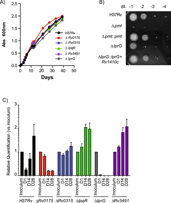
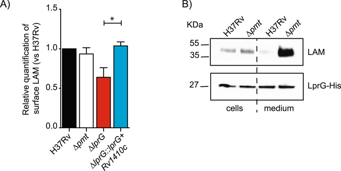
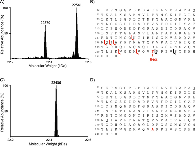
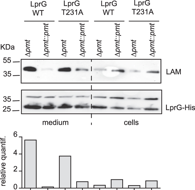
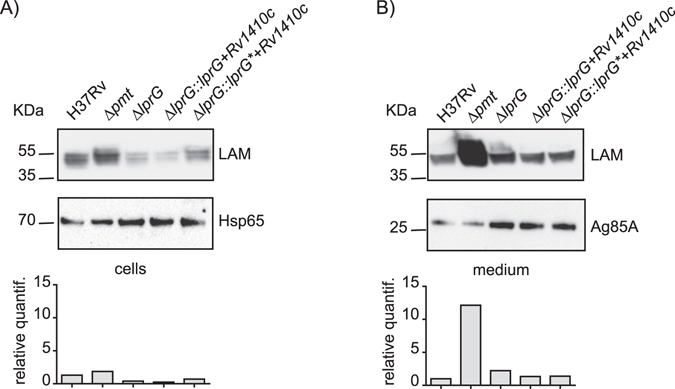
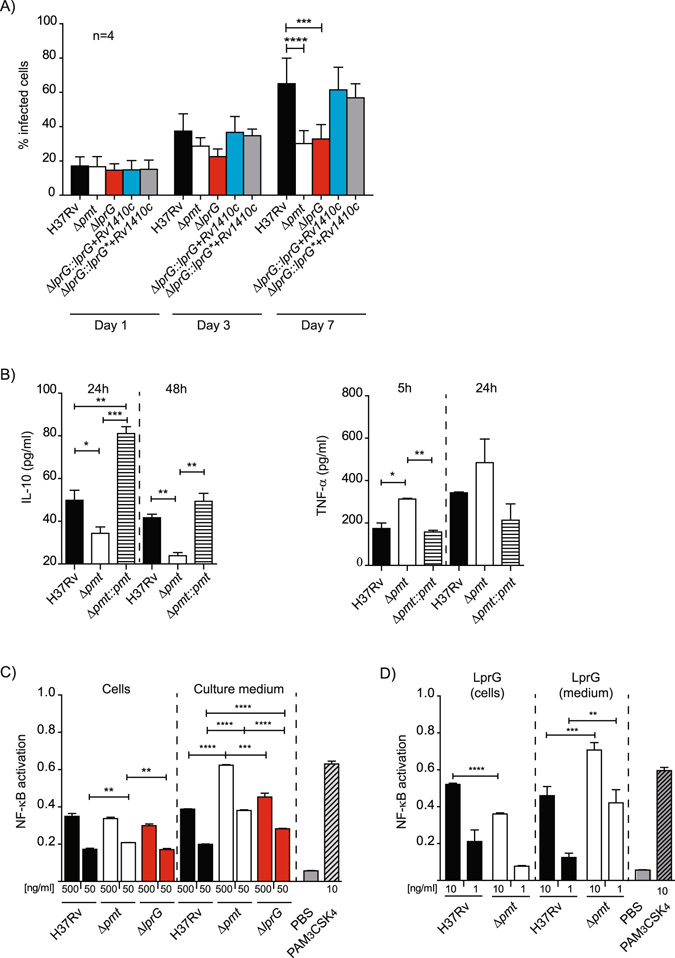
Protein O-mannosylation is crucial for the biology of Mycobacterium tuberculosis but the key mannosylated protein(s) involved and its(their) underlying function(s) remain unknown. Here, we demonstrated that the M. tuberculosis mutant (Δpmt) deficient for protein O-mannosylation exhibits enhanced release of lipoarabinomannan (LAM) in a complex with LprG, a lipoprotein required for LAM translocation to the cell surface. We determined that LprG is O-mannosylated at a unique threonine position by mass spectrometry analyses of the purified protein. However, although replacement of this amino acid by an alanine residue completely abolished LprG O-mannosylation, the increased release of the LAM/LprG complex was preserved. We found that the increased secretion of this complex is due to enhanced LAM production in the Δpmt M. tuberculosis and M. smegmatis mutants relative to their wild-type counterparts. This abnormal release of LAM/LprG has functional consequences on the induction of inflammatory responses and provides a possible explanation for the reduced virulence of the M. tuberculosis Δpmt mutant.


Conflict of interest statement

The authors declare that they have no competing interests.

Figures

Figure 1
Figure 1
Contribution of selected mannoproteins to M. tuberculosis growth in vitro and in vivo. (A) Growth of WT M. tuberculosis and mutants in liquid medium. Mycobacterial strains were inoculated at 106cfu/ml. Absorbance at 600 nm was followed over time for the various cultures. (B) Growth of WT M. tuberculosis and mutants on 7H11 Petri plates. Serial dilutions of adjusted liquid cultures of the various mutants were spotted on solid medium and incubated at 37 °C. (C) Growth of tagged-WT M. tuberculosis and tagged-mutants in BALB/c mice. Mice (5 for each time points) were infected with 6.102 cfu of a population containing similar concentration of each strains. At 1, 14 or 28 days post-infection, the bacterial population was recovered from the lung of the infected mice and grown for 7 days in liquid medium. Total DNA was extracted and the quantity of each mutant evaluated by qPCR using primers specific for the tags. The amount of each mutant relative to the inoculum is plotted.
Figure 2
Figure 2
Impact of protein O-mannosylation on LAM exposition to the bacterial cell surface and release of the LprG/LAM complex. (A) FACS analyses of LAM staining on the surface of the various M. tuberculosis strains. The bar graph values correspond to the median fluorescence value after normalization to H37Rv. Results are representative of two independent experiments. *P < 0.05. (B) Western-blot analyses of LAM associated with LprG in WT M. tuberculosis or Δpmt mutant. Similar amounts of LprG-His protein purified from the bacteria or culture medium were separated by SDS-PAGE. After transfer, the LAM or LprG-His molecules were revealed using the anti-LAM antibody CS-35 or an anti-His antibody. Blots are representative of at least three independent experiments.
Figure 3
Figure 3
MS analyses of LprG and LprG T231A glycosylation. (A) Deconvoluted ESI-HRMS spectrum of the purified recombinant LprG-His expressed in M. tuberculosis. (B) Peptide sequence of the molecular species observed in A reporting the fragment ions detected in the top-down electron transfer dissociation spectrum of the fragmentation of the 22,541 Da molecular mass ion precursor allowing the localization of the unique hexose on the T231. (C) Deconvoluted ESI-HRMS spectrum of purified recombinant LprG-His T231A expressed in M. smegmatis demonstrating the absence of glycosylation of the mutated protein. (D) Peptide sequence of the molecular species observed in C.
Figure 4
Figure 4
Impact of LprG O-mannosylation on the release of the LprG/LAM complex in M. smegmatis. Similar amounts of LprG-His or LprG-His T231A purified from either culture medium or bacterial cells were separated by SDS-PAGE, transferred onto membranes and revealed using anti-LAM or anti-His antibodies. The genetic backgrounds in which the protein was produced are indicated. The upper band revealed by the anti-His antibody is a non-specific cross-reacting protein. The signal intensity for LAM was quantified and plotted in the graph below the Western blot image. This blot is representative of three independent experiments.
Figure 5
Figure 5
Impact of protein O-mannosylation on LAM production. Crude extracts obtained from the culture medium (B) or bacterial cells (A) were separated by SDS-PAGE, transferred onto membranes, and analyzed using antibodies raised against LAM and either hsp65 (for cellular extracts) or Ag85A (for medium supernatants). The signal intensity for LAM was quantified and plotted in the graph below the western blot image. This blot is representative of two independent experiments.
Figure 6
Figure 6
Protein O-mannosylation affects the inflammatory response of infected cells. (A) Growth of WT, mutants and complemented strains in hMDM. hMDM were infected at an MOI 1 for 1 h with the indicated strains. Data are presented as the mean +/− SD of four independent experiments performed in duplicate. (B) Production of TNF-α and IL-10 by MHS cells infected by WT, Δpmt and complemented strains. MHS cells were infected at MOI 1:1 for 1 h and TNF-α and IL-10 were evaluated after 5 h or 24 h and 24 or 48 h respectively. Data are representative of two independent experiments performed in triplicate. (C) NK-κB activation in HEK-TLR2 cells incubated with crude extracts from WT and Δpmt M. tuberculosis strains. Activation of NF-κB, evaluated using SEAP activity, following incubation with 500 ng/ml or 50 ng/ml of crude extracts recovered from either bacterial cells or culture medium from the indicated M. tuberculosis strains. PBS 1× or PAM3CSK4 (10 ng/ml) were used as negative and positive controls respectively. Data are presented as the mean +/− SD of three independent experiments. (D) NF-κB activation in HEK-TLR2 cells incubated with LprG/LAM complex purified from bacterial cells or culture medium of WT or Δpmt M. tuberculosis strains. HEK-TLR2 cells were incubated with 10 ng/ml or 1 ng/ml of LprG-His and co-purified LAM. SEAP activity was quantified after 24 h. PBS 1× or PAM3CSK4 (10 ng/ml) were used as negative and positive controls respectively. Data are presented as the mean +/− SD of at least five independent experiments. *P < 0.05; **P < 0.01; ***P < 0.001; ****P < 0.0001.

References

    1. Cain JA, Solis N, Cordwell SJ. Beyond gene expression: the impact of protein post-translational modifications in bacteria. J. Proteomics. 2014;97:265–286. doi: 10.1016/j.jprot.2013.08.012. - DOI - PubMed
    1. Garbe T, et al. Expression of the Mycobacterium tuberculosis 19-kilodalton antigen in Mycobacterium smegmatis: immunological analysis and evidence of glycosylation. Infect. Immun. 1993;61:260–267. - PMC - PubMed
    1. Espitia C, Mancilla R. Identification, isolation and partial characterization of Mycobacterium tuberculosis glycoprotein antigens. Clin. Exp. Immunol. 1989;77:378–383. - PMC - PubMed
    1. Dobos KM, Swiderek K, Khoo K-H, Brennan PJ, Belisle JT. Evidence for glyosylation sites on the 45-kilodalton glycoprotein of Mycobacterium tuberculosis. Infect. Immun. 1995;63:2846–2853. - PMC - PubMed
    1. Herrmann JL, O’Gaora P, Gallagher A, Thole JER, Young DB. Bacterial glycoproteins: a link between glycosylation and proteolytic cleavage of a 19kDa antigen from Mycobacterium tuberculosis. EMBO J. 1996;15:3547–3554. - PMC - PubMed

Publication types

MeSH terms

LinkOut - more resources