Review
. 2017 Aug 10;170(4):605-635.
doi: 10.1016/j.cell.2017.07.029.

The PI3K Pathway in Human Disease

Affiliations
Review

The PI3K Pathway in Human Disease

David A Fruman et al. Cell. .

Abstract

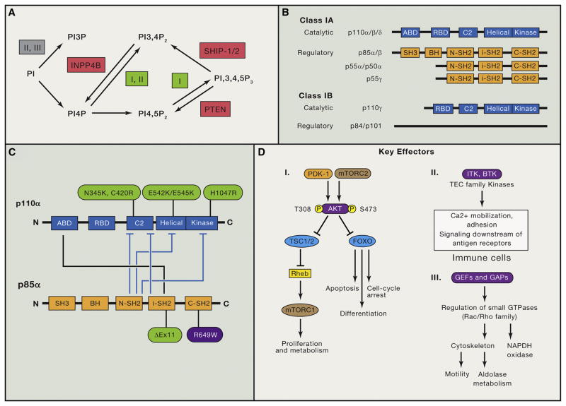
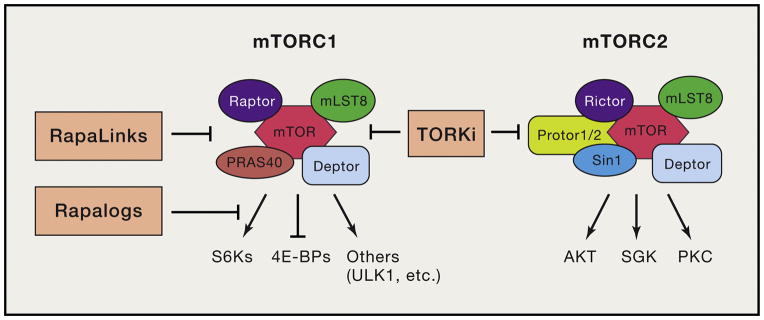
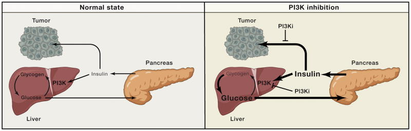
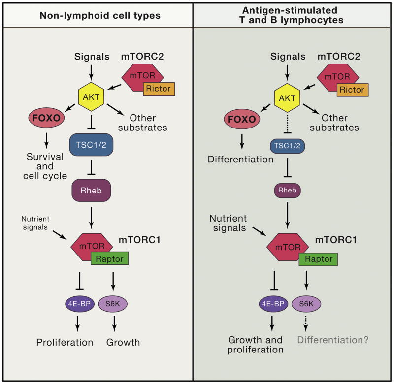
Phosphoinositide 3-kinase (PI3K) activity is stimulated by diverse oncogenes and growth factor receptors, and elevated PI3K signaling is considered a hallmark of cancer. Many PI3K pathway-targeted therapies have been tested in oncology trials, resulting in regulatory approval of one isoform-selective inhibitor (idelalisib) for treatment of certain blood cancers and a variety of other agents at different stages of development. In parallel to PI3K research by cancer biologists, investigations in other fields have uncovered exciting and often unpredicted roles for PI3K catalytic and regulatory subunits in normal cell function and in disease. Many of these functions impinge upon oncology by influencing the efficacy and toxicity of PI3K-targeted therapies. Here we provide a perspective on the roles of class I PI3Ks in the regulation of cellular metabolism and in immune system functions, two topics closely intertwined with cancer biology. We also discuss recent progress developing PI3K-targeted therapies for treatment of cancer and other diseases.


Figures

Figure 1
Figure 1. Overview of Phosphoinositides, Class I PI3K Protein Isoforms, p110α Activity Regulation, and PI3K Downstream Effectors
(A) Schematic overview of the major synthesis and degradation pathways for PtdIns-3-P (PI3P), PtdIns-3,4-P2 and PtdIns-3,4,5-P3. The classes of PI3K (I, II, or III) that mediate reactions are indicated. Lipid phosphatases are in red. INPP4B, inositol polyphosphate-4-phosphatase, type II. (B) Domain structure of class I PI3K catalytic and regulatory subunits. ABD, adaptor-binding domain; RBD, Ras-binding domain; BH, breakpoint cluster region homology. (C) Diagram of the intramolecular interactions between class IA catalytic and regulatory subunits (p110α and p85α are displayed as well studied examples). Tight binding of the ABD to iSH2 confers stability to p110α. The other contacts shown in blue block arrows diminish basal activity and are relieved upon regulatory subunits binding to pTyr. Cancer-associated activating mutations are shown in green. SHORT syndrome mutation in p85α (R649W) is in purple. (D) Brief summary of key PI3K effectors: PDK-1, AKT, TEC family kinases, and GEFs/GAPs for small GTPases. AKT has many other important substrates not shown here (Manning and Toker, 2017). The specific GEFs that mediate PI3K-dependent Rac activation to promote motility and aldolase release are not known.
Figure 2
Figure 2. Overview of mTORC1 and mTORC2 complexes, key substrates, and inhibitors
The processes inhibited by different classes of mTOR inhibitor are shown. First generation rapalogs are partial inhibitors of mTORC1 that inhibit phosphorylation of S6Ks more than 4E-BP1. Second generation TORKi fully inhibit mTORC1 and mTORC2. Third generation RapaLinks fully but selectively inhibit mTORC1, and also overcome single resistance mutations to rapalogs and TORKi.
Figure 3
Figure 3. Cartoon of systemic glucose homeostasis in the normal state (left) and upon PI3K inhibitor treatment (right)
In the normal state blood glucose levels are maintained in homeostasis through the actions of insulin, which stimulates glucose uptake and glycogen storage thereby keeping the system balanced. Changes in blood glucose levels (such as increases upon eating) stimulate commensurate changes in insulin release which drive either increased glucose uptake (when insulin levels are high) or gluconeogenesis (when insulin levels are low). When PI3K inhibitors are used they perturb insulin signaling in cells, thereby pushing the systemic balance to favor glucose release. This causes blood glucose levels to acutely increase, which in turn signals to the pancreas to release a bolus of insulin. As indicated by the cartoon these high insulin levels have the potential to reactivate insulin signaling both in metabolic tissues, which is critical in order for the system to come back to homeostasis, as well as in tumors, where insulin has the potential to reactivate PI3K signaling thereby undercutting the efficacy of the PI3K inhibitors.
Figure 4
Figure 4. Distinct wiring of PI3K/mTOR network in lymphocytes (T and B cells) compared to other commonly studied non-lymphoid cell types such as fibroblasts
In lymphocytes, FOXO transcription factors have prominent roles in differentiation. mTORC1 is tightly coupled to nutrient access and often uncoupled from PI3K/AKT activity. Downstream of mTORC1, the 4E-BP/eIF4E axis controls both growth and proliferation whereas in other cell types, S6Ks are crucial for cell growth. S6Ks might contribute to lymphocyte differentiation but this is unproven.
Figure 5
Figure 5. Asymmetric partitioning of PI3K/mTOR signaling during initial division of activated T and B cells results in distinct cell fates of daughter cells
This figure illustrates that the first division of activated lymphocytes produces two cells with differential levels of PI3K/mTOR signaling, which in turn drive distinct metabolic and differentiation programs. Top: CD8 T cells. Bottom: B cells.
Figure 6
Figure 6. Mechanisms of Adaptive, Primary, and Acquired Resistance to PI3K/mTOR Pathway Inhibitors
Adaptive resistance (in green, also labeled “1”) often involves upregulation of upstream regulators, including RTKs (HER3, INSR, IGF-1R), IRS-1, JAK2, or SRC by disruption of negative-feedback loops. Primary or acquired resistance (in red, “2”) can arise by expression or activation of kinases with downstream targets in common with AKT or mTORC1 (SGK1, PDK1, PIM1), constitutive activation of mTORC1 signaling (e.g., due to loss of TSC function), or loss of PTEN expression. Primary or acquired resistance (in orange, “3”) can also arise by PI3K isoform switching; selective inhibition of p110α can lead to substitution by p110β or vice versa. Finally, primary or acquired resistance can also arise by activation of heterologous pathways leading to common endpoints; for instance, MYC-dependent transcriptional activation or ERK activity (in pink, “4”).

References

    1. Adams DM, Trenor CC, Hammill AM, Vinks AA, Patel MN, Chaudry G, Wentzel MS, Mobberley-Schuman PS, Campbell LM, Brookbank C, et al. Efficacy and safety of sirolimus in the treatment of complicated vascular anomalies. Pediatrics. 2016;137:e20153257. - PMC - PubMed
    1. Ahmad S, Abu-Eid R, Shrimali R, Webb M, Verma V, Doroodchi A, Berrong Z, Samara R, Rodriguez PC, Mkrtichyan M, et al. Differential PI3Kδ Signaling in CD4(+) T-cell Subsets Enables Selective Targeting of T Regulatory Cells to Enhance Cancer Immunotherapy. Cancer Res. 2017;77:1892–1904. - PubMed
    1. Aksoy E, Taboubi S, Torres D, Delbauve S, Hachani A, Whitehead MA, Pearce WP, Berenjeno IM, Nock G, Filloux A, et al. The p110δ isoform of the kinase PI(3)K controls the subcellular compartmentalization of TLR4 signaling and protects from endotoxic shock. Nat Immunol. 2012;13:1045–1054. - PMC - PubMed
    1. Ali K, Bilancio A, Thomas M, Pearce W, Gilfillan AM, Tkaczyk C, Kuehn N, Gray A, Giddings J, Peskett E, et al. Essential role for the p110delta phosphoinositide 3-kinase in the allergic response. Nature. 2004;431:1007–1011. - PubMed
    1. Ali K, Soond DR, Piñeiro R, Hagemann T, Pearce W, Lim EL, Bouabe H, Scudamore CL, Hancox T, Maecker H, et al. Inactivation of PI(3)K p110δ breaks regulatory T-cell-mediated immune tolerance to cancer. Nature. 2014;510:407–411. - PMC - PubMed

Substances