

Variant histology, IgD and CD30 expression in low-risk pediatric nodular lymphocyte predominant Hodgkin lymphoma: A report from the Children's Oncology Group
- PMID: 28802087
- PMCID: PMC5699946
- DOI: 10.1002/pbc.26753
Variant histology, IgD and CD30 expression in low-risk pediatric nodular lymphocyte predominant Hodgkin lymphoma: A report from the Children's Oncology Group
Abstract
Background: Histologic prognostic factors have been described for nodular lymphocyte predominant Hodgkin lymphoma (NLPHL). This study examines histologic and immunophenotypic variants in a clinical trial for pediatric NLPHL.
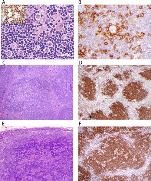
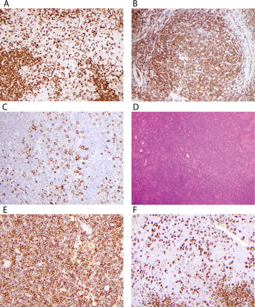
Procedure: One hundred sixty-eight cases of localized NLPHL were examined for histologic variants, CD30 and immunoglobulin D (IgD) expression, and outcome. Histologic types were scored categorically as 0 = 0, 1 ≤ 25%, and 2 > 25% of the sample.
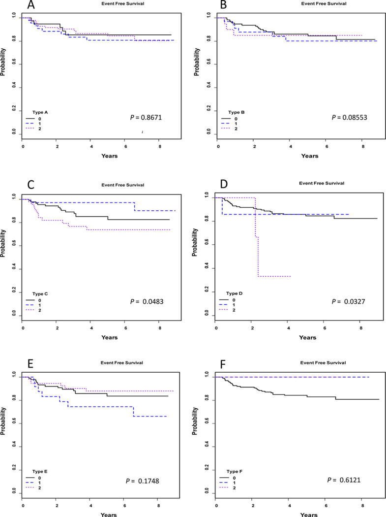
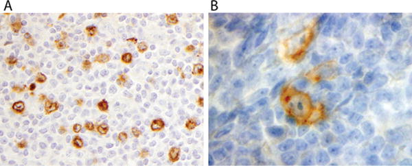
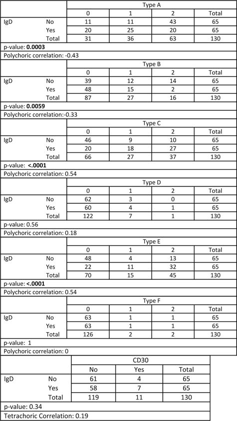
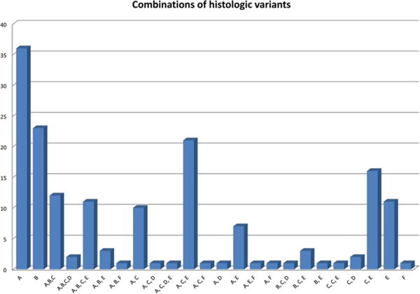
Results: Fifty-eight (35.1%) cases showed only typical nodular with or without serpiginous histology (types A and B). The remainder showed mixtures of histologies. The numbers of patients with score 2 are 85 (50.6%) type A, 21 (12.5%) type B, 46 (27.4%) with extranodular large B cells (type C), 3 with T-cell-rich nodular pattern (type D), 55 (32.7%) with diffuse T-cell-rich (type E) pattern, and 2 (1.2%) with diffuse B-cell pattern (type F). Higher level of types C (P = 0.048) and D (P = 0.033) resulted in lower event-free survival (EFS). Cytoplasmic IgD was found in 65 of 130 tested (50%), did not significantly associate with EFS but positively correlated with types C and E histology (P < 0.0001) and negatively correlated with types A (P = 0.0003) and B (P = 0.006). Seventeen (10%) expressed CD30, with no adverse effect.
Conclusions: Variant histology is common in pediatric NLPHL, especially types C and E, which are associated with IgD expression. Type C variant histology and possibly type D are associated with decreased EFS, but neither IgD nor CD30 are adverse features. Variant histology may warrant increased surveillance, but did not affect overall survival.
Keywords: Hodgkin; children; histology; lymphoma; pathology.
© 2017 Wiley Periodicals, Inc.
Conflict of interest statement
The authors declare that there is no conflict of interest.
Figures
References
-
- Swerdlow SH, Campo E, Harris NL, et al., editors. WHO Classification of Tumours of Haematopoietic and Lymphoid Tissues. IARC; Lyon: 2008.
-
- Anagnostopoulos I, Hansmann ML, Franssila K, et al. European Task Force on Lymphoma project on lymphocyte predominance Hodgkin disease: histologic and immunohistologic analysis of submitted cases reveals 2 types of Hodgkin disease with a nodular growth pattern and abundant lymphocytes. Blood. 2000;96(5):1889–1899. - PubMed
-
- Nicholas DS, Harris S, Wright DH. Lymphocyte predominance Hodgkin’s disease—an immunohistochemical study. Histopathology. 1990;16(2):157–165. - PubMed
-
- Poppema S, Delsol G, Pileri SA, et al. Nodular lymphocyte predominant Hodgkin lymphoma. In: Swerdlow SH, Campo E, Harris NL, et al., editors. WHO Classification of Tumours of Haematopoietic and Lymphoid Tissues. IARC; Lyon: 2008. pp. 323–325.
Publication types
MeSH terms
Substances
Grants and funding
LinkOut - more resources
Full Text Sources
Other Literature Sources
Medical
