Review
. 2017 Sep;37(3):527-546.
doi: 10.1016/j.cll.2017.06.006.

Cutaneous T-cell Lymphoma

Affiliations
Review

Cutaneous T-cell Lymphoma

Melissa Pulitzer. Clin Lab Med. 2017 Sep.

Abstract

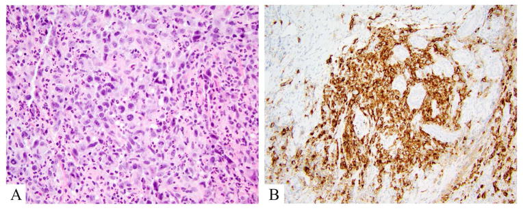
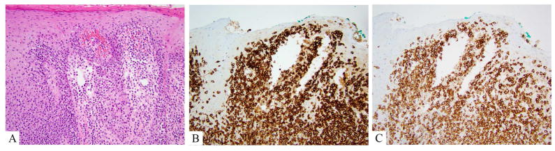
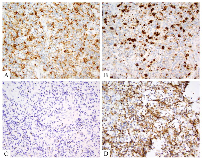
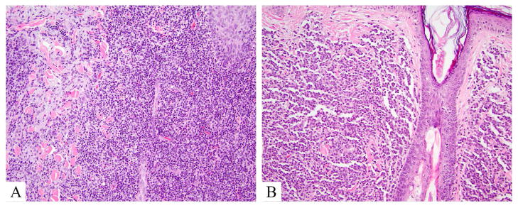
Cutaneous T-cell lymphomas comprise a heterogeneous group of diseases characterized by monoclonal proliferations of T lymphocytes primarily involving skin, modified skin appendages, and some mucosal sites. This article addresses the basic clinical, histologic, and immunohistochemical characteristics of this group of diseases, with additional attention to evolving literature on dermoscopy, reflectance confocal microscopy, flow cytometry, and molecular data that may increasingly be applied to diagnostic and therapeutic algorithms in these diseases. Select unusual phenotypes or diagnostic examples of classic phenotypes are demonstrated, and flags for consideration while making a pathologic diagnosis of cutaneous T-cell lymphoma are suggested.

Keywords: Anaplastic large cell lymphoma; Cutaneous lymphoma; Flow cytometry; Lymphomatoid papulosis; Mycosis fungoides; Reflective confocal microscopy; Sézary syndrome.


Figures

Figure 1
Figure 1
Mycosis fungoides, hematoxylin and eosin A. Patch lesion showing tagging of hyperchromatic lymphocytes out of proportion to spongiosis in a hyperkeratotic epidermis. B. Plaque lesion; hyperchromatic lymphocytes with cerebriform atypia are present in the epidermis as well as in the superficial dermis. C. Tumor lesion; lymphocytes mostly spare the epidermis and are present in sheets extending throughout the dermis, obscuring adnexal (hair) structures).
Figure 2
Figure 2
Mycosis fungoides, classic, immunohistochemistry. A. CD3 labels numerous lymphocytes extending into the mid and upper levels of the epidermis and tagging the dermoepidermal junction. B. CD7 shows complete negativity in the CD3+ cells. C. CD4 highlights the same population labeled by CD3. D. CD8 shows a rare scattered lymphocyte in the epidermis, but more notably highlights scattered reactive dermal cells.
Figure 3
Figure 3
Mycosis fungoides, aberrant gamma delta phenotype. A. Hematoxylin and eosin shows hyperchromatic atypical lymphocytes tagging the dermoepidermal junction, in mid layers of the epidermis and within papillary dermis. B. TCR beta is negative in the junctional lymphocytes, but positive in some small reactive papillary dermal lymphocytes. C. TCR gamma labels junctional lymphocytes, as does D, TCR delta
Figure 4
Figure 4
Tumor stage mycosis fungoides, showing large cell transformation. A, B. Hematoxylin and eosin show large Pautrier’s microabscesses comprised of cells that are 3–4x larger than intervening small reactive lymphocytes. Similar cells efface the dermis in sheets and large clusters. C. Ki-67 proliferation index is high in these lesion, probably 85% in this case. D. Only a rare CD30+ lymphocytes is noted, underscoring the oft absence of CD30 in cases of transformed mycosis fungoides.
Figure 5
Figure 5
Anaplastic large cell lymphoma, DUSP22 rearranged. A. Hematoxylin and eosin shows a mixed large and small cell population. Large cells are atypical with kidney bean shaped and donut shaped nuclei. B. CD30 labels >75% of lesional (large) atypical lymphocytes.
Figure 6
Figure 6
CD8+ aggressive epidermotropic T-cell lymphoma. A. Hematoxylin and eosin showing a florid front of monomorphic large atypical lymphocytes effacing the dermo-epidermal junction and involving the underlying dermis. B. CD7 is retained, strong and diffuse. C. CD8 is typically strong and diffuse.
Figure 7
Figure 7
Gamma delta T-cell lymphoma, immunohistochemistry. A, B, C. CD4, CD8, and TCR beta are negative in the majority of lesional lymphocytes. D. TCR delta is strongly and diffusely positive in lesional lymphocytes.
Figure 8
Figure 8
Indolent lymphoproliferative disorders, hematoxylin and eosin. A. CD4+ small/medium T-cell lymphoproliferative disorder shows a mixed cell morphology with histiocytes and variably sized lymphocytes, effacing epithelial structures. B. Primary cutaneous CD8+ acral lymphoma demonstrates sparing of epithelial structures by a monomorphic small cell population in dermis.

References

    1. Willemze R, et al. WHO-EORTC classification for cutaneous lymphomas. Blood. 2005;105(10):3768–85. - PubMed
    1. Swerdlow SH, EC, Harris NL, Jaffe ES. WHO Classification of Tumours of Haematopoietic and Lymphoid Tissues. 4. Lyon, France: IARC Press; 2008.
    1. Swerdlow SH, et al. The 2016 revision of the World Health Organization classification of lymphoid neoplasms. Blood. 2016;127(20):2375–90. - PMC - PubMed
    1. Beltraminelli H, et al. Primary cutaneous CD4+ small-/medium-sized pleomorphic T-cell lymphoma: a cutaneous nodular proliferation of pleomorphic T lymphocytes of undetermined significance? A study of 136 cases. Am J Dermatopathol. 2009;31(4):317–22. - PubMed
    1. Clark RA. Skin-resident T cells: the ups and downs of on site immunity. J Invest Dermatol. 2010;130(2):362–70. - PMC - PubMed