Review
. 2017 Nov:207:48-58.
doi: 10.1016/j.autneu.2017.07.008. Epub 2017 Jul 29.

Cardiac neuroanatomy - Imaging nerves to define functional control

Affiliations
Review

Cardiac neuroanatomy - Imaging nerves to define functional control

Peter Hanna et al. Auton Neurosci. 2017 Nov.

Abstract

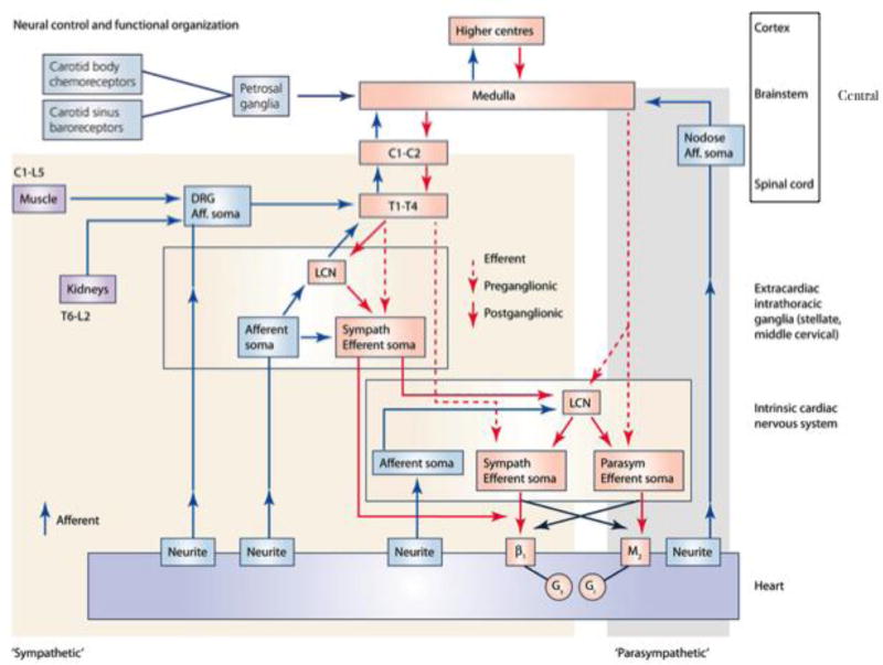
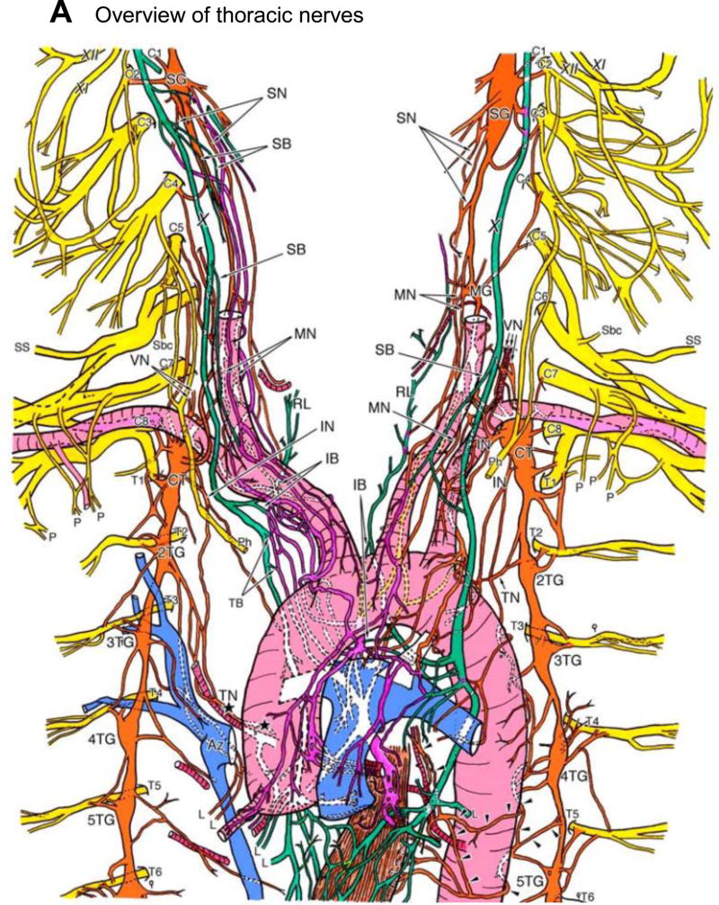
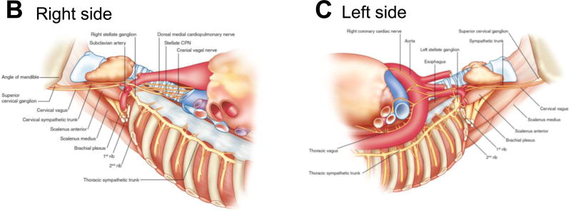
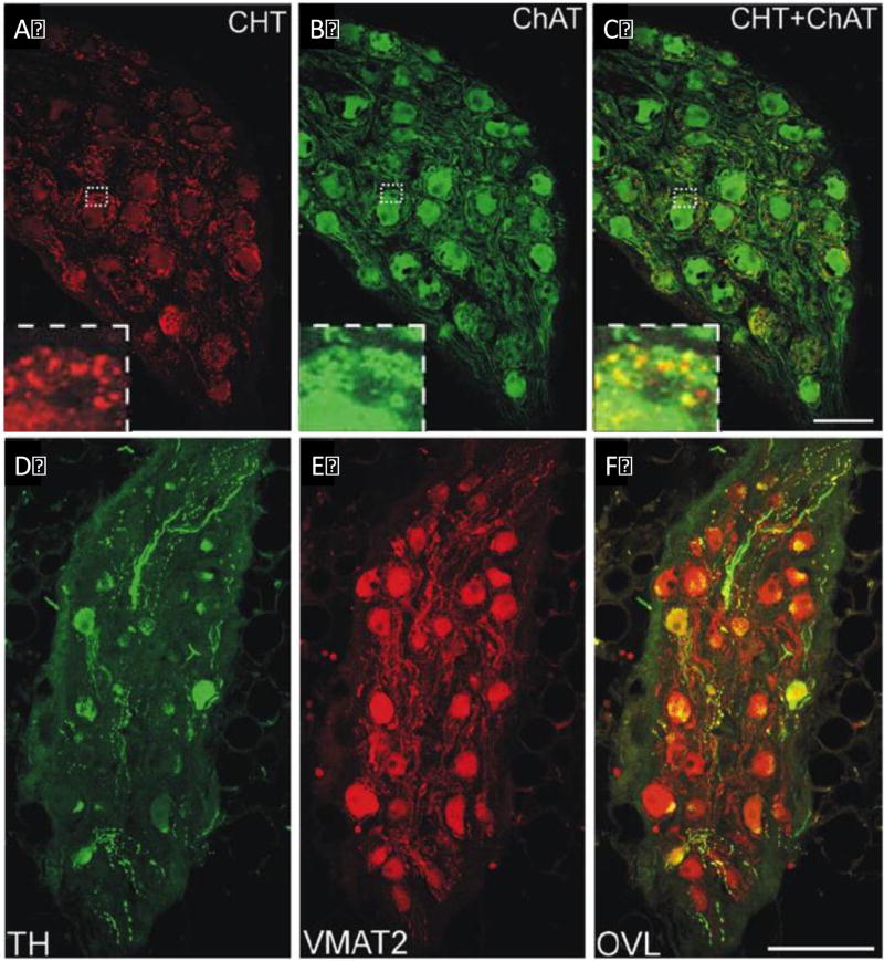
The autonomic nervous system regulates normal cardiovascular function and plays a critical role in the pathophysiology of cardiovascular disease. Further understanding of the interplay between the autonomic nervous system and cardiovascular system holds promise for the development of neuroscience-based cardiovascular therapeutics. To this end, techniques to image myocardial innervation will help provide a basis for understanding the fundamental underpinnings of cardiac neural control. In this review, we detail the evolution of gross and microscopic anatomical studies for functional mapping of cardiac neuroanatomy.

Keywords: Autonomic nervous system; Intracardiac nervous system; Myocardial innervation; Neurocardiology.


Figures

Figure 1
Figure 1
Neural control of the heart. Autonomic control of the heart is comprised of a series of nested feedback loops, from the intracardiac neurons (level 1) to the intrathoracic extra-cardiac neurons (levels 2) and the central nervous system (level 3). SG, sympathetic ganglion; DRG, dorsal root ganglion; ICNS, intracardiac nervous system. Adapted from Janig.
Figure 2
Figure 2
Neurohumoral control and functional organization of cardiac innervation. Aff, afferent; β1, β-adrenergic receptor; C, cervical; DRG, dorsal root ganglion; Gi, inhibitory G-protein; Gs, stimulatory G-protein; L, lumbar; LCN, local circuit neuron; M2, muscarinic receptor; T, thoracic. Adapted from Shivkumar et al.
Figure 3
Figure 3
A, Detailed illustration of the course of the autonomic cardiac nerves. The innervation of the heart via the right thoracic cardiac nerve and the inferior cardiac nerve is shown by the black stars. The arrowheads highlight the course of the left thoracic cardiac nerve. The sympathetic cardiac nerves, vagal cardiac branches, and the cardiac plexuses are colored in orange, green and purple, respectively. AI, anterior interventricular branch; Ao, aorta; Az, azygos vein; CB, circumflex branch; CC, common carotid artery; CT, cervicothoracic (stellate) ganglion; GV, great cardiac vein; IB, inferior (vagal) cardiac branch; IG, inferior cervical ganglion; IN, inferior cervical cardiac nerve; L, lung; LA, left atrium; LCA, left coronary artery; MG, middle cervical ganglion; MN, middle cardiac nerve; P, pectoral nerve; Ph, phrenic nerve; PT, pulmonary trunk; RA, right atrium; RCA, right coronary artery; RL, recurrent laryngeal nerve of vagus nerve; SB, superior (vagal) cardiac branch; Sbc, nerve to subclavian muscle; SG, superior cervical ganglion; SN, superior cardiac nerve; SS, suprascapular nerve; SVC, superior vena cava; TB, thoracic (vagal) cardiac branch; TG, thoracic ganglia; TN, thoracic cardiac nerve; VG, vertebral ganglion; VN, vertebral nerve; X, vagus nerve; XI, accessory nerve; XII, hypoglossal nerve. Adapted from Kawashima. Course of the right (B) and left (C) sympathetic trunks, vagus nerve and their major branches in an embalmed cadaver. Adapted from Janes et al. and Dilsizian and Narula.,
Figure 3
Figure 3
A, Detailed illustration of the course of the autonomic cardiac nerves. The innervation of the heart via the right thoracic cardiac nerve and the inferior cardiac nerve is shown by the black stars. The arrowheads highlight the course of the left thoracic cardiac nerve. The sympathetic cardiac nerves, vagal cardiac branches, and the cardiac plexuses are colored in orange, green and purple, respectively. AI, anterior interventricular branch; Ao, aorta; Az, azygos vein; CB, circumflex branch; CC, common carotid artery; CT, cervicothoracic (stellate) ganglion; GV, great cardiac vein; IB, inferior (vagal) cardiac branch; IG, inferior cervical ganglion; IN, inferior cervical cardiac nerve; L, lung; LA, left atrium; LCA, left coronary artery; MG, middle cervical ganglion; MN, middle cardiac nerve; P, pectoral nerve; Ph, phrenic nerve; PT, pulmonary trunk; RA, right atrium; RCA, right coronary artery; RL, recurrent laryngeal nerve of vagus nerve; SB, superior (vagal) cardiac branch; Sbc, nerve to subclavian muscle; SG, superior cervical ganglion; SN, superior cardiac nerve; SS, suprascapular nerve; SVC, superior vena cava; TB, thoracic (vagal) cardiac branch; TG, thoracic ganglia; TN, thoracic cardiac nerve; VG, vertebral ganglion; VN, vertebral nerve; X, vagus nerve; XI, accessory nerve; XII, hypoglossal nerve. Adapted from Kawashima. Course of the right (B) and left (C) sympathetic trunks, vagus nerve and their major branches in an embalmed cadaver. Adapted from Janes et al. and Dilsizian and Narula.,
Figure 4
Figure 4
A–B, Distribution of the intrinsic cardiac ganglionated plexuses (GP) in the heart. A, posterior; B, superior. Adapted from Dilsizian and Narula. C–E, Light photomicrographs of human intrinsic cardiac nerves, ganglia and neurons. C, Network of ganglia and nerves stained with methylene blue and dissected from the posteromedial left atrial ganglionated plexus. The ganglia appear as expansions along a nerve (box). D, Enlargement of boxed area in A illustrating a ganglion composed of approximately 150–200 nerve cell bodies. E, High-magnification micrograph of a multipolar neuron. Note the accumulation of lipofuscin granules (arrowheads). Scale bars 52.5mm in C, 250µm in D, 25µm in E. Adapted from Armour et al.
Figure 5
Figure 5
Confocal images of human intrinsic cardiac neurons expressing the cholinergic and adrenergic phenotypes. A–C, Confocal images of a ganglion that was double labeled to show CHT (A) and ChAT (B). CHT immunoreactivity (A, red) was prominent in varicose nerve fibers around intrinsic cardiac neurons and faint or absent in the neuronal cell bodies. In contrast, staining for ChAT (B, green) was notable in neuronal cell bodies and generally less intense in surrounding nerve processes. (C) Colocalization of CHT and ChAT (yellow) was noted in some cell bodies and nerve processes in the overlay image (CHT+ChAT). Inserts at lower left show boxed areas at higher magnification. All panels contain maximum projection images compiled from confocal scans that spanned 8µm. Scale bar = 100µm in A–C. D–F Confocal images of a section that was double labeled to show TH (D, green) and VMAT2 (E, red). (D) Few neurons and nerve fibers stained for TH. (B) Prominent staining for VMAT2 occurred in most neurons and many nerves fibers. (C) Overlap (OVL) of TH and VMAT2 images shows that a significant amount of the TH colocalizes (yellow) with VMAT2. All panels contain maximum projection images compiled from confocal scans that spanned 10µm. Scale bar = 150µm. Adapted from Hoover et al.
Figure 6
Figure 6
Confocal image of dorsal surface of mouse heart stained with pan-neuronal marker protein gene product 9.5 (PGP9.5) and demonstrating the rich innervation of the heart. CS, coronary sinus; LAA, left atrial appendage; LV, left ventricle; RAA, right atrial appendage; RV, right ventricle.
Figure 7
Figure 7
A, Activation recovery interval (ARI) maps at baseline and during right (RVNS) and left (LVNS) vagal nerve stimulation in a normal porcine heart. No significant regional differences in responses were found. Adapted from Yamakawa et al. B, ARI maps in control porcine hearts at baseline (BL) and during right (RSG), left (LSG) and bilateral (BSG) stellate ganglion stimulation. Myocardial regions are displayed in the BL control map. Adapted from Ajijola et al.
None
Effects of blue light photostimulation on heart rate and contractile force in Langendorff-perfused mouse hearts. A, Stability of force and heart rate shown over 15 minutes (top) and expressed as percent change (bottom, n = 8). B, Percent change from control phase after stimulating with blue light (grey, n = 10), administering isoproterenol (black, n = 5), or stimulating with blue light after administering propranolol (white, n = 5). C, Changes in force and heart rate after two rounds of blue light photostimulation (*) and addition of isoproterenol (+). D, Changes in force and heart rate after several rounds of blue light photostimulation (*) and response to photostimulation after administration of propranolol (+). **P<0.05 statistically different form baseline. Adapted from Wengrowski et al.

References

    1. Jänig W. Integrative Action of the Autonomic Nervous System: Neurobiology of Homeostasis. Cambridge University Press; 2008.
    1. Barron HV, Lesh MD. Autonomic nervous system and sudden cardiac death. J. Am. Coll. Cardiol. 1996;27:1053–1060. - PubMed
    1. Nolan J, et al. Prospective study of heart rate variability and mortality in chronic heart failure: results of the United Kingdom heart failure evaluation and assessment of risk trial (UK-heart) Circulation. 1998;98:1510–1516. - PubMed
    1. La Rovere MT, Bigger JT, Marcus FI, Mortara A, Schwartz PJ. Baroreflex sensitivity and heart-rate variability in prediction of total cardiac mortality after myocardial infarction. ATRAMI (Autonomic Tone and Reflexes After Myocardial Infarction) Investigators. Lancet Lond. Engl. 1998;351:478–484. - PubMed
    1. Floras JS. Sympathetic activation in human heart failure: diverse mechanisms, therapeutic opportunities. Acta Physiol. Scand. 2003;177:391–398. - PubMed

MeSH terms