Review
. 2017 Nov;74(21):3913-3925.
doi: 10.1007/s00018-017-2617-7. Epub 2017 Aug 12.

Melatonin, mitochondria, and the skin

Affiliations
Review

Melatonin, mitochondria, and the skin

Andrzej T Slominski et al. Cell Mol Life Sci. 2017 Nov.

Abstract

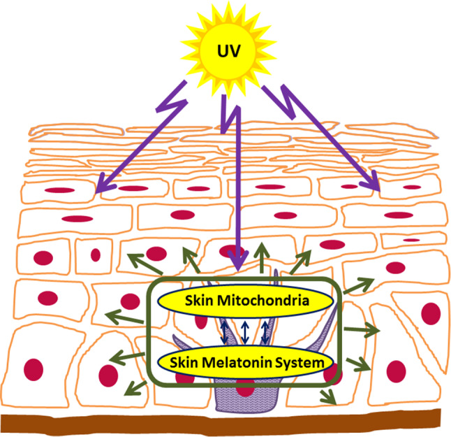
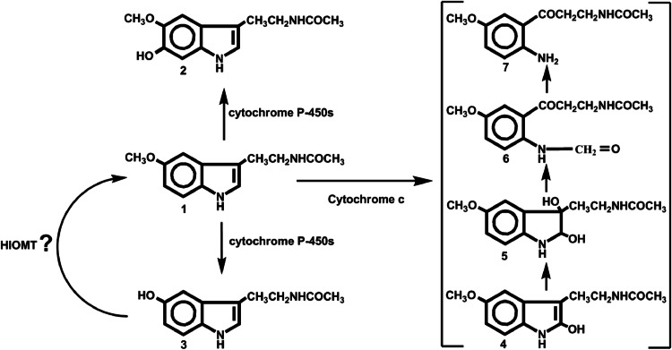
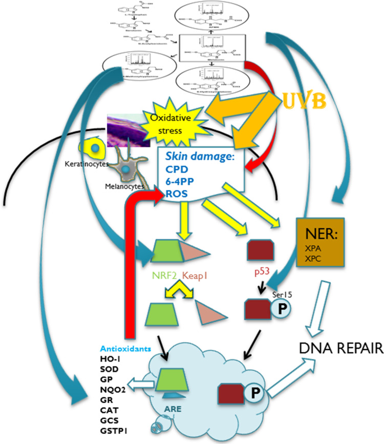
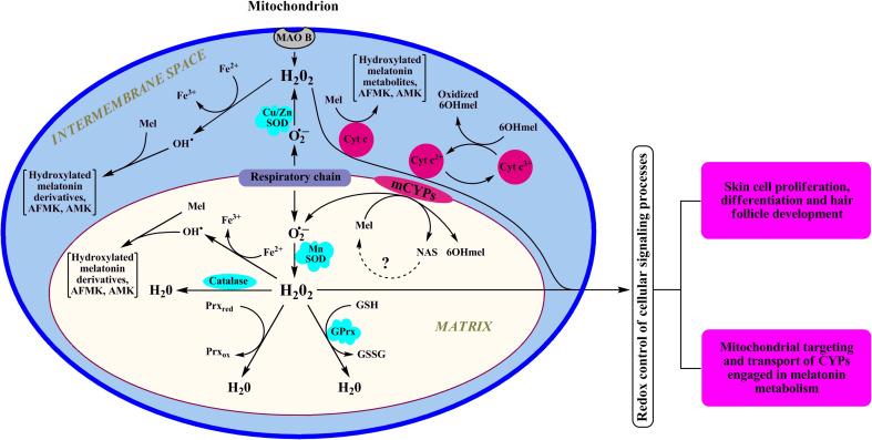
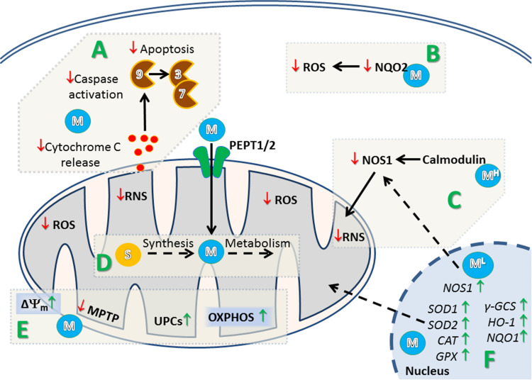
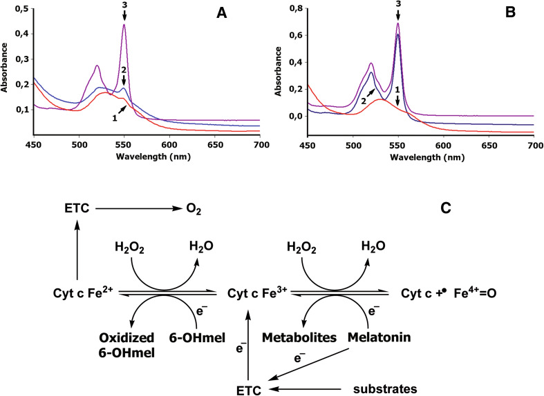
The skin being a protective barrier between external and internal (body) environments has the sensory and adaptive capacity to maintain local and global body homeostasis in response to noxious factors. An important part of the skin response to stress is its ability for melatonin synthesis and subsequent metabolism through the indolic and kynuric pathways. Indeed, melatonin and its metabolites have emerged as indispensable for physiological skin functions and for effective protection of a cutaneous homeostasis from hostile environmental factors. Moreover, they attenuate the pathological processes including carcinogenesis and other hyperproliferative/inflammatory conditions. Interestingly, mitochondria appear to be a central hub of melatonin metabolism in the skin cells. Furthermore, substantial evidence has accumulated on the protective role of the melatonin against ultraviolet radiation and the attendant mitochondrial dysfunction. Melatonin and its metabolites appear to have a modulatory impact on mitochondrion redox and bioenergetic homeostasis, as well as the anti-apoptotic effects. Of note, some metabolites exhibit even greater impact than melatonin alone. Herein, we emphasize that melatonin-mitochondria axis would control integumental functions designed to protect local and perhaps global homeostasis. Given the phylogenetic origin and primordial actions of melatonin, we propose that the melatonin-related mitochondrial functions represent an evolutionary conserved mechanism involved in cellular adaptive response to skin injury and repair.

Keywords: Homeostasis; Melatonin; Mitochondria; Photoprotection; Skin.


Figures

Fig. 1
Fig. 1
Skin melatonin–mitochondria axis counteracts the damage inflicted by solar radiation
Fig. 2
Fig. 2
Melatonin metabolism in mitochondria. 1 melatonin; 2 6-hydroxymelatonin; 3 N-acetylserotonin; 4 2-hydroxymelatonin; 5 2,3-dihydroxymelatonin; 6 N 1-acetyl-N 2-formyl-5-methoxykynuramine; 7 N 1-acetyl-5-methoxykynuramine
Fig. 3
Fig. 3
Melatonin and its metabolites act as radioprotectors on epidermal keratinocytes and melanocytes. Local melatoninergic system in skin includes sequential transformation of tryptophan to serotonin and melatonin. NAS is both precursor and metabolite of melatonin. Through indolic pathways, melatonin is hydroxylated to 6(OH)M or metabolized to 5-MT. Through kynuric pathway, melatonin is transformed to AFMK. UVB is absorbed in the epidermis inducing oxidative stress and direct DNA damage. Melatonin and its metabolites inhibit oxidative stress and DNA damage induced by UVB (red arrow). In response to oxidative stress, NRF2 is released from Keap (Kelch-like ECH-associated protein) and translocated to the nucleus. This process is stimulated by melatonin and its metabolites (blue arrow). NRF2 binds to ARE (anti-oxidant response element) and further activates detoxifying enzymes and proteins: melatonin and its metabolites also stimulate the production of anti-oxidants (blue arrow) which further reduces UVB-induced damage to melanocytes and keratinocytes (red arrow). UVB-induced DNA damage to the skin promotes p53 expression, which after phosphorylation accumulates in the nucleus and activates the DNA repair process. Melatonin and its metabolites stimulate phosphorylation of p53 at Ser-15 (blue arrow). Melatonin and its metabolites induce repair of DNA damaged by UVB by enhancing the NER core factors: complementation group C (XPC) and complementation group A (XPA)-DNA interactions (blue arrow). HO-1 heme oxygenase 1, SOD superoxide dismutase, GP glutathione peroxidase, NQO2 quinone reductase 2, GR glutathione reductase, CAT catalase, GCS glutamylcysteine synthetase, GSTP1 glutathione-S-transferase
Fig. 4
Fig. 4
Dynamic interaction between melatonin, melatonin metabolites, and mitochondrion redox homeostasis. In mitochondria, cytochrome c is a natural scavenger of H2O2 preventing its accumulation via mechanism linked to reverse electron transfer from succinate to NAD+ [88] or through “alternative electron leak pathway” [89]. The latter mechanism that requires ferrocytochrome is active under physiological conditions. However, if there is a block in electron transfer, ferricytochrome cannot be reduced and cytochrome c would lose its capability to scavenge H2O2. Reduction of ferricytochrome c by 6-hydroxymelatonin (6OHMel) allows use of the electrons removed from the 6-hydroxymelatonin for the detoxication of H2O2. In addition, when electron transport is disrupted, cytochrome c-dependent pseudoperoxidase reaction with melatonin could become dominant [62]. GPrx glutathione peroxidase, Prx peroxiredoxin, Mel melatonin, 6OHmel 6-hydroxymelatonin, MAO B monoamine oxidase B, SOD superoxide dismutase
Fig. 5
Fig. 5
Proposed mechanism of regulation of mitochondrial and cellular homeostasis in the skin by melatonin and its metabolites. A Melatonin (M) prevents initiation of mitochondrial pathway of apoptosis through inhibition of cytochrome c leakage from mitochondria and inhibition of activation of caspase 9, 3, and 7. B Melatonin (M) binds and inhibits generation of ROS by quinone reductase 2 (NQO2). C Low concentration of melatonin (1 nM, ML) triggers expression of nitric oxide synthase 1 (NOS1) resulting in elevation of reactive nitrosative species (RNS) and modulation of mitochondrial function. However, at higher concentration (>1 nM), melatonin (MH) interacts with calmodulin what results in inhibition of NOS1 and subsequent decrease in RNS. D Melatonin (M) could be synthesized from serotonin and further metabolized in mitochondria. It was also postulated that melatonin (M) can be transported to mitochondria by peptide transporter 1/2 (PEPT1/2). E Melatonin maintain mitochondrial membrane potential (Δψm) by inhibition of the mitochondrial permeability transition pore (MPTP), and stimulation of uncoupling proteins (UCPs), which results in an increase of oxidative phosphorylation (OXPHOS) and production of ATP. F: Through activation of MT1/2 receptors, melatonin (M) upregulates the expression of anti-oxidant genes in cells subjected to radiation. NOS1 nitric oxide synthase 1
Fig. 6
Fig. 6
Melatonin and 6-hydroxymelatonin as regulators of bioenergetics of mitochondria in physiologic or pathological conditions. Reduction of oxidized cytochrome c by melatonin (a) and 6-hydroxymelatonin (b). Melatonin (a) or 6-hydroxymelatonin (b) was added to 0.025 mM of an oxidized cyt c (trace 1) dissolved in 10 mM Tris–HCl buffer (pH 7.4), to a final concentrations of 0.5 mM (trace 2); sodium dithionite was added to produce total reduction of cyt c (trace 3). The absorbance spectrum was recorded 30 min after addition of melatonin or 6-hydroxymelatonin as described previously [62]. Trace 1 oxidized cytochrome c, trace 2 cytochrome c reduced by melatonin (a) or by 6-hydroxymelatonin (b); trace 3 total reduction of cytochrome c incubated with melatonin (a) or 6-hydroxymelatonin (b) by sodium dithionite. c Melatonin could donate an electron to the Complex I of the ETC in physiologic conditions [90]. Accumulation of oxoferryl cytochrome c (cyt c + ·FeIV = O) induced by high levels of H2O2 could impair the cytochrome c-mediated electron shuttle between complex III and complex IV. Interaction of melatonin with oxoferryl hemoprotein restores the normal redox cycle of cytochrome c, protecting mitochondrial energy homeostasis under oxidative stress. Melatonin metabolite, 6-hydroxymelatonin, effectively reduces oxidized cytochrome c (cyt c Fe3+), thereby supporting electron flux through the respiratory chain, even when cytochrome c is intensively oxidized by high levels of H2O2. Thus, melatonin and 6-hydroxymelatonin interactions with both oxoferryl and ferricytochrome c could play a significant role in bioenergetics of mitochondria in physiologic or pathological conditions

References

    1. Bolognia J. Dermatology. 2. Philadelphia: Mosby Elsevier; 2008.
    1. Slominski AT, Zmijewski MA, Skobowiat C, Zbytek B, Slominski RM, Steketee JD. Sensing the environment: regulation of local and global homeostasis by the skin’s neuroendocrine system. Adv Anat Embryol Cell Biol. 2012;212:1–115. doi: 10.1007/978-3-642-19683-6_1. - DOI - PMC - PubMed
    1. Slominski A, Wortsman J. Neuroendocrinology of the skin. Endocr Rev. 2000;21:457–487. - PubMed
    1. Elias PM. The skin barrier as an innate immune element. Semin Immunopathol. 2007;29:3–14. doi: 10.1007/s00281-007-0060-9. - DOI - PubMed
    1. Feingold KR, Schmuth M, Elias PM. The regulation of permeability barrier homeostasis. J Investig Dermatol. 2007;127:1574–1576. doi: 10.1038/sj.jid.5700774. - DOI - PubMed

Publication types

LinkOut - more resources