

Case Reports
doi: 10.1007/s00401-017-1763-1.
Epub 2017 Aug 12.
Brainstem angiocentric gliomas with MYB-QKI rearrangements
Affiliations
- PMID: 28803398
- PMCID: PMC6556888
- DOI: 10.1007/s00401-017-1763-1
Item in Clipboard
Case Reports
Brainstem angiocentric gliomas with MYB-QKI rearrangements
Acta Neuropathol.
2017 Oct.
No abstract available
Figures
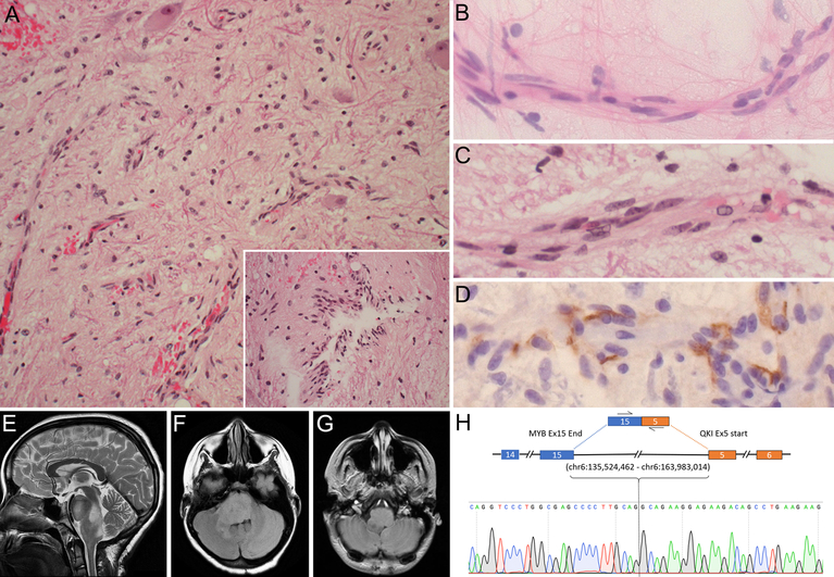
Case 1. Photomicrographs of representative H&E tissue section (A) showing low-cellularity glial tumor with growth along blood vessels or in a perpendicular orientation (inset), with entrapped neurons. Cytologic smear (B) and tissue section (C) highlighting longitudinal growth along blood vessels and elongated nuclei with finely speckled chromatin. D Immunohistochemistry for EMA showing surface and cytoplasmic positivity. MRI images showing a T2-hyperintense (E), FLAIR-hyperintense (F), T1-hypointense non-contrast enhancing (G), dorsally exophytic mass infiltrating pons and medulla. (H) Sanger sequencing of MYB-QKI fusion transcript. Initial magnification in A, 200x; inset, 400x; B-D, 630x
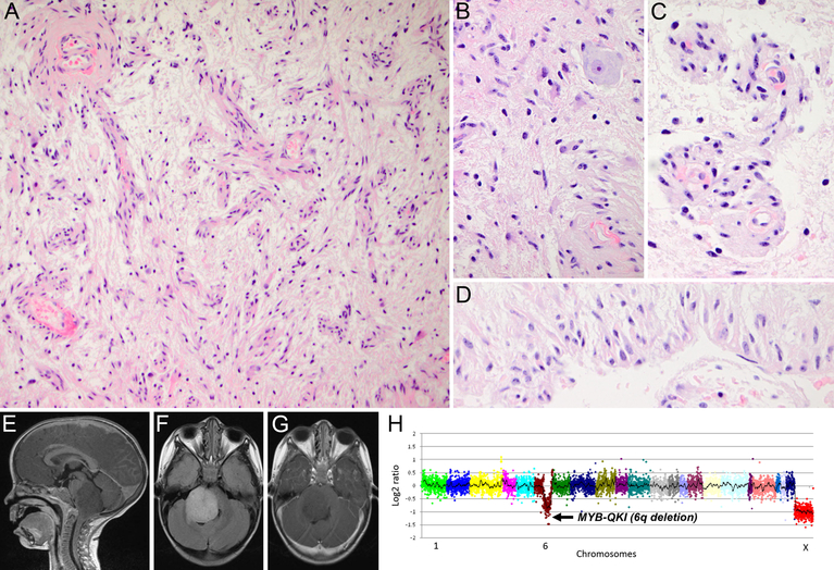
Case 2. Photomicrographs of representative H&E tissue sections showing low-cellularity glial tumor with angiocentric growth (A and C), entrapped neurons (B), and growth in a perpendicular orientation (D). MRI images showing a T1-hypointense (E), FLAIR-hyperintense (F), non-contrast enhancing (G), dorsally exophytic mass infiltrating pons and medulla. H Next-generation sequencing (OncoPanelTM) showing 6q deletion suggestive of MYB-QKI fusion. Initial magnification in A, 200x; B-D, 600x
References
-
- Burger PC, Jouvet A, Preusser M, Rosenblum MK, Ellison DW (2016) Angiocentric Glioma In: Louis DN, Ohgaki H, Wielster OD, Cavenee WK, Ellison DW, Figarella-Branger D, Perry A, Reifenberger G, Von Deimling A (eds) WHO Classification of Tumours of the Central Nervous System, revised 4th edn Internationnal Agency for Research on Cancer, Lyon, pp 119–120
Publication types
MeSH terms
Substances
Grants and funding
LinkOut - more resources
Full Text Sources
Other Literature Sources
