. 2017 Nov;108(11):2248-2264.
doi: 10.1111/cas.13354. Epub 2017 Sep 14.

Solasodine inhibits human colorectal cancer cells through suppression of the AKT/glycogen synthase kinase-3β/β-catenin pathway

Affiliations

Solasodine inhibits human colorectal cancer cells through suppression of the AKT/glycogen synthase kinase-3β/β-catenin pathway

Yu-Wen Zhuang et al. Cancer Sci. 2017 Nov.

Abstract

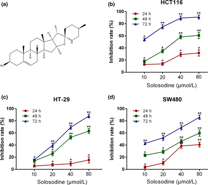
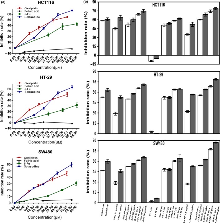
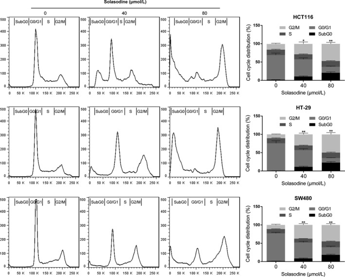
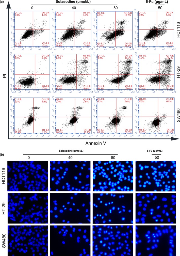
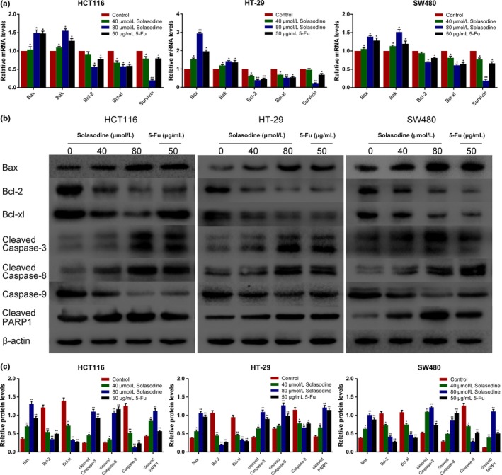
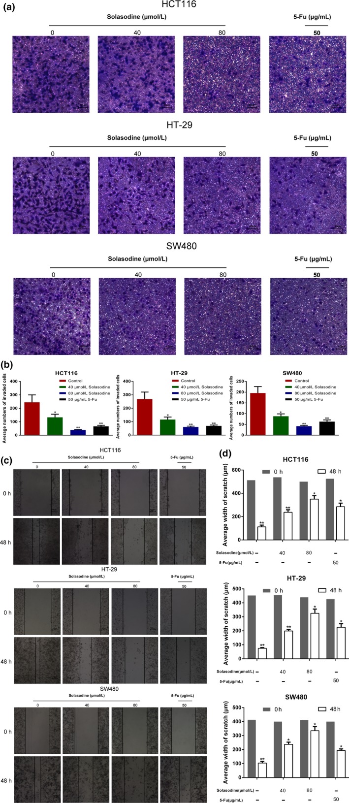
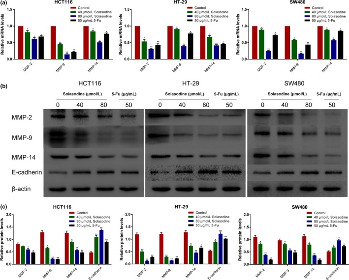
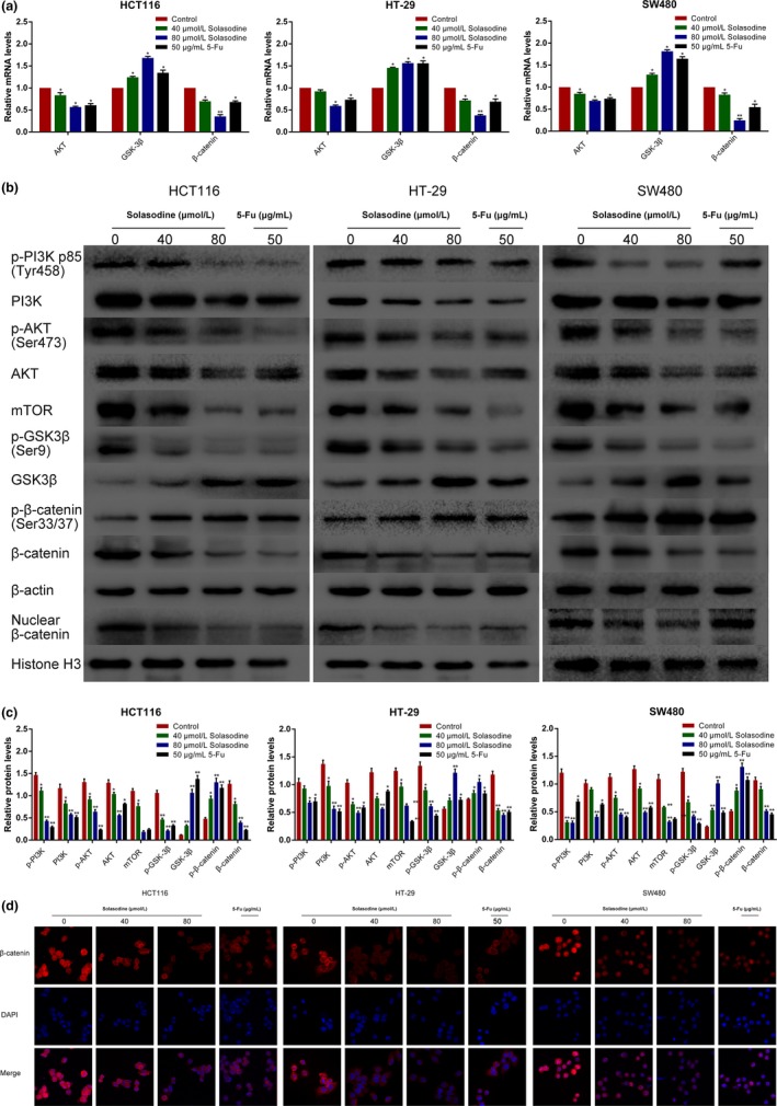
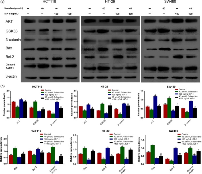
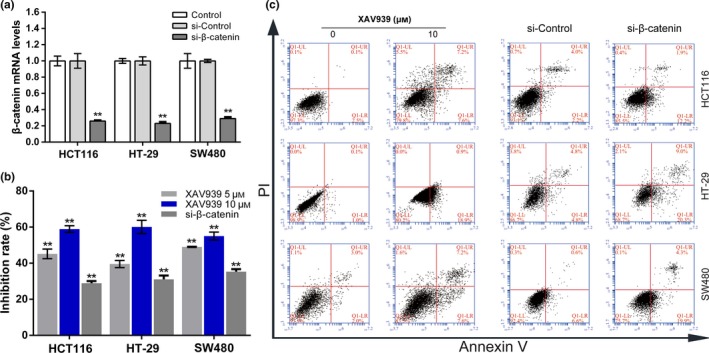
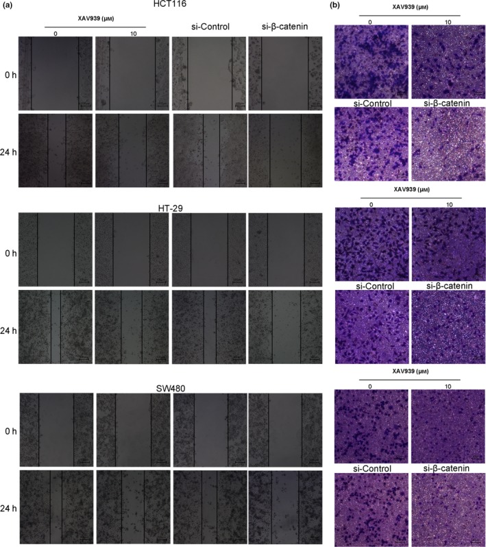
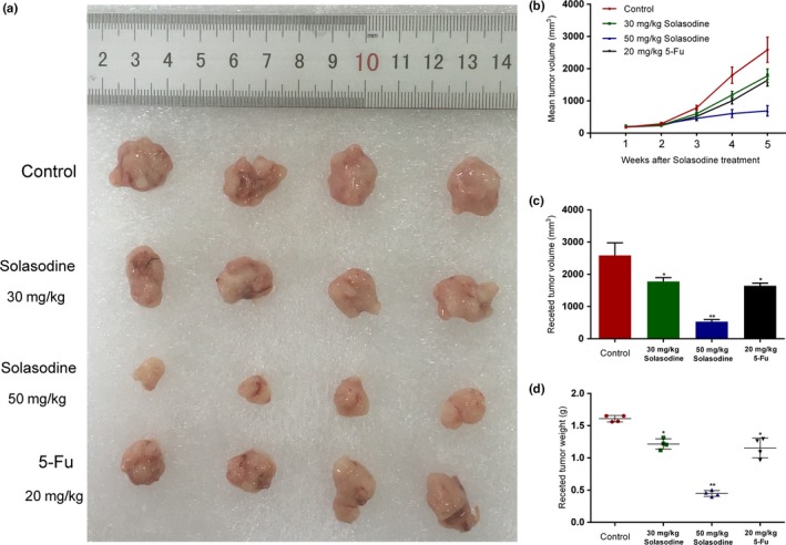
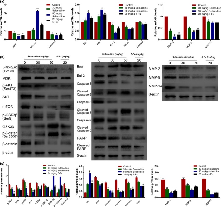
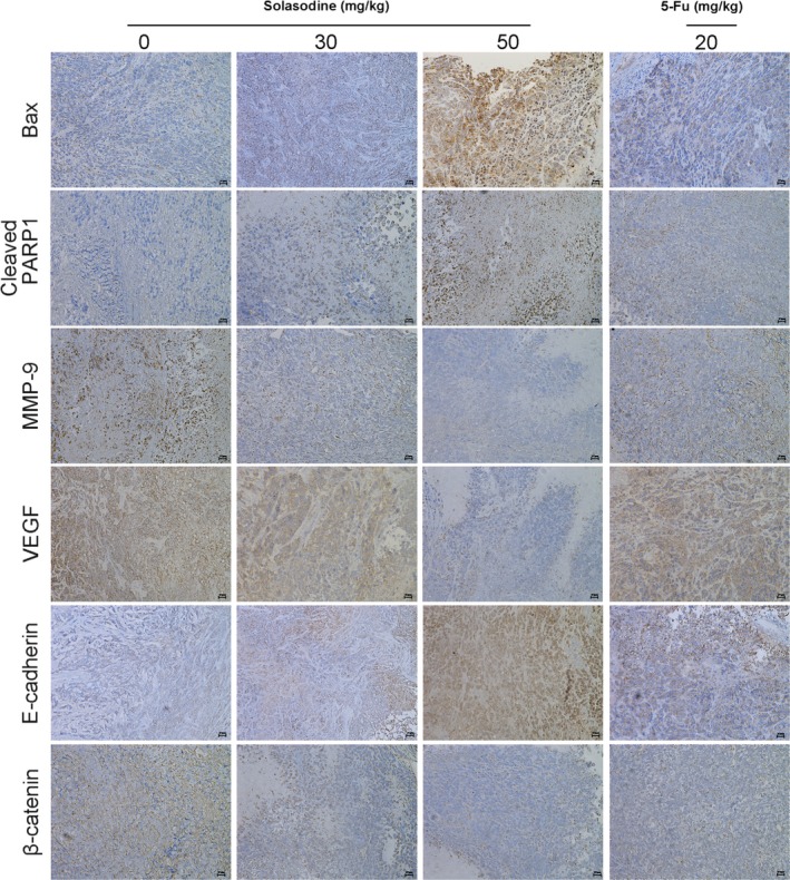
Solasodine is a main active component isolated from Solanum incanum L. that performs a wide range of functions containing anti-oxidant, anti-infection, and neurogenesis promotion. In this study, we explored the influence of solasodine on three types of human colorectal cancer (CRC) cell lines. The results show that solasodine prohibited CRC cell proliferation dose- and time-dependently and impeded CRC cell motility by downregulating MMPs. Solasodine was also found to fuel caspase-cascade reaction and increase the ratio between Bax and Bcl-2 so as to induce CRC cell apoptosis. When cells were pretreated with AKT activator (insulin-like growth factor-1) followed by solasodine, the solasodine-induced apoptosis was partially abrogated by insulin-like growth factor-1. Moreover, solasodine hindered tumor development and stimulated similar mechanisms in vivo. In general, our study provides the first evidence that solasodine has a suppressive effect on CRC cells and that this agent may be a novel therapeutic drug for CRC treatment.

Keywords: Apoptosis; colorectal cancer; metastasis; solasodine; β-Catenin.


Figures

Figure 1
Figure 1
Solasodine suppresses colorectal cancer cell growth. (a) Chemical structure of solasodine. (b–d) HCT116 (b), HT‐29 (c), and SW480 (d) cells were incubated with different concentrations of solasodine for 24, 48, and 72 h. Cell inhibitive rates were measured by MTT assay. The values were presented as mean ± SD of three independent experiments. *P < 0.05, **P < 0.01 versus control group.
Figure 2
Figure 2
Solasodine shows cytotoxicity against colorectal cancer cells and synergistic effects with established chemotherapeutic agents. (a,b) Three types of cells were treated with solasodine, 5‐fluorouracil (5‐Fu), oxaliplatin, and folinic acid alone or in combination for 48 h. Cell viability was tested by MTT assay. Values are shown as mean ± SD of three independent assays. *P < 0.05, **P < 0.01 versus control group.
Figure 3
Figure 3
Solasodine induces G2/M‐phase cell cycle arrest in colorectal cancer cells. Cells were incubated with RNase, treated with solasodine for 48 h, stained with propidium iodide, and then analyzed by flow cytometry. Data are expressed as the mean ± SD of three experiments. *P < 0.05, **P < 0.01 versus control.
Figure 4
Figure 4
Solasodine induces apoptosis in colorectal cancer cells. (a) Three types of colorectal cancer cells were treated with solasodine and then analyzed by annexin V/propidium iodide (AV/PI) staining. Lower left (LL) quadrants, living cells (AV negative/PI negative). Lower right (LR) quadrants, early apoptotic cells (AV positive/PI negative). Upper right (UR) quadrants, late apoptotic cells (AV positive/PI positive). Upper left (UL) quadrants, necrotic cells (AV negative/PI positive). (b) Variations in nuclear morphology were examined by Hoechst 33258 staining and captured by fluorescence microscopy. 5‐Fu, 5‐fluorouracil.
Figure 5
Figure 5
Solasodine regulates apoptosis‐associated genes. (a) RT–quantitative PCR analysis of apoptosis‐related mRNA. β‐Actin was selected as internal control. (b,c) Western blot analysis of apoptosis‐related proteins in all three cell lines. β‐Actin was chosen as loading control. Data are represented as the mean ± SD of three independent experiments. *P < 0.05, **P < 0.01 compared to control. 5‐Fu, 5‐fluorouracil; PARP1, poly (ADP‐ribose) polymerase 1.
Figure 6
Figure 6
Solasodine inhibits colorectal cancer cell invasion and migration. (a,b) Transwell assay was carried out to test changes in colorectal cancer cell invasive ability by solasodine. (c,d) Wound healing assay assessed the inhibitive role of solasodine in cell migration. The average width of each wound region was observed under an inverted microscope. Results are expressed as mean ± SD. *P < 0.05, **P < 0.01. 5‐Fu, 5‐fluorouracil.
Figure 7
Figure 7
Solasodine modifies invasion‐associated genes. (a) Analysis of mRNA levels of MMPs using RT–quantitative PCR. β‐Actin was elected as internal control. (b,c) Western blot analysis of invasion‐ and adhesion‐related proteins. β‐Actin was chosen as loading control. Data are shown as the mean ± SD. *P < 0.05, **P < 0.01 versus control group. 5‐Fu, 5‐fluorouracil.
Figure 8
Figure 8
Solasodine regulates the AKT/glycogen synthase kinase‐3β (GSK‐3β)/β‐catenin signaling and restrains nuclear translocation of β‐catenin. (a) RT–quantitative PCR analysis was carried out to test mRNA expression of AKT/GSK‐3β/β‐catenin elements. (b,c) Western blot analysis of AKT/GSK‐3β/β‐catenin‐related proteins. Data are shown as the mean ± SD. *P < 0.05, **P < 0.01 versus control group. (d) Immunofluorescence staining analysis of cytoplasmic and nuclear expression of β‐catenin. Magnification, ×200.5‐Fu, 5‐fluorouracil; mTOR, mammalian target of rapamycin.
Figure 9
Figure 9
Solasodine‐induced apoptosis is regulated by the AKT/glycogen synthase kinase‐3β (GSK‐3β)/β‐catenin pathway. (a, b) Western blot analysis was applied to test whether AKT/GSK‐3β/β‐catenin is associated with apoptotic protein expression regulated by solasodine and insulin‐like growth factor 1 (IGF‐1). Data are presented as the mean ± SD. *P < 0.05, **P < 0.01 versus control. # P < 0.05, ## P < 0.01 versus solasodine‐treated group. PARP1, poly (ADP‐ribose) polymerase 1.
Figure 10
Figure 10
Role of β‐catenin in proliferation and apoptosis in colorectal cancer cells. (a) Cells were transfected with β‐catenin siRNA and incubated for 48 h. β‐Catenin mRNA expression was determined using RTPCR. (b) MTT assay was used to study inhibitive rates of transfected cells or cells incubated with XAV939 for 24 h. (c) Transfected cells and cells treated with 10 μM XAV939 for 24 h were analyzed by annexin V/propidium iodide (PI) staining.
Figure 11
Figure 11
XAV939 inhibits colorectal cancer cell migration and invasion. (a) Wound healing assay tested the variation of cell migration and the average width of wound area was observed under an inverted microscope. (b) Transwell assay was undertaken to study the changes in cell invasion ability.
Figure 12
Figure 12
Solasodine confines tumor growth of mouse xenograft colorectal cancer in vivo. (a) Differences in excised tumor volume after the last treatment with solasodine. (b,c) Tumor volume was calculated after solasodine application. (d) Variations in tumor weight in the solasodine‐treated group compared to the control group. *P < 0.05, **P < 0.01. 5‐Fu, 5‐fluorouracil.
Figure 13
Figure 13
Solasodine induces apoptosis and prohibits AKT/glycogen synthase kinase‐3β (GSK‐3β)/β‐catenin signaling in HCT116 xenograft tumors. Tumors were abscised after the last solasodine i.p. injection and mRNA or protein was extracted. (a–c) RT–quantitative PCR and Western blot studies were undertaken to evaluate the levels of AKT/GSK‐3β/β‐catenin, apoptotic, and invasive molecules. 5‐Fu, 5‐fluorouracil; PARP, poly (ADP‐ribose) polymerase.
Figure 14
Figure 14
Solasodine varies apoptotic‐ and invasion‐relevant proteins and β‐catenin expression in mouse xenograft colorectal cancer. Immunohistochemical analysis of tumor specimens revealed the modifications between solasodine‐treated and control groups. 5‐Fu, 5‐fluorouracil; PARP1, poly (ADP‐ribose) polymerase 1; VEGF, vascular endothelial growth factor.

Similar articles

Cited by

References

    1. Ferlay J, Ervik M, Dikshit R et al Cancer incidence and mortality worldwide: sources, methods and major patterns in GLOBOCAN 2012. Int J Cancer 2015; 136: E359–86. - PubMed
    1. Ece E, Suayib Y. Maintenance strategy in metastatic colorectal cancer: a systematic review. Cancer Treat Rev 2016; 42: 82–90. - PubMed
    1. Kim KY, Cha IH, Ahn JB et al Estimating the adjuvant chemotherapy effect in elderly stage II and III colon cancer patients in an observational study. J Surg Oncol 2013; 107: 613–8. - PubMed
    1. Markowitz SD, Bertagnolli MM. Molecular origins of cancer: Molecular basis of colorectal cancer. N Engl J Med 2009; 361: 2449–60. - PMC - PubMed
    1. Goss KH, Groden J. Biology of the adenomatous polyposis coli tumor suppressor. J Clin Oncol 2000; 18: 1967–79. - PubMed

MeSH terms