. 2017 Aug 30;95(5):1056-1073.e5.
doi: 10.1016/j.neuron.2017.07.031. Epub 2017 Aug 10.

Reck and Gpr124 Are Essential Receptor Cofactors for Wnt7a/Wnt7b-Specific Signaling in Mammalian CNS Angiogenesis and Blood-Brain Barrier Regulation

Affiliations

Reck and Gpr124 Are Essential Receptor Cofactors for Wnt7a/Wnt7b-Specific Signaling in Mammalian CNS Angiogenesis and Blood-Brain Barrier Regulation

Chris Cho et al. Neuron. .

Erratum in

Abstract

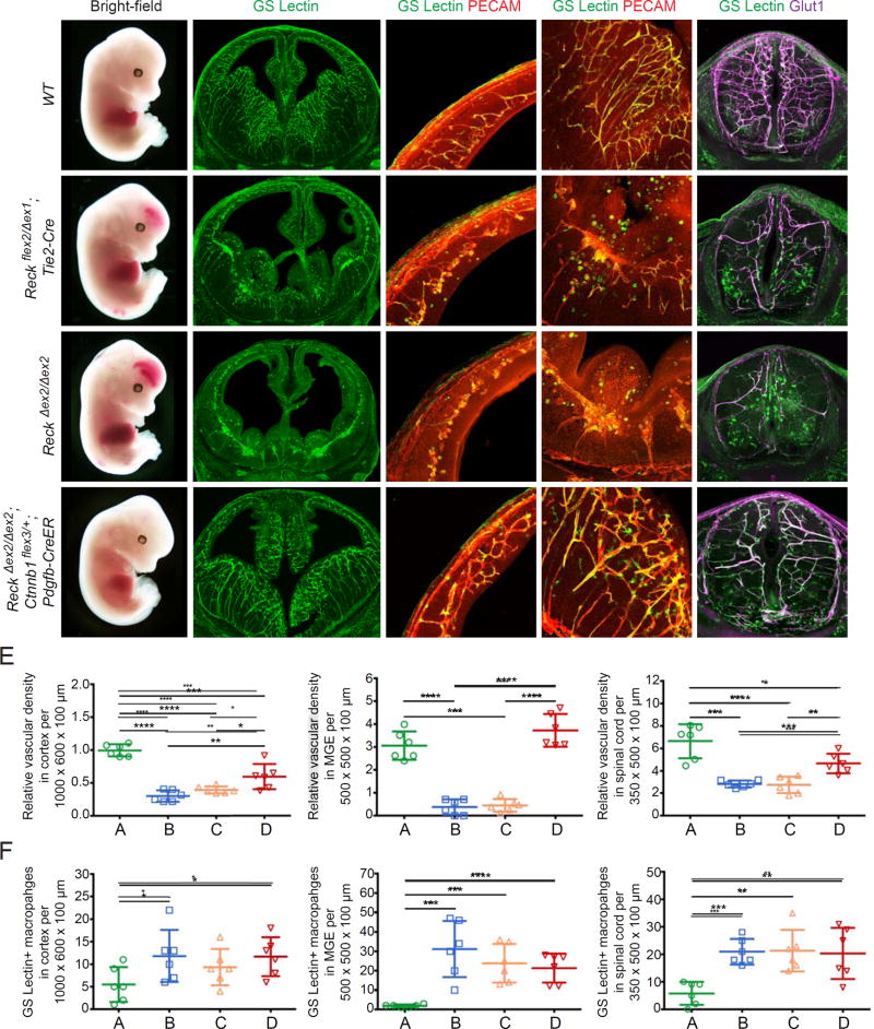
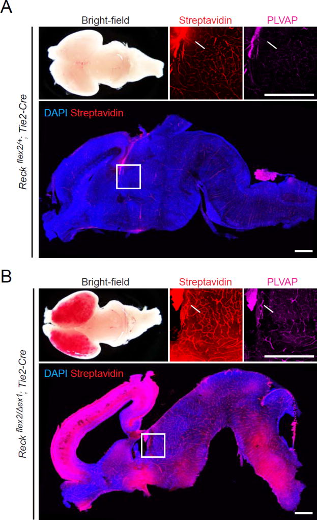
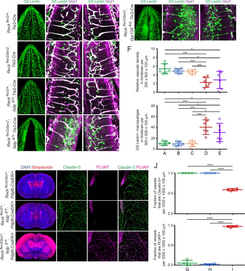
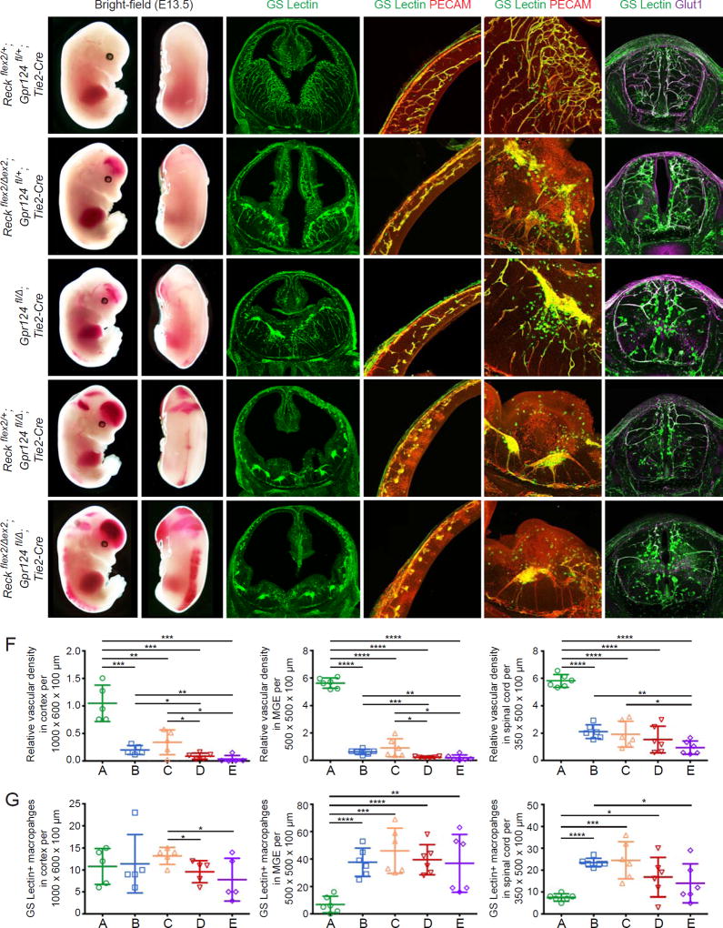
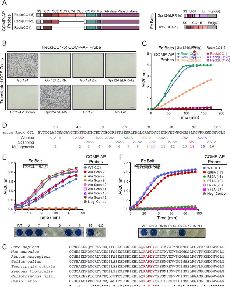
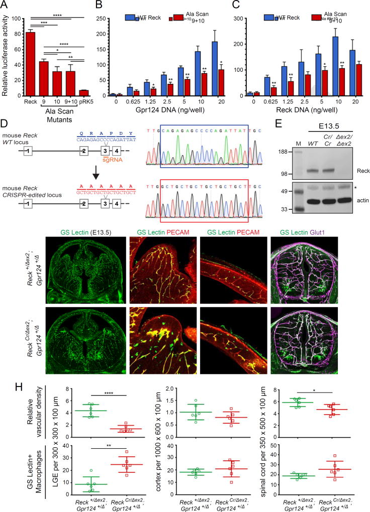
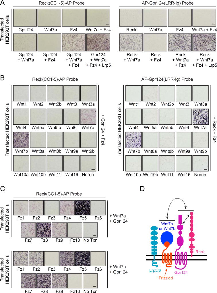
Reck, a GPI-anchored membrane protein, and Gpr124, an orphan GPCR, have been implicated in Wnt7a/Wnt7b signaling in the CNS vasculature. We show here that vascular endothelial cell (EC)-specific reduction in Reck impairs CNS angiogenesis and that EC-specific postnatal loss of Reck, combined with loss of Norrin, impairs blood-brain barrier (BBB) maintenance. The most N-terminal domain of Reck binds to the leucine-rich repeat (LRR) and immunoglobulin (Ig) domains of Gpr124, and weakening this interaction by targeted mutagenesis reduces Reck/Gpr124 stimulation of Wnt7a signaling in cell culture and impairs CNS angiogenesis. Finally, a soluble Gpr124(LRR-Ig) probe binds to cells expressing Frizzled, Wnt7a or Wnt7b, and Reck, and a soluble Reck(CC1-5) probe binds to cells expressing Frizzled, Wnt7a or Wnt7b, and Gpr124. These experiments indicate that Reck and Gpr124 are part of the cell surface protein complex that transduces Wnt7a- and Wnt7b-specific signals in mammalian CNS ECs to promote angiogenesis and regulate the BBB.

Keywords: CNS angiogenesis; Wnt reporter; blood-brain barrier; canonical Wnt signaling; mouse genetics; vascular endothelial cells.


Figures

Figure 1
Figure 1. Reck Activates Canonical Wnt Signaling to Promote CNS Angiogenesis
(A–D) E13.5 embryos are shown (column 1) alongside coronal sections of brain (columns 2–4) and cross-sections of spinal cord (column 5). The white line in (A) depicts the plane for each brain section. Boxed regions in cortex (a) and MGE (b) in column 2 are shown at higher magnification in columns 3 and 4, respectively. (B) Reckflex2/Δex1;Tie2-Cre vascular knockout embryos show hemorrhaging in the forebrain (arrow) and distal spinal cord (white arrowhead). Vascular density is reduced in the cortex, MGE, and spinal cord, accompanied by infiltration of GS Lectin+ macrophages. (C) ReckΔex2/Δex2 embryos display vascular defects similar to their Tie2-Cre conditional knockout counterparts in (B). (D) Beta-catenin was artificially stabilized in ECs of ReckΔex2/Δex2;Ctnnb1flex3/+;Pdgfb-CreER embryos following IP injection of 1.5 mg of tamoxifen in gestational day (G)10.5 females. At E13.5, the resulting embryos exhibit little or no hemorrhaging in the forebrain and spinal cord, and show a near-complete rescue of vascularization in these regions. (E) Quantification of the relative vascular density in cortex, MGE, and spinal cord for each genotype shown in (A–D). (F) Quantification of GS Lectin+ macrophages in cortex, MGE, and spinal cord for each genotype in (A–D). In this and subsequent figures, bars represent mean ± SD. Statistical significance, determined by the unpaired t-test, is represented by * (P<0.05), **(P<0.01), *** (P<0.001), and **** (P<0.0001). Scale bars, 200 µm.
Figure 2
Figure 2. Reck is Essential for the Development of the Blood-Brain Barrier
(A,B) Reckflex2/+;Tie2-Cre and Reckflex2/Δex1;Tie2-Cre P0 mice were injected IP with 2–3 mg of sulfo-NHS-biotin 30–45 minutes before sacrifice. Dorsal view (a) and sagittal section (d) of P0 brains, with the boxed region in (d) shown at higher magnification in (b) and (c). Left, anterior; right, posterior. Reckflex2/+;Tie2-Cre control mice (A) have an intact BBB with no biotin leakage, except in the choroid plexus (arrows in (b) and (c)). Reckflex2/Δex1;Tie2-Cre mice (B) show severe hemorrhaging in the cortex along with biotin leakage in cortical, subcortical, and hindbrain regions. PLVAP is widely up-regulated in the CNS vasculature in Reckflex2/Δex1;Tie2-Cre mice. Scale bars, 50 µm.
Figure 3
Figure 3. Reck- and Norrin-Mediated Wnt Signaling Pathways Function Redundantly to Regulate CNS Angiogenesis and BBB Integrity
(A–E) Coronal sections of E11.5 hindbrains (column 1). Boxed regions (a) and (b) are displayed at higher magnification in columns 2 and 3. In the hindbrain, Reckflex2/+;Tie2-Cre (A), Reckflex2/Δex2;Tie2-Cre (B), and Reckflex2/+;NdpΔ/Y;Tie2-Cre (C) animals show little or no vascular defects. Reckflex2/Δex2;NdpΔ/Y;Tie2-Cre (D) and Reckflex2/Δex2;Gpr124fl/Δ;Tie2-Cre (E) embryos show reduced vascular density with increased infiltration of GS Lectin+ macrophages. (F) Quantification of relative vascular density (top) and GS Lectin+ macrophages (bottom) in the hindbrain for each genotype in (A–E). (G–I) Coronal sections of P7 brains (column 1). Boxed regions are displayed at higher magnification (columns 2–4). Animals were injected IP with 50 µg of 4-HT at P3 and P4. Reckflex2/Δex1;Pdgfb-CreER (G) and Reckflex2/+;NdpΔ/Y;Pdgfb-CreER (H) animals show little or no biotin leakage into the brain parenchyma. Vessels are Claudin-5+;PLVAP, except in the choroid plexus where they are Claudin-5;PLVAP+ (upper left of each boxed region). Reckflex2/Δex1;NdpΔ/Y;Pdgfb-CreER (I) mice show extensive biotin leakage, most prominently in the cortex, with vessels in the brain parenchyma converted to Claudin-5;PLVAP+. (J) Quantification of the fraction of vessels that immunostain positive for Claudin-5 (top) and PLVAP (bottom) for each genotype in (G–I). Scale bars, 200 µm.
Figure 4
Figure 4. Genetic Evidence for Reck-Gpr124 Interaction in CNS Angiogenesis
(A–E) E13.5 embryos (columns 1 and 2), coronal sections of brain (columns 3–5), and cross-sections of spinal cord (column 6). Boxed regions of cortex (a) and MGE (b) in column 3 are shown at higher magnification in columns 4 and 5, respectively. Reckflex2/+;Gpr124fl/+;Tie2-Cre (A) embryos show normal vascular development, but ~2–3-fold greater numbers of GS Lectin+ macrophages compared to WT embryos (compare panel G to Fig. 1F). Reckflex2/Δex2;Gpr124fl/+;Tie2-Cre (B) and Gpr124fl/Δ;Tie2-Cre (C) embryos show hemorrhaging in the forebrain (arrow) and distal spinal cord (white arrowhead). Vascular density is reduced in the cortex, MGE, and spinal cord, and hypovascular regions show increased infiltration of GS Lectin+ macrophages. Reckflex2/+;Gpr124fl/Δ;Tie2-Cre (D) and Reckflex2/Δex2;Gpr124fl/Δ;Tie2-Cre (E) embryos show severe hemorrhaging in the forebrain (arrow) and along the length of the spinal cord (white arrowhead), as well as reduced vascular density in the cortex, MGE, and spinal cord. (F) Quantification of relative vascular density in cortex, MGE, and spinal cord for each genotype in (A–E). (G) Quantification of GS Lectin+ macrophages in cortex, MGE, and spinal cord for each genotype in (A–E). Scale bars, 200 µm.
Figure 5
Figure 5. Direct Binding of the Domains of Reck and Gpr124 Responsible for Canonical Wnt Signaling
(A) Diagram of COMP-AP probes (left) and Fc baits (right). (B) AP binding assay using COS-7 cells transfected with Gpr124, Gpr124 deletion mutants, or Gpr125, a close homologue of Gpr124. Live cells were incubated with the Reck(CC1–5)-COMP-AP probe shown in (A). No Txn, no transfection. (C) Cell-free binding assay using Gpr124(LRR-Ig) or Reck(CC1–5) (a negative control) Fc baits captured in Protein-G coated wells. Each bait was incubated with three COMP-AP probes: Reck(CC1), Reck(CC1–2), and Reck(CC1–5). After removing unbound probe, the bound probe was visualized with a colorimetric AP reaction. (D) Amino acid sequence of mouse Reck CC1, with alanine scanning mutants indicated. Alanine substitution blocks 2, 7, 10, 14, and 16 were produced at near-WT levels and 9 and 15 at reduced levels (Figure S7A and S7B). (E) Cell-free binding assay with the indicated alanine scanning mutants (top) and images of the AP reactions at the final time point (bottom). N.C., negative control. (F) Cell-free binding assay with the indicated single alanine substitutions (top) and images of the AP reactions at the final time point (bottom). See Figure S7A and S7B for protein yield. (G) Alignment and conservation of Reck CC1 across vertebrates, generated by Clustal Omega. Alignment symbols denote: (*) fully conserved residues; (:) residues with strongly similar properties; and (.) residues with weakly similar properties. Scale bars, 200 µm.
Figure 6
Figure 6. Reck-Gpr124 Complex Formation is Important for Wnt Activation and CNS Angiogenesis
(A) STF cells were transfected with: Wnt7a, Gpr124, and Reck WT or Ala Scan mutant plasmids. Luciferase assays were performed as described in ‘STAR Methods’. (B) As in (A), but showing the dose response curve for varying concentrations of Gpr124 DNA in the presence of constant WT Reck (blue) or Ala Scan 9+10 (red). (C) As in (A), but showing the dose response curve for varying concentrations of WT Reck (blue) or Ala Scan 9+10 (red) in the presence of constant WT Gpr124. (D) Diagram of the mouse Reck WT and CRISPR-edited alleles. The region corresponding to Ala Scan mutants 9 and 10 in Reck(CC1) is encoded by exon 3. (E) Reck protein abundance at E13.5 is unaltered in ReckCr/Cr embryos compared to a WT control, as determined by immunoblotting. The disappearance of the full-size Reck band in the ReckΔex2/Δex2 embryos confirms the specificity of the anti-Reck antibody. (*) denotes a non-specific band in the actin immunoblot. MW standards are shown at left. (F–G) E13.5 coronal sections of brain (columns 1–3) and cross-sections of spinal cord (column 4). Boxed regions in LGE (a) and cortex (b) are shown at higher magnification in columns 2 and 3, respectively. ReckCr/Δex2;Gpr124+/Δ mutant embryos (G) show reduced vascular density and glomeruloid tufts in the LGE, accompanied by infiltration of GS Lectin+ macrophages. Vascular density in the spinal cord is also reduced. (H) Quantification of relative vascular density (top) and the number of GS Lectin+ macrophages (bottom) in LGE, cortex, and spinal cord for the genotypes shown in (F–G). Values represent means ± SD. Scale bars, 200 µm.
Figure 7
Figure 7. Reck-Gpr124 Assembles into a Multi-Protein Complex with Wnt7a/Wnt7b and Frizzled
(A) AP binding assay using HEK293T cells transfected with Gpr124, Wnt7a, Fz4, and/or Lrp5 (left) or Reck, Wnt7a, Fz4, and/or Lrp5 (right). Live cells were incubated with Reck(CC1–5)-AP (left) or AP-Gpr124(LRR-Ig) (right). (B) AP binding assay using HEK293T cells transfected with Gpr124 and Fz4, along with each of the 19 Wnts and Norrin. Live cells were incubated with Reck(CC1–5)-AP (left) or AP-Gpr124(LRR-Ig) (right). (C) AP binding assay using HEK293T cells transfected with Gpr124 and Wnt7a (top) or Wnt7b (bottom), toegther with each of the 10 Fz receptors or a no transfection control. Live cells were incubated with Reck(CC1–5)-AP. (D) Model of the multi-protein signaling complex, consisting of Reck, Gpr124, Wnt7a or Wnt7b, Frizzled, and Lrp5 or Lrp6. The straight double-headed arrow indicates a direct interaction between Reck(CC1) and Gpr124(LRR-Ig). The curved double-headed arrow indicates a direct interaction between Reck/Gpr124 and Fz/(Wnt7a or Wnt7b) sub-complexes. Modified from Zhou and Nathans (2014). Scale bars, 200 µm.

References

    1. Anderson KD, Pan L, Yang XM, Hughes VC, Walls JR, Dominguez MG, Simmons MV, Burfeind P, Xue YZ, Wei Y, et al. Angiogenic sprouting into neural tissue requires Gpr124, an orphan G protein-coupled receptor. Proc. Natl. Acad. Sci. USA. 2011;108:2807–2812. - PMC - PubMed
    1. Ben-Zvi A, Lacoste B, Kur E, Andreone BJ, Mayshar Y, Yan H, Gu C. Mfsd2a is critical for the formation and function of the blood-brain barrier. Nature. 2014;509:507–511. - PMC - PubMed
    1. Chandana EP, Maeda Y, Ueda A, Kiyonari H, Oshima N, Yamamoto M, Kondo S, Oh J, Takahashi R, Yoshida Y, et al. Involvement of the Reck tumor suppressor protein in maternal and embryonic vascular remodeling in mice. BMC Dev. Biol. 2010;10:84. - PMC - PubMed
    1. Chang CK, Hung WC, Chang HC. The Kazal motifs of RECK protein inhibit MMP-9 secretion and activity and reduce metastasis of lung cancer cells in vitro and in vivo. J. Cell Mol. Med. 2008;12:2781–2789. - PMC - PubMed
    1. Chang J, Mancuso M, Maier C, Liang X, Yuki K, Yang L, Kwong JW, Wang J, Rao V, Vallon M, et al. Gpr124 is essential for blood-brain barrier integrity in central nervous system disease. Nat. Med. 2017;23:450–460. - PMC - PubMed

MeSH terms