. 2017 Aug 17;67(4):566-578.e10.
doi: 10.1016/j.molcel.2017.07.013. Epub 2017 Aug 10.

Myc Regulates Chromatin Decompaction and Nuclear Architecture during B Cell Activation

Affiliations

Myc Regulates Chromatin Decompaction and Nuclear Architecture during B Cell Activation

Kyong-Rim Kieffer-Kwon et al. Mol Cell. .

Abstract

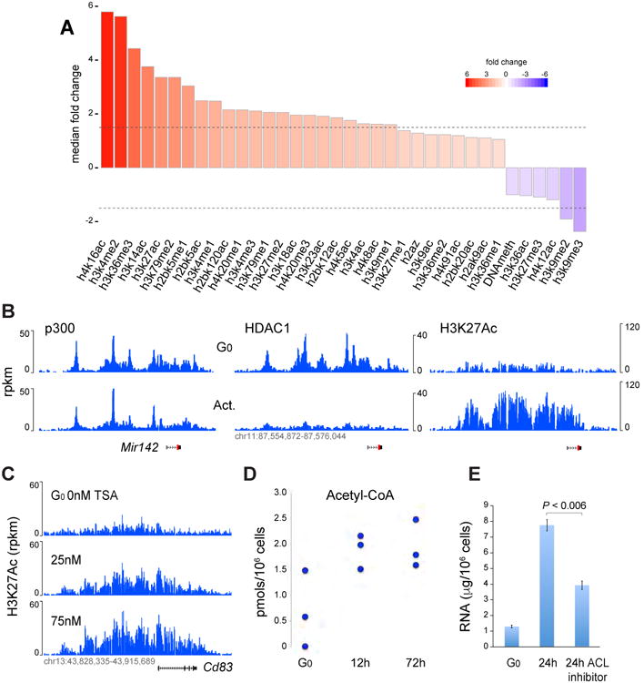
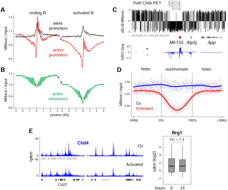
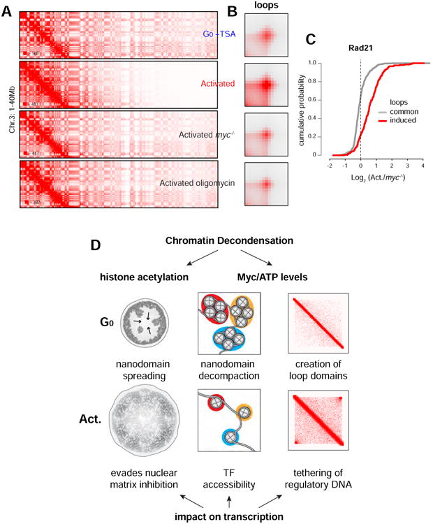
50 years ago, Vincent Allfrey and colleagues discovered that lymphocyte activation triggers massive acetylation of chromatin. However, the molecular mechanisms driving epigenetic accessibility are still unknown. We here show that stimulated lymphocytes decondense chromatin by three differentially regulated steps. First, chromatin is repositioned away from the nuclear periphery in response to global acetylation. Second, histone nanodomain clusters decompact into mononucleosome fibers through a mechanism that requires Myc and continual energy input. Single-molecule imaging shows that this step lowers transcription factor residence time and non-specific collisions during sampling for DNA targets. Third, chromatin interactions shift from long range to predominantly short range, and CTCF-mediated loops and contact domains double in numbers. This architectural change facilitates cognate promoter-enhancer contacts and also requires Myc and continual ATP production. Our results thus define the nature and transcriptional impact of chromatin decondensation and reveal an unexpected role for Myc in the establishment of nuclear topology in mammalian cells.

Keywords: B cells; CTCF; Histone acetylation; chromatin remodeling; cohesin; immune response; myc; nanoscopy; nuclear architecture; transcriptome amplification.


Conflict of interest statement

Conflict of Interest Statement: The authors declare no conflicts of interest.

Figures

Figure 1
Figure 1. Extent and regulation of epigenome amplification during lymphocyte activation
(A) Genome-wide fold changes in 34 epigenetic marks, including CpG methylation, between G0 and 24h activated B cell chromatin. Dotted lines represent +/- 1.5 fold change. (B) p300 (left), HDAC1 (middle), and H3K27Ac ChIP-Seq profiles at the Mir142 locus in resting and activated B cells. (C) H3K27Ac at Cd83 locus in resting B cells untreated (top track) or treated with increasing concentrations of TSA for 4h (lower tracks). (D) Acetyl-CoA production during B cell activation. Concentration is provided as pmols/106 cells and was normalized per cell size (see Methods). (E) RNA synthesis in G0 cells or 24h activated B cells in the presence or absence of the ACL inhibitor Medica16. Data are represented as mean +/- SEM.
Figure 2
Figure 2. Global remodeling of chromatin during B cell activation
(A) Nucleosome profiles at silent (black lines) and transcribed (red lines) promoters in G0 (left) and activated (right) B cells as determined by MNase-Seq. (B) Same analysis as in panel A for enhancer domains. Arrows in A and B indicate remodeling in activated B cells extending beyond regulatory elements. (C) Nucleosome remodeling across the mir155 locus. MNase-Seq signals from G0 samples were subtracted from those of activated counterparts. The schematics depict genes with black boxes and mir155 enhancers with red boxes. (D) Extent of nucleosome remodeling in heterochromatin and euchromatin compartments during B cell activation. (E) Chromatin occupancy of Chd4 and Brg1 in G0 and 24h activated B cells. Data are represented as mean +/- SEM.
Figure 3
Figure 3. Chromatin decondensation during B cell activation involves spreading and decompaction of nucleosome nanodomains
(A) STORM images of H2B distribution in representative G0 and 24h activated B cells. Scale bars = 1μm. (B) Violin plot showing the nuclear area (percentage) occupied by H2B during B cell activation. Number of cells analyzed: 28 (0h), 29 (6h), 20 (24h). (C) Close up view of H2B signals in a B cell nucleus (left), its decompaction via STORM software (center), and a zoomed in section of the same image (right) showing nucleosome nanodomains, nanodomain centroids, and nearest neighbor distances. Crosses within nanodomains represent single molecule localizations. Scale bars = 500nm, and 50nm as indicated. (D) Bar graph shows localizations per nanodomains in resting (blue) and activated (red) B cells. Number of cells analyzed: 35 (G0) and 33 (activated). B and D data are represented as mean +/- SEM.
Figure 4
Figure 4. Pathways that regulate chromatin decondensation
(A) Representative distribution of chromatin (H2B) in G0 follicular (upper), B-1 (middle), and MZ (lower) B cells. (B-C) Box plots portraying the extent of nanodomain spreading and decompaction respectively in B cell subsets. G0 and activated B cell data reproduced from Figure 3. Cells analyzed: 28 and 35 (G0), 20 and 33 (activated), 21 and 30 (MZ), 20 and 34 (B-1). (D) H2B nanodomain spreading in controls samples and in the presence of TSA (left, G0), Medica16 (middle, 6h), and myc deficiency (right, 24h). Cells analyzed: 28 (G0), 11 (G0 TSA), 29 (6h control), 11 (6h Medica), 20 (24h control), 12 (24h myc-/-. (E) H2B nanodomain decompaction (single molecule localizations/nanodomain) in the presence of TSA, myc-/-, and oligomycin. G0 and activated B cell data reproduced from Figure 3. Cells analyzed: 35 (G0), 17 (G0 TSA), 33 (24h activated), 42 (24h activated myc-/-), and 7 (oligomycin). Data are represented as mean +/- SEM.
Figure 5
Figure 5. Chromatin decondensation reduces target search time and residence time for transcription factors
(A) Mice knockin for JunD- or CTCF-HaloTags were used to track single molecule dynamics. Volume rendering of three dimensional movement of JunD-HaloTag molecules (black dots) superimposed to single-molecule trajectories. For all SMT experiments the JF549 dye was used. (B) Bar graph shows the number of collisions (non-specific binding) at accessible chromatin sites in the different cell types and conditions indicated. (C) Micrographs depict 2D tracking at long exposure times of JunD-HaloTag molecules, which are visible as bright spots (stably bound) or diffusing signals (fast-moving). (D) FRAP analysis (data points) and model (lines) of CTCF-HaloTag in G0 (blue) and activated (red) B cells. (E) FRAP analysis of TSA treated G0 (grey), and myc-/- (green) or oligomycin (black) treated activated B cells. Number of cells analyzed 21 G0, 18 G0 + TSA, 18 aB, and 8 aB + oligomycin, 7 aB myc-/-. For all FRAP experiments the TMR dye was used. B, D, and E data are represented as mean +/- SEM.
Figure 6
Figure 6. B cell activation doubles the number of loops and loop domains which facilitate the tethering of regulatory DNA
(A) Hi-C contact matrices for chromosome 11 (1-29Mb close-up) in G0, activated B cells, and ES cells. Juicebox parameters were balanced normalized at 100Kb resolution with a color range of 150 (G0 and activated) or 75 (ES cells). (B) Hi-C contact probability as a function of distance (In(s)) in G0 (blue), activated B cells (red), or ES cells (black line). (C) Closeup view of a representative genomic locus that acquires loop domains during B cell activation (D) Venn diagram shows number of loops in G0 and activated B cells. (E) Aggregate peak analysis (APA) plots display the average Hi-C signal at chromatin loops that are shared or gained during activation. (F) ChIP-Seq profiles of CTCF and Rad21 from the genomic locus displayed in panel C. (G) Cumulative plot showing Rad21 levels (activated/G0) at anchors for loops that are common or that are induced during B cell activation. (H) Box plot shows mRNA fold change (activated/G0) at contact domains displaying low (left) or high (right) increase in intra-domain interactions (see Figure S7E). Data are represented as mean +/- SEM.
Figure 7
Figure 7. Nuclear architecture in mammalian cells requires Myc and continual ATP production
(A) Hi-C contact matrices for chromosome 3 (1-40Mb close-up) in G0 + TSA, activated B cells, activated myc-/- B cells, and activated B cells + oligomycin. (B) APA plots showing contacts at loops in the cell types described in panel A. (C) Cumulative plot showing Rad21 levels (activated activated/myc-/-) at anchors for loops that are common or that are induced during B cell activation. (D) Global chromatin decondensation during lymphocyte activation is mediated by histone acetylation and the Myc-ATP pathway. Acetylation enables the spreading of chromatin nanodomains from the nuclear membrane to the entire nucleoplasm, an activity that has been shown to evade the transcriptional inhibitory impact of the nuclear matrix. The Myc-ATP pathway on the other hand facilitates the decompaction of chromatin nanodomains into a mononucleosome fiber, which in turn imparts DNA target accessibility to transcription factors. Architecturally, Myc and continual energy input also mediate the creation of loops and loop domains by increasing cohesin recruitment. The resulting intra-domain interactions facilitate in turn transcription by augmenting the tethering of regulatory DNA.

References

    1. Abrahamsson S, Chen J, Hajj B, Stallinga S, Katsov AY, Wisniewski J, Mizuguchi G, Soule P, Mueller F, Dugast Darzacq C, et al. Fast multicolor 3D imaging using aberration-corrected multifocus microscopy. Nat Methods. 2013;10:60–63. - PMC - PubMed
    1. Andres S, Pyl PT, Huber W. HTSeq — A Python framework to work with high-throughput sequencing data 2014 - PMC - PubMed
    1. Blair D, Dufort FJ, Chiles TC. Protein kinase Cbeta is critical for the metabolic switch to glycolysis following B-cell antigen receptor engagement. Biochem J. 2012;448:165–169. - PubMed
    1. Boggs PT, Donaldson JR, Byrd RH, Schnabel RB. Algorithm 676: software for weighted orthogonal distance regression. ACM Transactions on Mathematical Software (TOMS) 1989;15
    1. Bunting KL, Soong TD, Singh R, Jiang Y, Beguelin W, Poloway DW, Swed BL, Hatzi K, Reisacher W, Teater M, et al. Multi-tiered Reorganization of the Genome during B Cell Affinity Maturation Anchored by a Germinal Center-Specific Locus Control Region. Immunity. 2016;45:497–512. - PMC - PubMed

MeSH terms