. 2017 Nov 1;25(11):2502-2512.
doi: 10.1016/j.ymthe.2017.07.010. Epub 2017 Jul 20.

Epidermal E-Cadherin Dependent β-Catenin Pathway Is Phytochemical Inducible and Accelerates Anagen Hair Cycling

Affiliations

Epidermal E-Cadherin Dependent β-Catenin Pathway Is Phytochemical Inducible and Accelerates Anagen Hair Cycling

Noha S Ahmed et al. Mol Ther. .

Abstract

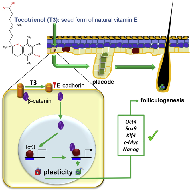
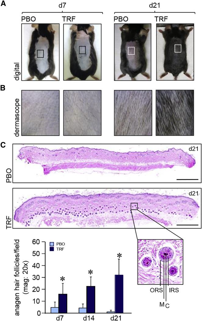
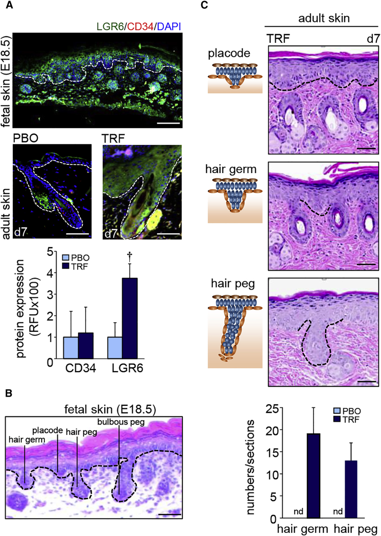
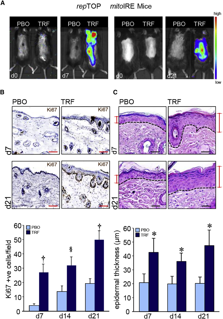
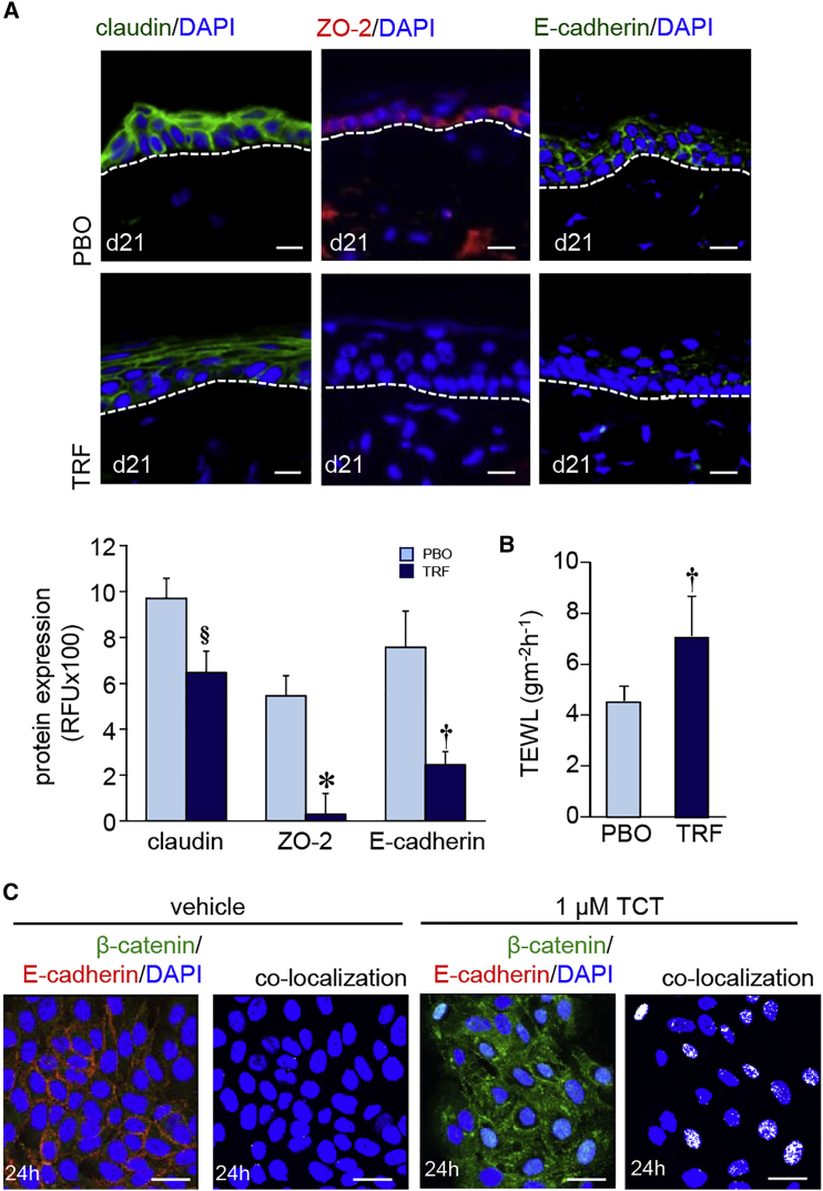
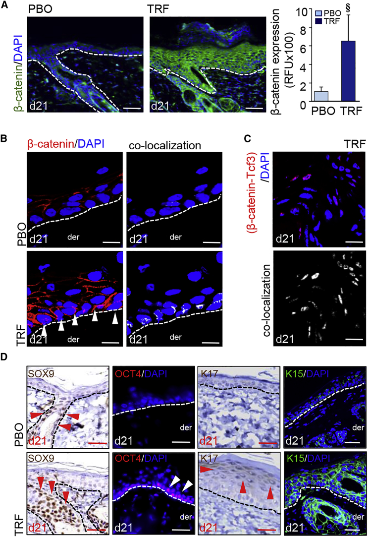
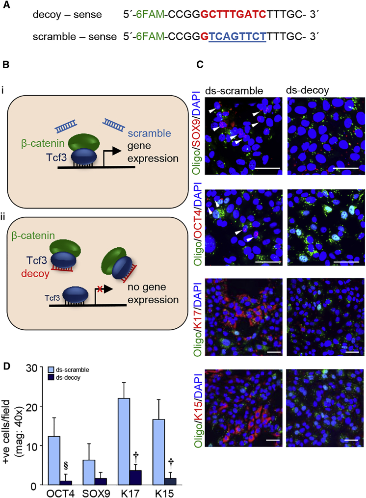
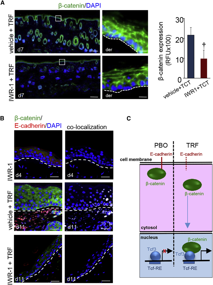
Unlike the epidermis, which regenerates continually, hair follicles anchored in the subcutis periodically regenerate by spontaneous repetitive cycles of growth (anagen), degeneration (catagen), and rest (telogen). The loss of hair follicles in response to injuries or pathologies such as alopecia endangers certain inherent functions of the skin. Thus, it is of interest to understand mechanisms underlying follicular regeneration in adults. In this work, a phytochemical rich in the natural vitamin E tocotrienol (TRF) served as a productive tool to unveil a novel epidermal pathway of hair follicular regeneration. Topical TRF application markedly induced epidermal hair follicle development akin to that during fetal skin development. This was observed in the skin of healthy as well as diabetic mice, which are known to be resistant to anagen hair cycling. TRF suppressed epidermal E-cadherin followed by 4-fold induction of β-catenin and its nuclear translocation. Nuclear β-catenin interacted with Tcf3. Such sequestration of Tcf3 from its otherwise known function to repress pluripotent factors induced the plasticity factors Oct4, Sox9, Klf4, c-Myc, and Nanog. Pharmacological inhibition of β-catenin arrested anagen hair cycling by TRF. This work reports epidermal E-cadherin/β-catenin as a novel pathway capable of inducing developmental folliculogenesis in the adult skin.

Keywords: E-cadherin; anagen; hair follicles; stem cells; β-catenin.


Figures

None
Graphical abstract
Figure 1
Figure 1
Induction of Epidermal Anagen Hair Cycling in Skin (A) Photomicrographs of mouse dorsal skin at day 7 and day 21 showing induction of anagen hair cycling in TRF-treated shaved skin. (B) Dermascopic images from insets in (A) of the dorsal mouse skin at day 7 and day 21. TRF induced anagen hair on day 21. (C) Photomicrograph of formalin-fixed, paraffin-embedded H&E-stained sections showing more anagen hair follicles with dermal papillae reaching the subcutaneous fat layer in TRF-treated sections at day 21. Scale bars, 500 μm. Inset: close-up of a hair follicle with outer root sheath (ORS), inner root sheath (IRS), cortex (C), and medulla (M). Hair follicles were quantified from H&E-stained sections. Data are mean ± SD (n = 6). *p < 0.001.
Figure 2
Figure 2
Developmental Hair Folliculogenesis (A) LGR6 and CD34 immunostaining counterstained with nuclear DAPI. Upper panel: LGR6 (green) and CD34 (red) in fetal skin. Scale bar, 100 μm. Bottom panel: Adult skin. Scale bars, 50 μm. Epidermal and dermal junction is marked by white dashed lines. Fluorescence is plotted as relative florescence units (RFUs). Data are mean ± SD (n = 3). p < 0.01. (B) H&E-stained section showing development of hair follicle from the fetal skin epidermis. Scale bar, 50 μm. (C) TRF treatment on adult skin showed morphological characteristics similar to those of murine fetal skin. Schematic diagrams are shown in the left-hand panels, while actual regions are marked with dashed lines in the H&E-stained sections in the right-hand panels. Scale bars, 50 μm. The number of “hair germ” and “hair peg” features were quantified from H&E-stained sections and expressed graphically (n = 6). nd, not detected.
Figure 3
Figure 3
Keratinocyte Proliferation in Hair Folliculogenesis (A) IVIS image from repTOPmitoIRE showing cell proliferation in animals treated with TRF or placebo (PBO) on days 7 and 21. (B) Immunohistochemical localization of Ki67 in paraffin sections of mouse skin showing the abundance of Ki67+ cells in mice treated (days 7 and 21) with TRF compared to PBO. Scale bars, 50 μm. Ki67+ cells were quantified and plotted graphically. Data are mean ± SD (n = 3) §p < 0.05; p < 0.01. (C) Photomicrograph of H&E-stained paraffin sections showing epidermal thickening in mice treated (days 7 and 21) with TRF. The dermal and epidermal junctions are each marked by a dashed line. Scale bars, 20 μm. Epidermal thickness was quantified and plotted graphically. Data are mean ± SD (n = 4). *p < 0.001.
Figure 4
Figure 4
Epidermal Junctional Proteins in Murine Skin Folliculogenesis (A) TRF lowered (day 21) the expression of claudin (green), ZO-2 (red), and E-cadherin (green) in murine skin. Sections were counterstained with DAPI. Dermal-epidermal junction is indicated by dashed white line. Scale bars, 20 μm. Abundance of junctional proteins in (A) were quantified and expressed graphically as mean ± SD (n = 6). §p < 0.05; p < 0.01; *p < 0.001. (B) Trans-epidermal water loss (TEWL) was measured from the dorsal skin of mice after topical application of TRF or PBO for 21 days. Data are mean ± SD (n = 6). p < 0.01. (C) Keratinocytes (HaCaT cells) treated with pure tocotrienol (1 μM, 24 hr) showed lower expression of E-cadherin (red) in the cell membrane and increased nuclear translocation of β-catenin (green). Scale bars, 20 μm.
Figure 5
Figure 5
Induction of β-Catenin and Nuclear Translocation (A) TRF-induced (day 21) β-catenin (green) in murine epidermis. Counterstained with DAPI (blue). Scale bars, 50 μm. Fluorescence intensity was plotted graphically. Data are mean ± SD (n = 3). §p < 0.05. (B) Confocal microscopy showing TRF-induced translocation (day 21) of β-catenin into the nucleus. Counterstained with DAPI. Dermal (der) and epidermal junction indicated by a white dashed line. Scale bars, 10 μm. (C) Proximity ligation assay (PLA) showed β-catenin-Tcf3 co-localization in the nucleus in TRF treated adult skin. Scale bars, 10 μm. (D) Robust expression of SOX9, OCT4, K15, and K17 in TRF-treated (day 21) adult skin epidermis. The arrowhead indicates positive signals: red for DAB staining and white for fluorescent staining. Scale bars, 20 μm.
Figure 6
Figure 6
β-Catenin-Tcf3 Interaction in Induction of Pluripotency (A) Design of FAM-labeled TCF decoy and eight-base mismatch scramble control. (B) Theoretical scheme of Tcf3 decoy strategy. (i) Normal β-catenin signaling. β-catenin/Tcf3 complex binding facilitates gene expression. (ii) β-catenin may preferentially bind to the Tcf3 decoy region, which competitively inhibits target gene activation. (C) Expression of SOX9, OCT4, K17, and K15 in Tcf3 scramble (0.1 μM) and decoy (0.1 μM) transfected cells after pure tocotrienol treatment (1 μM, 24 hr). The arrowhead indicates positive signals. Scale bars, 50 μm. (D) The number of positive cells per field was quantified and plotted graphically. Data are mean ± SD (n = 3). §p < 0.05; p < 0.01.
Figure 7
Figure 7
β-Catenin Inhibition Arrested Inducible Anagen Hair Cycling (A) IWR-1, a β-catenin inhibitor, significantly attenuated TRF-induced epidermal β-catenin (green) expression in the adult skin. Counterstained with DAPI (blue). Scale bars, 500 μm. β-Catenin signal was quantified and plotted graphically. Data are mean ± SD (n = 3; p < 0.01). The dermal (der) and epidermal junctions are marked by a dashed line in the inset pictures. Scale bars, 20 μm. (B) IWR-1 inhibited TRF-induced translocation of β-catenin to the nucleus. Scale bar, 50 μm. (C) Proposed schematic diagram of TRF-induced hair folliculogenesis.

Similar articles

Cited by

References

    1. Mistriotis P., Andreadis S.T. Hair follicle: a novel source of multipotent stem cells for tissue engineering and regenerative medicine. Tissue Eng. Part B Rev. 2013;19:265–278. - PMC - PubMed
    1. Schneider M.R., Schmidt-Ullrich R., Paus R. The hair follicle as a dynamic miniorgan. Curr. Biol. 2009;19:R132–R142. - PubMed
    1. Millar S.E. Molecular mechanisms regulating hair follicle development. J. Invest. Dermatol. 2002;118:216–225. - PubMed
    1. Fuchs E. Skin stem cells: rising to the surface. J. Cell Biol. 2008;180:273–284. - PMC - PubMed
    1. McGowan K.M., Coulombe P.A. Onset of keratin 17 expression coincides with the definition of major epithelial lineages during skin development. J. Cell Biol. 1998;143:469–486. - PMC - PubMed

MeSH terms