. 2017 Jul 25;7(4):34-46.
eCollection 2017.

Bioresorbable scaffold as a dermal substitute

Affiliations

Bioresorbable scaffold as a dermal substitute

Lenon Cardoso et al. Int J Burns Trauma. .

Abstract

Introduction: Bioresorbable polymers are often used in medical procedures. Since they are biocompatible, this class of materials is a viable alternative for cases in which tissue regeneration is strongly compromised. Bioresorbable synthetic polymers may be used as membranes to support and guide cell growth through the process of tissue repair.

Objective: To assess the efficiency of a porous bioresorbable membrane Poly (L-co-DL lactic acid)-co-trimethylene carbonate, PL-co-DLA-co-TMC, as a dermal substitute associated with partial skin graft in rats.

Methods: A 1.5×1.5 cm defect was created on the backs of 40 Wistar rats. The rats were divided into a control group, in which the defects were filled with partial skin graft, and a treated group, in which a membrane associated with the graft was implemented. The animals were sacrificed 7 days or 60 days after the procedure and the results were evaluated by macroscopic and microscopic analysis.

Results: The polymer was biocompatible and allowed better regeneration of the dermis with less shrinkage, unlike what occurs in second intention healing. Compared to the control group, the treated group showed a thicker and wider dermis with the presence of skin appendages, suggesting partial graft integration and better healing. The skin graft acted as a biological protection of the wound.

Conclusion: The study material was shown to act as a biocompatible dermal substitute and promoted less scarring of the dermis. Further studies should be conducted to improve the methodology of the surgical procedure.

Keywords: Cutaneous wound; bioresorbable polymer; dermal substitute; graft; in vivo study.


Conflict of interest statement

None.

Figures

Figure 1
Figure 1
Process for obtaining the porous membrane by solvent evaporation with the addition of sucrose as a leaching agent.
Figure 2
Figure 2
Obtaining the graft still fixed at its lower end.
Figure 3
Figure 3
A. Defect of 1.5×1.5 cm created on the back of the animal. B. Control group: filling the defect with partial skin graft and sutures.
Figure 4
Figure 4
A. Filling the defect with the membrane. B. After skin grafting secured with sutures.
Figure 5
Figure 5
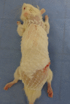
Animal in the immediate postoperative curative with three layers.
Figure 6
Figure 6
Electron micrographs obtained by SEM of the polymer membrane PL-co-co-TMC-DLA (50:50). A. Surface of the membrane. B. Fracture surface of the membrane.
Figure 7
Figure 7
Control group animals sacrificed 7 days after the procedure. Note the heterogeneous graft integration between defects in animals.
Figure 8
Figure 8
Treated animals sacrificed 7 days after the procedure. Note that the graft did not show signs of good integration compared to the control group.
Figure 9
Figure 9
Histological analysis of the control group 7 days after surgery (10×). A. Note the presence of young collagen organized with delicate collagen bundles parallel to the epidermal surface vessels, fibroblasts, mast cells, and mononuclear infiltrate. B. Surface crust and acute inflammation in the area has not yet re-epithelized.
Figure 10
Figure 10
Histological analysis of the treatment groups 7 days after surgery (10×). A. Dense acute inflammatory infiltrate. B. Segment of skin showing ischemic necrosis.
Figure 11
Figure 11
Animals in the control group sacrificed 60 days after the procedure. Note the homogeneous appearance of the skin between animals and integration of the graft to the recipient bed.
Figure 12
Figure 12
Treated animals 60 days after the procedure. There is delineation of neighboring grafted skin with good elasticity of the graft area.
Figure 13
Figure 13
Histological analysis of the control group 60 days after surgery (10×). A. The skin graft is thinner in the surrounding dermis and panniculus carnosus in the absence of skin depth. B. The normal range of the grafted area.
Figure 14
Figure 14
Histological analysis of the treatment group after 60 days of surgery (10×). A. Note the thick dermis tissue organization. B. Presence of skin scarring in the area of attachment.
Figure 15
Figure 15
Graph illustrating the comparison of dermal thickness between the control group and the treated group. Values represent the mean ± standard deviation. The means were statistically different. (P<0.05).
Figure 21
Figure 21
Graph illustrating the comparison of the collagen’s area in the control group and the treated group. Values represent the mean ± standard deviation. The means were statistically different. (P<0.05).
Figure 16
Figure 16
20× magnification. Type I collagen demonstrating the appearance of normal skin.
Figure 17
Figure 17
40× magnification. The short and interwoven type 1 collagen bundles are highlighted.
Figure 18
Figure 18
10× magnification. Presence of collagen type 1 in parallel beams on the surface.
Figure 19
Figure 19
10× magnification. Transition area between the dermis and scar region.
Figure 20
Figure 20
10× magnification. It is possible to note the area of transition between the area of the scar and the normal dermis. There is fibrosis near the hypodermis.

References

    1. Ferreira MC, Paggiaro AO, Isaac C, Teixeira Neto N, Santos GB. Substitutos cutâneos: conceitos atuais e proposta de classificação. Rev Bras Cir Plást. 2011;26:696–702.
    1. Ferreira MC, Tuma P Jr, Carvalho VF, Kamamoto F. Complex wounds. Clinics (São Paulo) 2006;61:571–8. - PubMed
    1. Leung JJ, Fish J. Skin Grafts. UTMJ. 2009;86:61–4.
    1. Burke JF, Yannas IV, Quinby WC Jr, Bondoc CC, Jung WK. Successful use of a physiologically acceptable artificial skin in the treatment of extensive burn injury. Ann Surg. 1981;194:413–28. - PMC - PubMed
    1. Wainwright DJ, Bury SB. Acellular dermal matrix in the management of the burn patient. Aesthet Surg J. 2011;31(Suppl):13S–23S. - PubMed

LinkOut - more resources