Review
. 2017;5(4):323-341.
doi: 10.1007/s40336-017-0235-x. Epub 2017 Jun 27.

Hybrid intraoperative imaging techniques in radioguided surgery: present clinical applications and future outlook

Affiliations
Review

Hybrid intraoperative imaging techniques in radioguided surgery: present clinical applications and future outlook

S L Bugby et al. Clin Transl Imaging. 2017.

Abstract

Purpose: This review aims to summarise the hybrid modality radioguidance techniques currently in clinical use and development, and to discuss possible future avenues of research. Due to the novelty of these approaches, evidence of their clinical relevance does not yet exist. The purpose of this review is to inform nuclear medicine practitioners of current cutting edge research in radioguided surgery which may enter standard clinical practice within the next 5-10 years. Hybrid imaging is of growing importance to nuclear medicine diagnostics, but it is only with recent advances in technology that hybrid modalities are being investigated for use during radioguided surgery. These modalities aim to overcome some of the difficulties of surgical imaging while maintaining many benefits, or providing entirely new information unavailable to surgeons with traditional radioguidance.

Methods: A literature review was carried out using online reference databases (Scopus, PubMed). Review articles obtained using this technique were citation mined to obtain further references.

Results: In total, 2367 papers were returned, with 425 suitable for further assessment. 60 papers directly related to hybrid intraoperative imaging in radioguided surgery are reported on. Of these papers, 25 described the clinical use of hybrid imaging, 22 described the development of new hybrid probes and tracers, and 13 described the development of hybrid technologies for future clinical use. Hybrid gamma-NIR fluorescence was found to be the most common clinical technique, with 35 papers associated with these modalities. Other hybrid combinations include gamma-bright field imaging, gamma-ultrasound imaging, gamma-β imaging and β-OCT imaging. The combination of preoperative and intraoperative images is also discussed.

Conclusion: Hybrid imaging offers new possibilities for assisting clinicians and surgeons in localising the site of uptake in procedures such as in sentinel node detection.

Keywords: Hybrid imaging; Intraoperative imaging; Multimodality imaging; Radioguided surgery.


Conflict of interest statement

S L Bugby, J E Lees and A C Perkins developed the Hybrid Gamma Camera discussed in this review (“Gamma-bright field imaging Current status”) and declare that they have no other conflicts of interest. This is a review article—this article does not contain any studies with human or animal subjects performed by any of the authors.

Figures

Fig. 1
Fig. 1
Comparison of preoperative images. In a patient with breast cancer, oblique planar lymphoscintigraphy (a) 2 h post-injection showed one sentinel node (SN) in the left axilla. The portable hybrid camera was placed above the lymphatic field to obtain an overview image at a distance of approximately 15 cm (b). Standard portable gamma camera imaging showed the injection site (IS) and SN in the same relation to each other, but without any anatomical references (c). Combined optical and γ-imaging visualised the image field of view and anatomical SN location in the left axilla (d). In the fused image, the SN is visualised on top of the red laser pointer cross indicating an accurate image fusion (Figure reproduced with permission from [21]—Hellingman et al. A new portable hybrid camera for fused optical and scintigraphic imaging: first clinical experiences. Clinical Nuclear Medicine (http://journals.lww.com/nuclearmed), 41(1):e39–e43, 2016. Promotional and commercial use of the material in print, digital or mobile device format is prohibited without the permission from the publisher Wolters Kluwer. Please contact healthpermissions@wolterskluwer.com for further information)
Fig. 2
Fig. 2
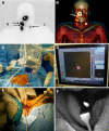
Combined preoperative lymphatic mapping and intraoperative radio- and fluorescence-guided sentinel node biopsy. a Early static anterior preoperative lymphoscintigram 10 min after infraorbital peritumoral injection of ICG–99mTc-nanocolloid showing the injection site (T) with lymphatic drainage to two sentinel nodes in the neck on the right (R) side and a third one on the left (L) side (arrows). b 3D SPECT/CT image 2 h post-injection providing additional anatomical information with visualisation of a lymphatic duct (arrow) originating from the injection site (T). c, d Intraoperatively, the radioactive component of the hybrid tracer in the left sentinel node is visualised using a portable gamma camera, and its laser pointer guides placement of the incision. e, f A near-infrared fluorescence camera is used to visualise the fluorescent component of the hybrid tracer in the same (non-blue) sentinel node (Figure reproduced with permission from [40])
Fig. 3
Fig. 3
Freehand SPECT–ultrasound in action during sentinel lymph node aspiration biopsy. a Freehand SPECT acquisition using a handheld gamma camera as nuclear detector, in a breast cancer patient. b Placement of the needle for aspiration biopsy based on freehand SPECT–US image. c B-mode image of axilla of patient showing at least one lymph node. d Freehand SPECT–US combination highlights the radioactive SLN by making it more prominent (Figure reproduced with permission from [19]—Wendler. Intraoperative 3D nuclear imaging and its hybrid extensions)
Fig. 4
Fig. 4
Wide local excision specimen from a patient with a grade 3, ER-/HER2-, no special type (NST) carcinoma. a Cerenkov image; b Greyscale photographic image overlaid with Cerenkov signal. An increased signal from the tumour is visible (white arrows); the mean radiance is 871 ± 131 photons/s/cm2/sr and the mean tumour to background ratio is 3.22. Both surgeons measured the posterior margin (outlined in blue) as 2 mm (small arrow); a cavity shaving would have been performed if the image had been available intraoperatively. The medial margin (outlined in green) measured >5 mm by both surgeons. Pathology ink prevented assessing the lateral margin; a phosphorescent signal is visible (open arrows). c Specimen radiography image. The absence of one surgical clip to mark the anterior margin, and the odd position of the superior margin clip (white arrow) prevented reliable margin assessment. d Combined histopathology image from two adjacent pathology slides on which the posterior margin (bottom of image) and part of the primary tumour are visible (open arrows). The distance from the posterior margin measured 3 mm microscopically (two headed arrow). The medial margin is >5 mm (not present in image) (Figure reproduced with permission from research originally published in JNM [96] © by the Society of Nuclear Medicine and Molecular Imaging Inc.)
Fig. 5
Fig. 5
One set of images obtained from the right ovary (abnormal) of a patient. a Positron distribution map; b one representative OCT image obtained from a sequence of co-registered OCT images; c corresponding 40× H&E histology. Red circle corpus albicans; red arrow collagen; purple arrow congested vessels; yellow arrow dermoid tumour. The OCT image size is 2 mm (depth) × 5 mm (lateral) (height × width); the histology size is 2 mm × 2.6 mm (height × width); the white scale bar is 0.5 mm (Figure reproduced with permission from [87])

Similar articles

Cited by

References

    1. Povoski SR, Neff RL, Mojzisik CM, O’Malley DM, Hinkle GH, Hall NC, Murrey DA, Jr, Knopp MV, Martin EW., Jr A comprehensive overview of radioguided surgery using gamma detection probe technology. World J Surg Oncol. 2009;7:11. doi: 10.1186/1477-7819-7-11. - DOI - PMC - PubMed
    1. Krag DN, Weaver DL, Alex JC, Fairbank JT. Surgical resection and radiolocalization of the sentinel lymph node in breast cancer using a gamma probe. Surg Oncol. 1993;2(6):335–340. doi: 10.1016/0960-7404(93)90064-6. - DOI - PubMed
    1. Giammarile F, Alazraki N, Aarsvold JN, Audisio RA, Glass E, Grant SF, Kunikowska J, Leidenius M, Moncayo VM, Uren RF, Oyen WJG, Valdés Olmos RA, Vidal Sicart S. The EANM and SNMMI practice guideline for lymphoscintigraphy and sentinel node localization in breast cancer. Eur J Nucl Med Mol Imaging. 2013;40(12):1932–1947. doi: 10.1007/s00259-013-2544-2. - DOI - PubMed
    1. Chen SL, Iddings DM, Scheri RP, Bilchik AJ. Lymphatic mapping and sentinel node analysis: current concepts and applications. CA Cancer J Clin. 2006;56(5):292–309. doi: 10.3322/canjclin.56.5.292. - DOI - PubMed
    1. Garcia-Parra R, Clinthorne N, Wang L, Picchio M, Piert M. Performance of beta- and high-energy gamma probes for the detection of cancer tissue in experimental surgical resection beds. Ann Nucl Med. 2011;25(7):486–493. doi: 10.1007/s12149-011-0492-0. - DOI - PubMed

LinkOut - more resources