Clinical Trial
. 2017:2017:4636809.
doi: 10.1155/2017/4636809. Epub 2017 Jul 18.

Open Wedge High Tibial Osteotomy with Distal Tubercle Osteotomy Lessens Change in Patellar Position

Affiliations
Clinical Trial

Open Wedge High Tibial Osteotomy with Distal Tubercle Osteotomy Lessens Change in Patellar Position

Hoon Park et al. Biomed Res Int. 2017.

Abstract

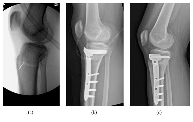
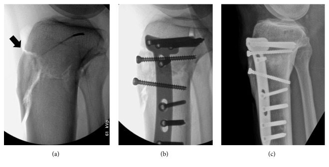
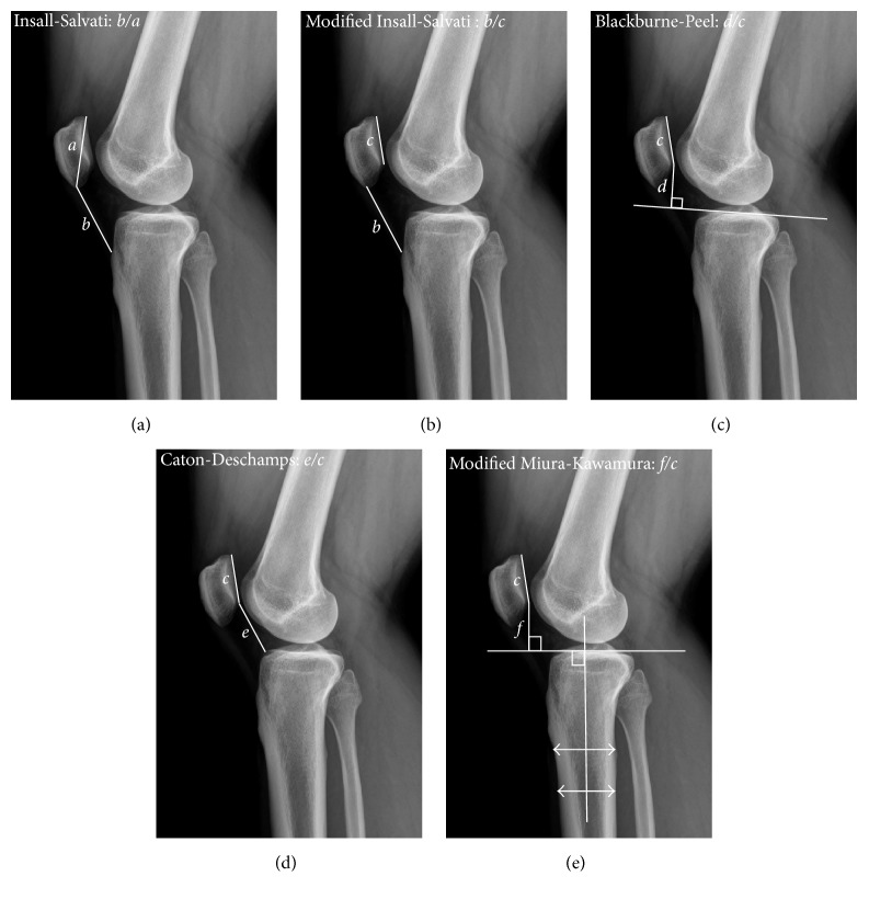
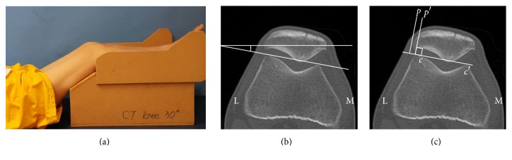
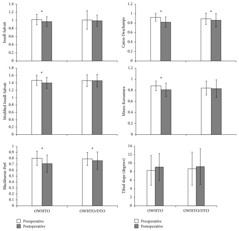
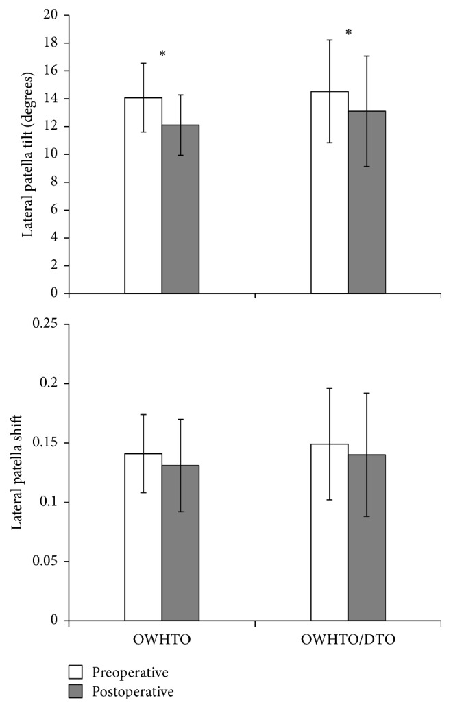
The purpose of this study was to investigate the change in patellar position after open wedge high tibial osteotomy (OWHTO) with distal tubercle osteotomy (DTO), comparing outcomes of conventional OWHTO in young adults with proximal tibia varus deformity but no arthritic manifestations. Thirty-three patients (mean age, 31.8 years) subjected to OWHTO/DTO were matched with 30 patients (mean age, 33.5 years) undergoing conventional OWHTO. Patellar position, as measured in pre- and postoperative standing lateral radiographs, was compared. Patellar height was assessed via Insall-Salvati ratio, modified Insall-Salvati ratio, Blackburne-Peel (BP) index, Caton-Deschamps (CD) index, and modified Miura-Kawamura index. Computed tomography was used to measure lateral patellar tilt and shift. In the OWHTO group, all patellar height indices decreased significantly following surgery. Although mean values of BP and CD indices decreased significantly in the OWHTO/DTO group, other determinants of patellar height showed no significant postoperative differences. Significant postoperative declines in average lateral patellar tilt were also evident in both groups, but pre- and postoperative lateral patellar shift did not differ significantly. OWHTO/DTO can be performed without significant changes in patellar height. The results obtained support that OWHTO/DTO is suitable for relatively young patients with proximal tibia vara but no arthritic change.


Figures

Figure 1
Figure 1
Lateral radiographs of open wedge high tibial osteotomy with distal tubercle osteotomy: (a) biplanar retrotubercle osteotomy (intraoperative view); (b) locking plate fixation at osteotomy site (immediately postoperative); and (c) union of the osteotomy site (postoperative month 3).
Figure 2
Figure 2
Images of patient with tibial tuberosity fracture: (a) fracture of proximal tibial tuberosity (arrow) in C-arm image; (b) fixation via two additional cannulated screws; and (c) healed fracture (postoperative month 3).
Figure 3
Figure 3
Radiographic diagrams showing five indices measured to assess patellar height: (a) Insall-Salvati; (b) modified Insall-Salvati; (c) Blackburne-Peel; (d) Caton-Deschamps; and (e) modified Miura-Kawamura. a, diagonal patellar length; b, patellar length; c, length of the patellar articular surface; d, perpendicular distance between the proximal tibial articular surface and the inferior tip of patellar articular surface; e, distance from the anterosuperior border of the tibial plateau to the inferior tip of the patellar articular surface; f, distance from a line perpendicular to the tibial diaphyseal line to the inferior tip of the patellar articular surface.
Figure 4
Figure 4
Methods of assessing lateral patellar tilt (LPT) and lateral patellar shift (LPS): (a) patient positioned for computed tomography imaging with knee in 30 degrees of flexion; (b) LPT, defined as the angle between a line intersecting the widest bony structure of the patella and a line tangential to the anterior femoral condylar surfaces; (c) LPS, defined as the ratio of distances pp′/cc′, where cc′ is the distance between the summits of the medial and lateral femoral condyles and pp′ is the distance between the summit of the lateral femoral condyle and the point where a line from the lateral edge of the patella perpendicular to the line that passed through the summits of the femoral condyles crosses that line. L, lateral; M, medial.
Figure 5
Figure 5
Patellar height indices and tibial slope pre- and postoperatively, charted by operative group. OWHTO, open wedge high tibial osteotomy; DTO; distal tubercle osteotomy. Significant change after surgery (p < 0.05).
Figure 6
Figure 6
Lateral patellar tilt and lateral patellar shift pre- and postoperatively, charted by operative group. OWHTO, open wedge high tibial osteotomy; DTO, distal tubercle osteotomy. Significant change after surgery (p < 0.05).

Similar articles

Cited by

References

    1. Strecker W. Planning Analysis of Knee-Adjacent Deformities. European Journal of Trauma and Emergency Surgery. 2007;33(6):662–668. doi: 10.1007/s00068-007-5175-0. - DOI - PubMed
    1. Rossi R., Bonasia D. E., Amendola A. The Role of High Tibial Osteotomy in the Varus Knee. American Academy of Orthopaedic Surgeon. 2011;19(10):590–599. doi: 10.5435/00124635-201110000-00003. - DOI - PubMed
    1. Marti R. K., Verhagen R. A. W., Kerkhoffs G. M. M. J., Moojen T. M. Proximal tibial varus osteotomy: Indications, technique, and five to twenty-one-year results. Journal of Bone and Joint Surgery - Series A. 2001;83(2):164–170. doi: 10.2106/00004623-200102000-00002. - DOI - PubMed
    1. El-Azab H., Glabgly P., Paul J., Imhoff A. B., Hinterwimmer S. Patellar height and posterior tibial slope after open- and closed-wedge high tibial osteotomy: A radiological study on 100 patients. American Journal of Sports Medicine. 2010;38(2):323–329. doi: 10.1177/0363546509348050. - DOI - PubMed
    1. Kaper B. P., Bourne R. B., Rorabeck C. H., MacDonald S. J. Patellar infera after high tibial osteotomy. Journal of Arthroplasty. 2001;16(2):168–173. doi: 10.1054/arth.2001.20538. - DOI - PubMed

Supplementary concepts

LinkOut - more resources