. 2018 Jan;22(1):261-276.
doi: 10.1111/jcmm.13316. Epub 2017 Aug 14.

Mesenchymal stem cells deliver exogenous miR-21 via exosomes to inhibit nucleus pulposus cell apoptosis and reduce intervertebral disc degeneration

Affiliations

Mesenchymal stem cells deliver exogenous miR-21 via exosomes to inhibit nucleus pulposus cell apoptosis and reduce intervertebral disc degeneration

Xiaofei Cheng et al. J Cell Mol Med. 2018 Jan.

Abstract

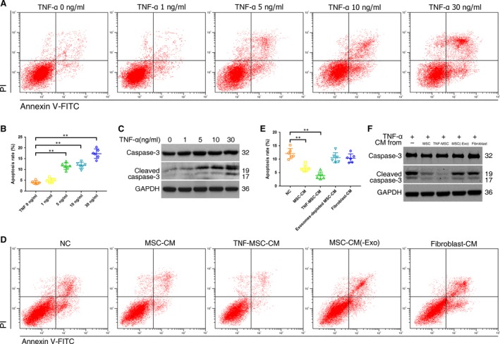
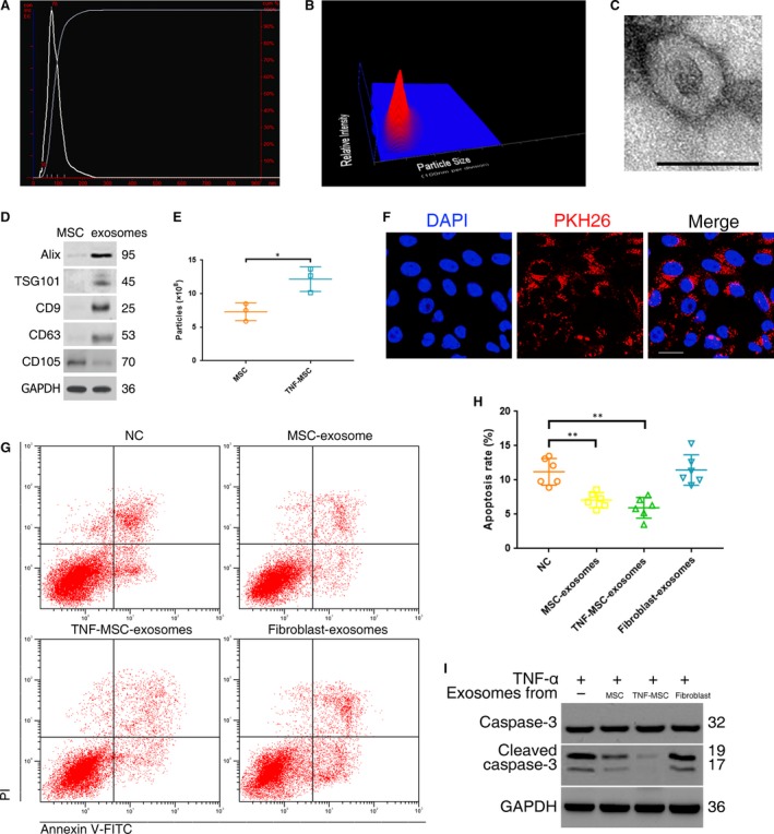
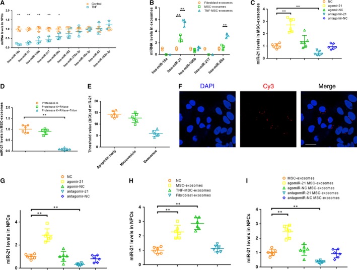
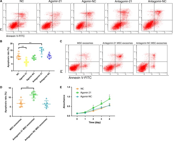
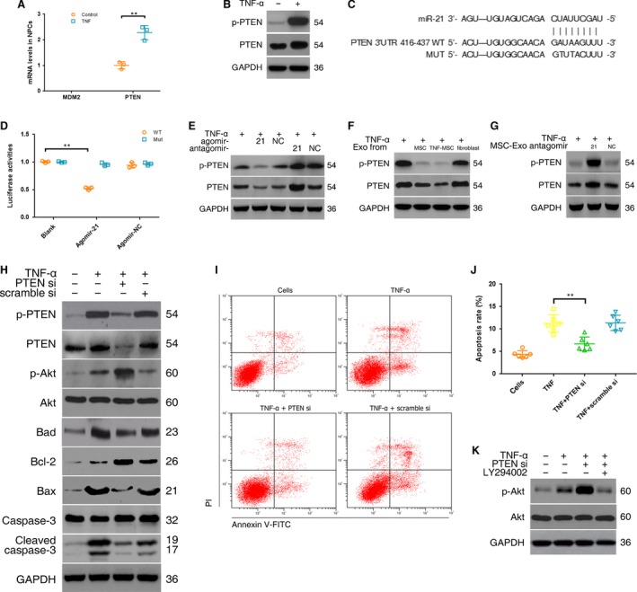
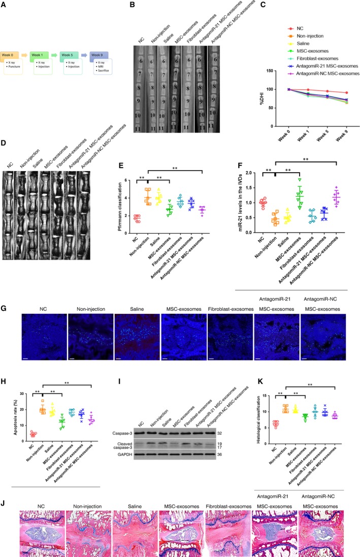
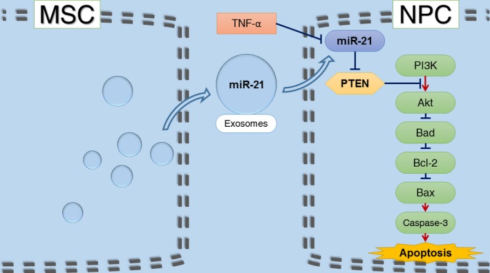
Although mesenchymal stem cells (MSCs) transplantation into the IVD (intervertebral disc) may be beneficial in inhibiting apoptosis of nucleus pulposus cells (NPCs) and alleviating IVD degeneration, the underlying mechanism of this therapeutic process has not been fully explained. The purpose of this study was to explore the protective effect of MSC-derived exosomes (MSC-exosomes) on NPC apoptosis and IVD degeneration and investigate the regulatory effect of miRNAs in MSC-exosomes and associated mechanisms for NPC apoptosis. MSC-exosomes were isolated from MSC medium, and its anti-apoptotic effect was assessed in a cell and rat model. The down-regulated miRNAs in apoptotic NPCs were identified, and their contents in MSC-exosomes were detected. The target genes of eligible miRNAs and possible downstream pathway were investigated. Purified MSC-exosomes were taken up by NPCs and suppressed NPC apoptosis. The levels of miR-21 were down-regulated in apoptotic NPCs while MSC-exosomes were enriched in miR-21. The exosomal miR-21 could be transferred into NPCs and alleviated TNF-α induced NPC apoptosis by targeting phosphatase and tensin homolog (PTEN) through phosphatidylinositol 3-kinase (PI3K)-Akt pathway. Intradiscal injection of MSC-exosomes alleviated the NPC apoptosis and IVD degeneration in the rat model. In conclusion, MSC-derived exosomes prevent NPCs from apoptotic process and alleviate IVD degeneration, at least partly, via miR-21 contained in exosomes. Exosomal miR-21 restrains PTEN and thus activates PI3K/Akt pathway in apoptotic NPCs. Our work confers a promising therapeutic strategy for IVD degeneration.

Keywords: apoptosis; exosomes; intervertebral disc degeneration; mesenchymal stem cells; miR-21; nucleus pulposus cells; phosphatase and tensin homolog.


Figures

Figure 1
Figure 1
Effect of TNF‐α on NPC apoptosis and anti‐apoptotic function of MSC‐conditioned medium (CM). (A) NPCs were treated with 0, 1, 5, 10 or 30 ng/ml of TNF‐α for 12 hrs. Representative dot plots of apoptosis flow cytometry detection were shown after Annexin V‐FITC/propidium iodide (PI) dual staining. (B) The percentage of the apoptotic cells was 4.0% ± 0.7% (0 ng/ml), 5.1% ± 1.0% (1 ng/ml), 11.6% ± 1.5% (5 ng/ml), 12.0% ± 1.4% (10 ng/ml) and 17.4% ± 2.2% (30 ng/ml) (** P < 0.01 by Kruskal–Wallis test). (C) Western blot analysed caspase‐3 and cleaved caspase‐3 in NPCs after treatment of different TNF‐α concentrations. (D) Anti‐apoptotic activities of MSCCM, CM of TNF‐α (5 ng/ml) pretreated MSCs (TNFMSCCM), exosomes‐depleted MSCCM (MSCCM(‐Exo)) and fibroblast‐CM were assessed on TNF‐α‐treated NPCs. Representative dot plots of cell apoptosis were shown. (E) MSCCM and TNFMSCCM reduced NPC apoptosis rates from 12.1% ± 1.9% to 6.7% ± 1.2% and 4.0% ± 1.3%, respectively (** P < 0.01 by Kruskal–Wallis test). Neither exosomes‐depleted MSCCM nor fibroblast‐CM remarkably reduced the number of dead cells (10.6% ± 1.9%; 10.4% ± 1.6%). (F) Western blot analysed caspase‐3 and cleaved caspase‐3 in NPCs after treatment of different CMs.
Figure 2
Figure 2
Exosomes secreted by MSCs were taken up by NPCs and suppressed NPC apoptosis. (A) Purified particles were analysed by nanoparticle tracking analysis (NTA). Sizes of the particles were between 30 and 200 nm. Modal and mean size was 78 nm and 87 nm, respectively. Concentration was 6.85 × 10⁸ particles/ml. (B) 3D heat map of the size distribution of the particles. (C) Transmission electron micrograph of purified particles. The image showed small vesicles of approximately 100 nm in diameter. Scale bar = 100 nm. (D) Expression of exosomes markers (ALIX, TSG101, CD9, CD63) and MSC marker (CD105) detected by Western blot. The protein expression of exosomes markers was detectable in MSC‐exosomes but not MSCs. (E) NTA counted the production of particles derived from MSCs and those from MSCs pretreated with 5 ng/ml TNF‐α (TNFMSC) (n = 3; *P < 0.05 by t‐test). (F) PKH26‐labelled MSC‐exosomes showed red fluorescence in the cytoplasm of NPCs. Scale bar = 25 μm. (G) Anti‐apoptotic activities of MSC‐exosomes, exosome secreted by TNFMSC (TNFMSC‐exosomes) and fibroblast‐exosomes were assessed on TNF‐α‐treated NPCs. Representative dot plots of apoptosis flow cytometry detection were shown. (H) The percentage of apoptotic NPCs could be decreased to 7.1% ± 1.1% by MSC‐exosomes or to 6.0% ± 1.5% by TNFMSC‐exosomes compared with the control (11.2% ± 1.9%), while fibroblast‐derived exosomes did not cause significant change of apoptosis rate (11.5% ± 2.2%) (**P < 0.01 by Kruskal–Wallis test). (I) Western blot analysed caspase‐3 and cleaved caspase‐3 in NPCs after treatment of different exosomes.
Figure 3
Figure 3
MSC‐exosomes were enriched in miR‐21, which could be transferred into NPCs (A) qRTPCR revealed that five miRNAs were significantly down‐regulated in TNF‐α‐treated NPCs compared with non‐treated cells (n = 3; *P < 0.05, **P < 0.01 versus the control by t‐test, t′‐test or Mann–Whitney U‐test). (B) qRTPCR showed that miR‐21 contents in MSC‐exosomes were significantly greater than that in fibroblast‐derived exosomes (n = 3; **P < 0.01 by anova or Kruskal–Wallis test). (C) Overexpression or knockdown of miR‐21 was confirmed by qRTPCR in exosomes derived from MSCs transfected with a miR‐21 agonist or antagonist (**P < 0.01 by Kruskal–Wallis test). (D) qRTPCR showed that miR‐21 within lipid‐bilayered exosomes was protected from RNase degradation (**P < 0.01 by anova test). (E) Quantitative analysis of miR‐21 was performed by qRTPCR on the pellets of extracellular vesicles isolated from MSC supernatant by differential centrifugation. (F) Exosomes were collected from the supernatant of MSCs transfected with cy3‐labeled agomir‐21. NPCs incubated with these exosomes exhibited a granular fluorescent pattern within the cytoplasm. Scale bar = 25 μm (G) qRTPCR showed that miR‐21 levels in NPCs were up‐regulated by the miR‐21 agonist and down‐regulated by the miR‐21 antagonist (**P < 0.01 by Kruskal–Wallis test). (H) qRTPCR showed that the decreased levels of miR‐21 in TNF‐α‐treated NPCs were rescued by incubation with MSC‐exosomes or exosomes from TNF‐α (5 ng/ml) pretreated MSCs (TNFMSC‐exosomes) but not with fibroblast‐exosomes (**P < 0.01 by Kruskal–Wallis test). (I) qRTPCR showed that overexpression or knockdown of intracellular miR‐21 levels was confirmed on MSC‐exosomes treated apoptotic NPCs when exposed to miR‐21‐overexpressing or miR‐21‐deficient exosomes (**P < 0.01 by Kruskal–Wallis test).
Figure 4
Figure 4
miR‐21 in MSC‐exosomes alleviated TNF‐α induced NPC apoptosis. (A) Effect of miR‐21 on NPC apoptosis was evaluated using the flow cytometry analysis. Representative dot plots of cell apoptosis were shown. (B) The agomir‐21 significantly alleviated NPC apoptosis (apoptosis rate 6.6% ± 2.0%), while the antagomir‐21 exacerbated apoptosis (apoptosis rate 14.9% ± 2.5%; **P < 0.01 by Kruskal–Wallis test). (C) Anti‐apoptotic activities of miR‐21‐depleted MSC‐exosomes were detected using the flow cytometry analysis. Representative dot plots of cell apoptosis were shown. (D) The percentage of the apoptotic cells was increased after knockdown of miR‐21 in MSC‐exosomes (apoptosis rate 11.4% ± 1.6% versus control 6.9% ± 1.9%; **P < 0.01 by Kruskal–Wallis test). (E) CCK‐8 proliferation assay showed that cell proliferation was significantly increased in agomir‐21‐transfected NPCs compared with non‐transfected or agomir‐NC‐transfected cells (* P < 0.05 versus NC or agomir‐NC by Kruskal–Wallis test).
Figure 5
Figure 5
Delivery of miR‐21 in MSC‐exosomes inhibited NPC apoptosis by targeting PTEN through PI3K‐Akt pathway. (A) The mRNA expression of MDM2 and PTEN in NPCs was detected using qRTPCR. PTEN expression was significantly increased following TNF‐α treatment (n = 3; **P < 0.01 by Mann–Whitney U‐test). (B) Western blot analysed PTEN protein levels in NPCs after TNF‐α treatment. (C) 3′‐UTR region of human PTEN mRNA was found to harbour a binding site for hsa‐miR‐21. (D) Luciferase reporter assay found that agomir‐21 exclusively decreased luciferase activity of the wild‐type reporter plasmids (n = 3; **P < 0.01 by anova test). Fluorescence intensity of firefly and Renilla was (3.80 ± 0.24) × 105 and(7.20 ± 0.83) × 106, respectively, for agomir‐21 cotransfection, and (1.08 ± 0.09) × 106 and(9.48 ± 0.44) × 106, respectively, for agomir‐NC cotransfection. (E) Western blot showed that the agomir‐21 significantly reduced while the antagomir‐21 increased the expression levels of PTEN protein compared with the control in TNF‐α‐treated NPCs. (F) Western blot showed that MSC‐exosomes (Exo) lowered PTEN protein levels in TNF‐α‐treated NPCs. (G) Western blot showed that miR‐21‐deficient MSC‐exosomes lost the ability of MSC‐exosomes to suppress PTEN expression. (H) Western blot showed that PTEN‐siRNA (PTEN si) remarkably increased expression of phospho‐Akt and Bcl‐2, and decreased expression of Bad, Bax and cleaved caspase‐3. (I) Effect of PTEN‐siRNA on TNF‐α induced NPC apoptosis was evaluated using flow cytometry analysis. Representative dot plots of cell apoptosis were shown. (J) PTEN‐siRNA significantly decreased the percentage of the apoptotic cells compared with the control (6.7% ± 1.5% versus 11.3% ± 1.9%; **P < 0.01 by Kruskal–Wallis test). (K) Western blot showed that the PI3K inhibitor (LY294002) markedly decreased phospho‐Akt protein expression.
Figure 6
Figure 6
Intradiscal injection of MSC‐exosomes alleviated the NPC apoptosis and IVD degeneration in a rat model. (A) A flow diagram of the experiments in vivo. (B) Radiographs of the indicated groups were obtained 9 weeks after needle puncture. Co6/7, Co8/9 and Co10/11 were punctured with Co7/8 and Co9/10 left intact. (C) Changes in disc height index (DHI) of the indicated groups after needle puncture. The DHI was measured at week 0, 1, 5, 9 time point. A significant decrease of the %DHI was observed in all puncture groups at 1 week after surgery (P < 0.01by t test). At each time point after puncture, a significant decrease of %DHI was noted in all puncture groups compared with the negative control group (P < 0.01 by Kruskal–Wallis test). No significant difference was found in the %DHI between all puncture groups. (D) MRIs of the indicated groups were obtained 9 weeks after needle puncture. Co6/7, Co8/9 and Co10/11 were punctured with Co7/8 and Co9/10 left intact. (E) The change of MRI grade in the indicated groups at 9 weeks after needle puncture. The degree of disc degeneration by MRI grade was significantly lower in the MSC‐exosomes group than in the non‐injection group (**P < 0.01 by Kruskal–Wallis test). (F) qRTPCR showed that the decreased levels of miR‐21 in the punctured IVDs were rescued by the injection of MSC‐exosomes but not fibroblast‐exosomes (**P < 0.01 by Kruskal–Wallis test). (G) TUNEL staining of IVDs in the indicated groups at 9 weeks after needle puncture. Blue fluorescence (DAPI) indicating total cells; Green fluorescence (FITC) indicating TUNEL‐positive cells. Scale bar = 150 μm (H) A significant decrease in the apoptosis rate was noted in the MSC‐exosomes group compared with the non‐injection group (**P < 0.01 by Kruskal–Wallis test). (I) Western blot analysed the levels of caspase‐3 and cleaved caspase‐3 in the IVDs in the indicated groups at 9 weeks after needle puncture. (J) HE staining of IVDs in the indicated groups at 9 weeks after needle puncture. (magnification ×40). (K) A significant decrease in the degeneration grade was noted in the MSC‐exosomes group compared with the non‐injection group (** P < 0.01 by Kruskal–Wallis test).
Figure 7
Figure 7
Schematic of our working hypothesis. MSC‐derived exosomes prevent NPCs from TNF‐α‐induced apoptotic process via miR‐21 contained in exosomes. Exosomal miR‐21 restrains PTEN and thus activates PI3K/Akt pathway in apoptotic NPCs.

References

    1. Disease GBD, Injury I, Prevalence C. Global, regional, and national incidence, prevalence, and years lived with disability for 310 diseases and injuries, 1990‐2015: a systematic analysis for the Global Burden of Disease Study 2015. Lancet. 2016; 388: 1545–602. - PMC - PubMed
    1. Risbud MV, Shapiro IM. Role of cytokines in intervertebral disc degeneration: pain and disc content. Nat Rev Rheumatol. 2014; 10: 44–56. - PMC - PubMed
    1. Johnson ZI, Schoepflin ZR, Choi H, et al Disc in flames: Roles of TNF‐alpha and IL‐1beta in intervertebral disc degeneration. Eur Cell Mater. 2015; 30: 104–16; discussion 16‐7. - PMC - PubMed
    1. Kepler CK, Markova DZ, Hilibrand AS, et al Substance P stimulates production of inflammatory cytokines in human disc cells. Spine. 2013; 38: E1291–9. - PubMed
    1. Wang B, Wang D, Yan T, et al MiR‐138‐5p promotes TNF‐α‐induced apoptosis in human intervertebral disc degeneration by targeting SIRT1 through PTEN/PI3K/Akt signaling. Exp Cell Res. 2016; 345: 199–205. - PubMed

Publication types

MeSH terms