. 2017 Aug 12;9(8):248.
doi: 10.3390/toxins9080248.

Lengths of the C-Terminus and Interconnecting Loops Impact Stability of Spider-Derived Gating Modifier Toxins

Affiliations

Lengths of the C-Terminus and Interconnecting Loops Impact Stability of Spider-Derived Gating Modifier Toxins

Akello J Agwa et al. Toxins (Basel). .

Abstract

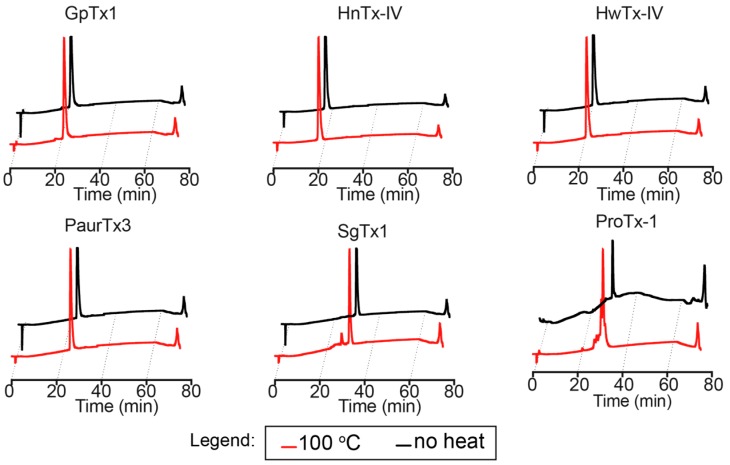
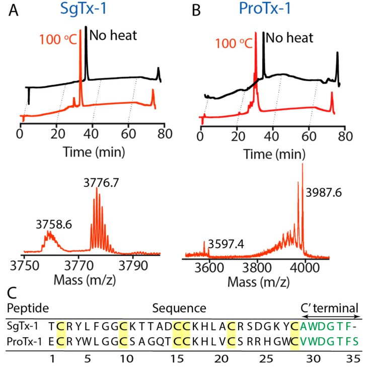
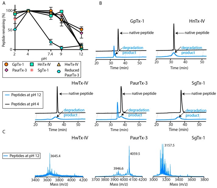
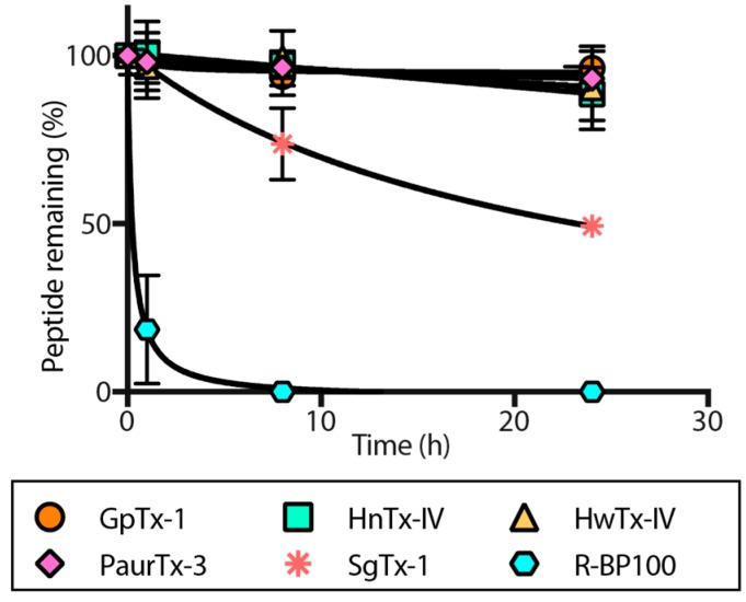
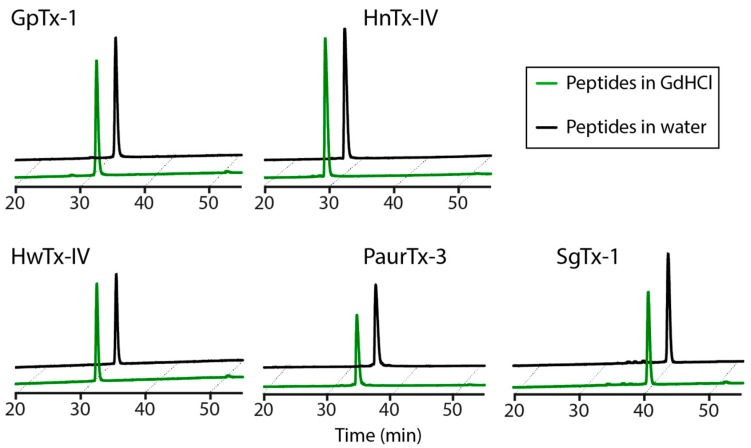
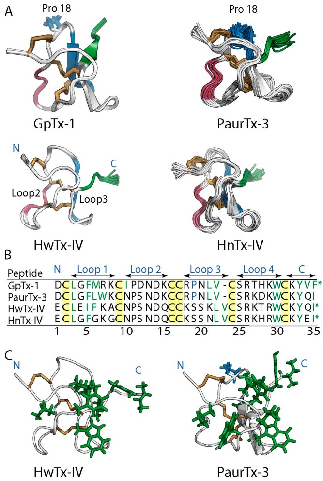
Spider gating modifier toxins (GMTs) are potent modulators of voltage-gated ion channels and have thus attracted attention as drug leads for several pathophysiological conditions. GMTs contain three disulfide bonds organized in an inhibitory cystine knot, which putatively confers them with high stability; however, thus far, there has not been a focused study to establish the stability of GMTs in physiological conditions. We examined the resistance of five GMTs including GpTx-1, HnTx-IV, HwTx-IV, PaurTx-3 and SgTx-1, to pH, thermal and proteolytic degradation. The peptides were stable under physiological conditions, except SgTx-1, which was susceptible to proteolysis, probably due to a longer C-terminus compared to the other peptides. In non-physiological conditions, the five peptides withstood chaotropic degradation, and all but SgTx-1 remained intact after prolonged exposure to high temperature; however, the peptides were degraded in strongly alkaline solutions. GpTx-1 and PaurTx-3 were more resistant to basic hydrolysis than HnTx-IV, HwTx-IV and SgTx-1, probably because a shorter interconnecting loop 3 on GpTx-1 and PaurTx-3 may stabilize interactions between the C-terminus and the hydrophobic patch. Here, we establish that most GMTs are exceptionally stable, and propose that, in the design of GMT-based therapeutics, stability can be enhanced by optimizing the C-terminus in terms of length, and increased interactions with the hydrophobic patch.

Keywords: NaV1.7; nuclear magnetic resonance; pain; rational drug design; serum stability; spider venom.


Conflict of interest statement

The authors declare no conflicts of interest.

Figures

Figure 1
Figure 1
Conserved structural features of spider-derived GMTs. (A) ribbon representations of GpTx-1 [17], HnTx-IV (PDB ID: 1NIY) [19], HwTx-IV (PDB ID: 2M4X) [21], PaurTx-3 (PDB ID: 5WE3, this study) and SgTx-1 (PDB ID: 1LA4) [18]. The backbones of the GMTs are shown in white and the disulfide bridges forming the inhibitory cystine knot are shown in brown. GpTx-1, HnTx-IV and PaurTx-3 each have two anti-parallel β-sheets, SgTx-1 has three, and HwTx-IV has no anti-parallel β-sheets. Locations of N- and C-termini and of Cys I–VI (highlighted in yellow) are identified on HwTx-IV for clarity; (B) surface representations of the GMTs showing the conserved hydrophobic patch and charged ring of spider toxins, where hydrophobic residues are green, positively charged residues are blue and negatively charged residues are red. The disulfide bridges are buried within the hydrophobic patch; (C) sequences of the peptides are shown (aligned to cysteine residues of HwTx-IV and HnTx-IV) with cysteines highlighted in yellow, residues making up interconnecting loops are identified with arrows, N- and C-termini are shown and * denotes amidated C-terminal.
Figure 2
Figure 2
Solution NMR structure of PaurTx-3 (PDB ID: 5WE3, this study). The 20 best conformers selected from lowest energy and best MolProbity scores are shown [35]. (A) Backbone of the structures is red and disulfide bridges are yellow with N- and C-termini and cysteines I-VI labeled; (B) two antiparallel β-sheets are shown in red, the residues forming the hydrophobic patch are shown in green and the turns on the structures are shown in blue with proline residues in position 11 and 18.
Figure 3
Figure 3
Reversible thermal denaturation of GMTs. 1D 1H solution NMR was used to monitor the structural changes to the five peptides at temperatures ranging from 20 to 80 °C. GpTx-1, HwTx-IV and PaurTx-3 maintained narrow peaks up to 80 °C, although there was some loss in peak intensity in the amide region of the spectra. SgTx-1 and HnTx-IV showed both a loss in intensity and broadening of peaks at 80 °C. NOE correlations confirm that the appearance of two peaks in the 10 ppm region of the 1D 1H spectra of PaurTx-3 are a result of the separation of the ε1 proton on the imidazole rings of Trp 29 and Trp 7.
Figure 4
Figure 4
Analytical RP-HPLC traces of the GMTs following heating to 100 °C. All of the peptides except SgTx-1 and ProTx-1 showed remarkable stability after the thermal assault.
Figure 5
Figure 5
Irreversible thermal degradation of SgTx-1 and ProTx-1. Analytical RP-HPLC was used to examine the peptides after heating to 100 °C and subsequent cooling to room temperature. Degradation products were further characterized using MALDI-MS (A) SgTx-1 lost 18 Da from the parent peptide; (B) ProTx-1 underwent more extensive degradation, showing a 389.4 Da loss; (C) sequences of SgTx-1 and ProTx-1 are shown with cysteines highlighted in yellow and C-terminal residues shown in green.
Figure 6
Figure 6
(A) pH dependent hydrolysis of the peptides was monitored at pH 2, 4, 7.4, 9 and 12 using analytical RP-HPLC following a 24 h incubation at 37 °C. Reduced PaurTx-3 was used as a control. Data points are relative to amount of peptide at pH 4 and error bars are ± SE for n = 3; (B) comparisons of the analytical RP-HPLC traces of the peptides at pH 4 and pH 12 are shown and (C) MALDI-MS spectra for HwTx-IV, PaurTx-3 and SgTx-1 at pH 12 are also shown.
Figure 7
Figure 7
Proteolytic degradation of GMTs. The five GMTs were incubated in human serum and degradation was monitored using RP-HPLC at 0 h, 1 h, 8 h and 24 h. R-BP100 was used as a control. Data points are relative to amount of peptide at 0 h and error bars are ± SE for n = 3.
Figure 8
Figure 8
GMT stability in 6 M GdHCl following 16 h incubation at 25 °C, as monitored using RP-HPLC.
Figure 9
Figure 9
Structural features of the loops, turns and C-termini of GMTs. (A) ribbon representations of 20 structures of PaurTx-3 (PDB ID: 5W3E, this study), HwTx-IV (PDB ID: 2M4X) [21], and HnTx-IV (PDB ID: 1NIY) [19], and 10 structures of GpTx-1 [17], are shown where loop 2 is highlighted in red, loop 3 is in blue and the C-terminal is in green (labels on HwTx-IV for clarity). The side chain of Pro 18 is shown for GpTx-1 and PaurTx-3 in blue; (B) sequences of the peptides are also shown and the residues forming each loop and the C-terminal are identified by arrows. Sequences of hydrophobic amino acid residues and residues forming the hydrophobic patch are colored green and * denotes amidated C-terminal; (C) green sticks show the side chains of hydrophobic residues on HwTx-IV and PaurTx-3.

References

    1. Smith J.J., Lau C.H.Y., Herzig V., Ikonomopoulou M.P., Rash L.D., King G.F. Therapeutic applications of spider-venom peptides. In: King G.F., editor. Venoms to Drugs: Venom as a Source for the Development of Human Therapeutics. Royal Society of Chemistry; London, UK: 2015. pp. 221–244.
    1. Escoubas P., Bosmans F. Spider peptide toxins as leads for drug development. Expert Opin. Drug Discov. 2007;2:823–835. doi: 10.1517/17460441.2.6.823. - DOI - PubMed
    1. Catterall W.A., Cestele S., Yarov-Yarovoy V., Yu F.H., Konoki K., Scheuer T. Voltage-gated ion channels and gating modifier toxins. Toxicon. 2007;49:124–141. doi: 10.1016/j.toxicon.2006.09.022. - DOI - PubMed
    1. Vetter I., Deuis J.R., Mueller A., Israel M.R., Starobova H., Zhang A., Rash L.D., Mobli M. NaV1.7 as a pain target—From gene to pharmacology. Pharmacol. Ther. 2017;172:73–100. doi: 10.1016/j.pharmthera.2016.11.015. - DOI - PubMed
    1. Osteen J.D., Herzig V., Gilchrist J., Emrick J.J., Zhang C., Wang X., Castro J., Garcia-Caraballo S., Grundy L., Rychkov G.Y., et al. Selective spider toxins reveal a role for the NaV1.1 channel in mechanical pain. Nature. 2016;534:494–499. doi: 10.1038/nature17976. - DOI - PMC - PubMed

Publication types

LinkOut - more resources