. 2017 Oct;49(10):1529-1538.
doi: 10.1038/ng.3933. Epub 2017 Aug 14.

Mutations in KEOPS-complex genes cause nephrotic syndrome with primary microcephaly

Daniela A Braun  1 Jia Rao  1 Geraldine Mollet  2   3 David Schapiro  1 Marie-Claire Daugeron  4 Weizhen Tan  1 Olivier Gribouval  2   3 Olivia Boyer  2   3   5 Patrick Revy  3   6 Tilman Jobst-Schwan  1 Johanna Magdalena Schmidt  1 Jennifer A Lawson  1 Denny Schanze  7 Shazia Ashraf  1 Jeremy F P Ullmann  8   9 Charlotte A Hoogstraten  1 Nathalie Boddaert  3   10   11 Bruno Collinet  4   12   13 Gaëlle Martin  2   3 Dominique Liger  4 Svjetlana Lovric  1 Monica Furlano  2   3   14 I Chiara Guerrera  15 Oraly Sanchez-Ferras  16 Jennifer F Hu  17 Anne-Claire Boschat  18 Sylvia Sanquer  19   20 Björn Menten  21 Sarah Vergult  21 Nina De Rocker  21 Merlin Airik  1 Tobias Hermle  1 Shirlee Shril  1 Eugen Widmeier  1   22 Heon Yung Gee  1   23 Won-Il Choi  1 Carolin E Sadowski  1 Werner L Pabst  1 Jillian K Warejko  1 Ankana Daga  1 Tamara Basta  4 Verena Matejas  24 Karin Scharmann  25   26 Sandra D Kienast  25   26 Babak Behnam  27   28 Brendan Beeson  29 Amber Begtrup  30 Malcolm Bruce  29 Gaik-Siew Ch'ng  31 Shuan-Pei Lin  32   33 Jui-Hsing Chang  34 Chao-Huei Chen  35 Megan T Cho  30 Patrick M Gaffney  36 Patrick E Gipson  37 Chyong-Hsin Hsu  34 Jameela A Kari  38 Yu-Yuan Ke  35 Cathy Kiraly-Borri  39 Wai-Ming Lai  40 Emmanuelle Lemyre  41 Rebecca Okashah Littlejohn  42   43 Amira Masri  44 Mastaneh Moghtaderi  45 Kazuyuki Nakamura  46 Fatih Ozaltin  47   48   49 Marleen Praet  50 Chitra Prasad  51 Agnieszka Prytula  52 Elizabeth R Roeder  42   43 Patrick Rump  53 Rhonda E Schnur  30 Takashi Shiihara  46 Manish D Sinha  54 Neveen A Soliman  55   56 Kenza Soulami  57 David A Sweetser  58 Wen-Hui Tsai  59 Jeng-Daw Tsai  33   34   60   61 Rezan Topaloglu  47 Udo Vester  62 David H Viskochil  63 Nithiwat Vatanavicharn  64 Jessica L Waxler  58 Klaas J Wierenga  65 Matthias T F Wolf  66 Sik-Nin Wong  67 Sebastian A Leidel  25   26   68 Gessica Truglio  8 Peter C Dedon  69   70 Annapurna Poduri  8   9 Shrikant Mane  71 Richard P Lifton  71   72 Maxime Bouchard  16 Peter Kannu  73 David Chitayat  73 Daniella Magen  74 Bert Callewaert  21 Herman van Tilbeurgh  4 Martin Zenker  7 Corinne Antignac  2   3   75 Friedhelm Hildebrandt  1
Affiliations

Mutations in KEOPS-complex genes cause nephrotic syndrome with primary microcephaly

Daniela A Braun et al. Nat Genet. 2017 Oct.

Abstract

Galloway-Mowat syndrome (GAMOS) is an autosomal-recessive disease characterized by the combination of early-onset nephrotic syndrome (SRNS) and microcephaly with brain anomalies. Here we identified recessive mutations in OSGEP, TP53RK, TPRKB, and LAGE3, genes encoding the four subunits of the KEOPS complex, in 37 individuals from 32 families with GAMOS. CRISPR-Cas9 knockout in zebrafish and mice recapitulated the human phenotype of primary microcephaly and resulted in early lethality. Knockdown of OSGEP, TP53RK, or TPRKB inhibited cell proliferation, which human mutations did not rescue. Furthermore, knockdown of these genes impaired protein translation, caused endoplasmic reticulum stress, activated DNA-damage-response signaling, and ultimately induced apoptosis. Knockdown of OSGEP or TP53RK induced defects in the actin cytoskeleton and decreased the migration rate of human podocytes, an established intermediate phenotype of SRNS. We thus identified four new monogenic causes of GAMOS, describe a link between KEOPS function and human disease, and delineate potential pathogenic mechanisms.


Conflict of interest statement

Competing interests statement

M.T.C., A.B., and R.E.S. are employees of GeneDx, Gaithersburg, MD, USA.

The other authors declare that they have no competing financial interests.

Figures

Figure 1
Figure 1. Whole exome sequencing identifies recessive mutations in the 4 KEOPS complex encoding genes LAGE3, OSGEP, TP53RK, TPRKB in 30 families with GAMOS 2–5
(a–d) Left or upper panels show clinical phenotype of individuals with recessive mutations, right or lower panels show exon structures and secondary structures of human LAGE3 (a), OSGEP (b), TP53RK (c), and TPRKB (d) cDNA. Positions of start codons and of stop codons are indicated. Predicted secondary structure is indicated for beta strand and α-helix conformation as triangle and zigzag lines, respectively. (a) Clinical features of individual B60 with a hemizygous mutation of LAGE3: Renal histology showing FSGS, transmission electron microscopy (TEM) showing podocyte foot process effacement (arrowheads), and MRI showing microcephaly with polymicrogyria and diffuse cerebellar atrophy. (b) Clinical features of individuals ‘DC’, B80, B58, and B377 with mutations of OSGEP: Renal histology of individual ‘DC’ showing FSGS, and TEM of B80 showing foot process effacement (arrow heads). Cranial imagining of individuals B58 and B377 showing microcephaly, reduced gyration, and diffuse cortical atrophy. Clinical photograph of B58 showing severe deformation of the forehead. (c) Clinical features of individual B77_22 with compound heterozygous mutations of TP53RK: Cranial imaging showing microcephaly and polymicrogyria. (d) Clinical features of individual B1144 with a homozygous mutation of TPRKB: Renal histology showing FSGS and TEM showing podocyte foot process effacement (arrow heads). Brain MRI showing microcephaly and pachygyria. HEMI, hemizygous; het, heterozygous; HOM, homozygous.
Figure 2
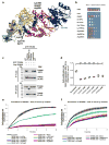
Figure 2. TP53RK mutations abrogate interaction with TPRKB, and GAMOS mutations fail to rescue cell proliferation rate decreased by knockdown of OSGEP or TP53RK
(a) Structural modeling of KEOPS mutations. The three-dimensional representation of the human KEOPS complex is based on structural information of subcomplexes from archeae and yeast. Subunits are represented in different colors: LAGE3 (green), OSGEP (yellow), TP53RK (red) and TPRKB (blue). The residues corresponding to missense mutations identified in patients with GAMOS2–5 are mapped as black spheres and their corresponding positions are written in green (LAGE3), black (OSGEP), red (TPR53K) and blue (TPRKB). A model of a nucleotide bound in the active site of OSGEP is represented as cyan spheres. (b) Overexpression of human OSGEP in a Δkae1 (orthologous yeast gene) yeast strain partially rescues the Δkae1 associated growth defect. Each horizontal panel corresponds to tenfold serial dilution of the cell suspension. The extent of growth complementation was assessed by comparison with full complementation (KAE1) and no complementation (none). Overexpressed alleles are indicated on the left. Mutants OSGEP alleles fall into two classes: hypomorphic alleles (Lys198Arg, Arg247Gln, Arg280Cys, Arg325Gln) and amorphic alleles (Ile14Phe, Ile111Thr, Cys110Arg). (c) Co-immunoprecipitation of FLAG-tagged full-length wildtype TPRKB with GFP-tagged wildtype TP53RK or mutant TP53RK cDNA constructs that reflect the GAMOS mutations. Constructs reflecting the TP53RK mutants p.Lys60Serfs*61 and p.Thr81Arg abrogate interaction with TPRKB. (d) A colorimetric BrdU assay demonstrates that human podocytes with stable knockdown of OSGEP, TP53RK, and TPRKB exhibit reduced cell proliferation rates. Cells are at passage 3 after transduction, * indicates p<0.05, **indicates p<0.01 calculated by one-way ANOVA. (e–f) Proliferation rate of human immortalized podocytes was assayed using the xCELLigence® system. Cells are at passage 4 after stable transduction with shRNA targeting OSGEP and TP53RK. (e) OSGEP knockdown reduced podocyte proliferation rate (red), which was rescued by stable overexpression of mouse wildtype, full-length Osgep (green), but not of mutant constructs reflecting the mutations identified in individuals with GAMOS. (f) TP53RK knockdown reduced podocyte proliferation rate (red), which was rescued by stable overexpression of mouse wildtype, full-length Tp53rk (green), but not of mutant Tp53rk constructs reflecting the mutations identified in individuals with GAMOS.
Figure 3
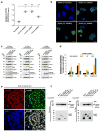
Figure 3. Knockdown of OSGEP, TP53RK, or TPRKB in human podocytes induces DNA damage response signaling (DDR) and subsequently apoptosis
(a) In a colorimetric assay, knockdown of OSGEP, TP53RK, or TPRKB in human podocytes increases caspase-3 activity indicating apoptotic cell death. Cells are at passage 4 after transduction, **indicates p<0.01 calculated by one-way ANOVA. (b) Staining with an antibody against phosphorylated histone H2A.X (γH2AX) was increased in human podocytes 7 days after stable shRNA knockdown of OSGEP, TP53RK, or TPRKB (passage 3) as compared to scrambled control cells indicating induction of DDR. DAPI stains DNA (blue). Scale bar 10μm. Quantification of 100 cells for each condition is shown in Suppl. Fig. 16A. (c) Compared to scrambled shRNA transfected control cells, knockdown of OSGEP in human podocytes increases γH2AX and CDK inhibitor p21 on days 5, 7, and 9 after knockdown. Strong increase of the proapoptotic factor BAX and cleaved caspase-3 is only observed on days 7 and 9. (d) Densitometry of the intensity of immunoblot signals of γH2AX, p21, BAX, and cleaved caspase-3 in human immortalized podocytes cell lines was measured using ImageJ. Absolute values were normalized to beta actin (loading controls). Intensities of scrambled control cells (black) were normalized as 1. Data points are presented as mean of two OSGEP shRNAs and standard deviation. Knockdown of OSGEP results in an increase of γH2AX and p21 on days 5,7, and 9 after knockdown. In contrast, BAX and cleaved caspase-3 only show strong induction on days 7 and 9 after transduction demonstrating that the activation of DDR (γH2AX) precedes the induction of apoptosis (BAX, cleavage of caspase-3) upon knockdown of OSGEP. The decrease of signals for γH2AX, p21, and BAX on day 9 is likely explained by apoptotic cell death. (f) Staining of renal rat sections (p1) with antibodies against PARP1 and TP53RK demonstrates that both proteins colocalize to PARP1 positive nuclear foci in renal glomeruli. DAPI stains DNA (blue). Scale bars are 7.5 μm. (g) Upon overexpression Flag-tagged LAGE3, OSGEP, TP53RK, and TPRKB immunoprecipitate endogenous PARP1 in HEK293T cells.
Figure 4
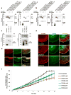
Figure 4. OSGEP and TP53RK colocalize with the ARP2/3 complex at lamellipodia of podocytes, and knockdown disrupts the actin cytoskeleton and impairs cell migration
(a–b) Upon co-overexpression in HEK293T cells GFP-tagged OSGEP (a) and TP53RK (b) interact with 4 FLAG-tagged proteins of the ARP2/3 complex, namely ARPC1B, ARPC2, ACTR2, and ACTR3. (c) Half-endogenous co-immunoprecipitation of GFP-tagged OSGEP and TP53RK confirms interaction with endogenous ARP2 in HEK293T cells. (d) Upon EGF stimulation (100 ng/ml, 20 min) to induce lamellipodia formation, OSGEP (red) and TP53RK (red) co-localize with ARP2 (green) and ARP3 (green) at lamellipodia of differentiated human podocytes. The panels show a 100 x magnification of lamellipodia. Scale bars are 7.5 μm. (e) Upon EGF stimulation (100 ng/ml, 20 min) of differentiated podocytes, actin stress fibers (labelled red with α-ACTN4 antibody) radiate as actin fans to support the sub-lamellipodia network (white bracket). Note that in OSGEP and TP53RK knockdown cell lines the formation of the actin fan supporting lamellipodia is severely disrupted. Scale bars are 7.5 μm. Cells are at passage 3 after stable transduction with shRNA targeting OSGEP or TP53RK, respectively. (f) Cell migration rate of human immortalized podocytes was assessed using the IncuCyte™ system to measure wound closure as representing podocyte migration rate in real-time. 45,000 cells were seeded for each condition. Experiments are performed 5 days after stable transduction with shRNA against KEOPS genes (passage 3). Note that compared to control cells (black line), knockdown of OSGEP (red lines), TP53RK (green lines), and TPRKB (blue lines) with two different shRNAs for each gene reduced podocyte migration rate in vitro.

Comment in

References

REFERENCES for Online Methods

    1. Chaki M, et al. Exome capture reveals ZNF423 and CEP164 mutations, linking renal ciliopathies to DNA damage response signaling. Cell. 2012;150:533–48. - PMC - PubMed
    1. Li H, et al. The Sequence Alignment/Map format and SAMtools. Bioinformatics. 2009;25:2078–9. - PMC - PubMed
    1. Seelow D, Schuelke M, Hildebrandt F, Nurnberg P. HomozygosityMapper--an interactive approach to homozygosity mapping. Nucleic Acids Res. 2009;37:W593–9. - PMC - PubMed
    1. Hildebrandt F, et al. A Systematic Approach to Mapping Recessive Disease Genes in Individuals from Outbred Populations. PloS Genetics. 2009;5:31000353. - PMC - PubMed
    1. MacArthur DG, et al. Guidelines for investigating causality of sequence variants in human disease. Nature. 2014;508:469–76. - PMC - PubMed

References

    1. Vodopiutz J, et al. WDR73 Mutations Cause Infantile Neurodegeneration and Variable Glomerular Kidney Disease. Hum Mutat. 2015;36:1021–8. - PMC - PubMed
    1. Jinks RN, et al. Recessive nephrocerebellar syndrome on the Galloway-Mowat syndrome spectrum is caused by homozygous protein-truncating mutations of WDR73. Brain. 2015;138:2173–90. - PMC - PubMed
    1. Colin E, et al. Loss-of-function mutations in WDR73 are responsible for microcephaly and steroid-resistant nephrotic syndrome: Galloway-Mowat syndrome. Am J Hum Genet. 2014;95:637–48. - PMC - PubMed
    1. Hildebrandt F, et al. A Systematic Approach to Mapping Recessive Disease Genes in Individuals from Outbred Populations. PloS Genetics. 2009;5:31000353. - PMC - PubMed
    1. Chaki M, et al. Exome Capture Reveals ZNF423 and CEP164 Mutations, Linking Renal Ciliopathies to DNA Damage Response Signaling. Cell. 2012;150:533–548. - PMC - PubMed

MeSH terms

Supplementary concepts