. 2017 Sep 26;17(19):3221-3233.
doi: 10.1039/c7lc00623c.

Microfluidic modeling of the biophysical microenvironment in tumor cell invasion

Affiliations

Microfluidic modeling of the biophysical microenvironment in tumor cell invasion

Yu Ling Huang et al. Lab Chip. .

Abstract

Tumor cell invasion, whether penetrating through the extracellular matrix (ECM) or crossing a vascular endothelium, is a critical step in the cancer metastatic cascade. Along the way from a primary tumor to a distant metastatic site, tumor cells interact actively with the microenvironment either via biomechanical (e. g. ECM stiffness) or biochemical (e.g. secreted cytokines) signals. Increasingly, it is recognized that the tumor microenvironment (TME) is a critical player in tumor cell invasion. A main challenge for the mechanistic understanding of tumor cell-TME interactions comes from the complexity of the TME, which consists of extracellular matrices, fluid flows, cytokine gradients and other cell types. It is difficult to control TME parameters in conventional in vitro experimental designs such as Boyden chambers or in vivo such as in mouse models. Microfluidics has emerged as an enabling tool for exploring the TME parameter space because of its ease of use in recreating a complex and physiologically realistic three dimensional TME with well-defined spatial and temporal control. In this perspective, we will discuss designing principles for modeling the biophysical microenvironment (biological flows and ECM) for tumor cells using microfluidic devices and the potential microfluidic technology holds in recreating a physiologically realistic tumor microenvironment. The focus will be on applications of microfluidic models in tumor cell invasion.


Conflict of interest statement

Conflict of interest

There are no conflicts to declare.

Figures

Figure 1
Figure 1
Important biophysical parameters in the tumor microenvironment (TME): intramural (blood and lymph) flows, interstitial flow, and the architectural support of extracellular matrices (ECM). Important biochemical parameters: cytokine gradients, nutrients, and oxygen, and multiple other cell types (stromal, immune, and endothelial cells).
Figure 2
Figure 2
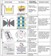
Modeling interstitial flows in tumor cell invasion studies. A, Modified Boyden chamber platform. Tumor cell embedded biomatrix is introduced into a Boyden Chamber insert, which is placed in a well. The gravitational pressure, provided by the fluid level difference between the fluid within the insert and that in the surrounding well, drives the interstitial flow. The invasion rate is marked by the number of cells transmigrated through the porous membrane at the bottom of the insert. B,C,D: Three different microfluidic platforms for modeling interstitial flows. B. In this device, lines of pillars with circular cross section of diameter 500 μm are used to confine collagen. Interstitial flow is driven by gravity along the horizontal direction. Numerically simulated flow field is shown in the right panel of B. C: In this device, lines of square pillars, each with a cross section of 250 μm x 250 μm, are used to confine collagen. Gravity driven flow is introduced horizontally across collagen matrices. D: In this device, contact lines with cross section of 10 μm x 5 μm are used to confine collagen within the cell channel. Interstitial flow is introduced using a syringe pump in the horizontal flow channel. Image on the right side of panel A is reproduced from reference , with permission from Elsevier. Images in B are reproduced from reference with permission from the Royal Society of Chemistry. Images in C are reproduced from references , with permission from the Proceedings of the National Academy of Sciences, and images in D are reproduced from reference with permission from the Royal Society of Chemistry.
Figure 3
Figure 3
Illustration of a microfluidic model recreating the biophysical microenvironment for tumor cell invasion studies. ECM or tumor cell embedded ECM (pink) is introduced into three wall-less channels confined by contact lines (yellow rectangular, not to scaled). Blood vessel (red) and lymphatic vessel (green) are formed by growing a layer of blood and lymphatic EC cells respectively. Blood and lymph flows are introduced into two vascular vessels (direction marked by x), and interstitial flow is introduced horizontally (direction marked by an arrow).

Similar articles

Cited by

References

    1. Chambers AF, Groom AC, MacDonald IC. Nat Rev Cancer. 2002;2:563–572. - PubMed
    1. Steeg PS. Nat Med. 2006;12:895–904. - PubMed
    1. Wirtz D, Konstantopoulos K, Searson PC. Nat Rev Cancer. 2011;11:512–522. - PMC - PubMed
    1. Paszek MJ, Weaver VM. J Mammary Gland Biol Neoplasia. 2004;9:325–342. - PubMed
    1. Paszek MJ, Zahir N, Johnson KR, Lakins JN, Rozenberg GI, Gefen A, Reinhart-King CA, Margulies SS, Dembo M, Boettiger D, Hammer DA, Weaver VM. Cancer cell. 2005;8:241–254. - PubMed

Publication types

LinkOut - more resources