

NANOCI-Nanotechnology Based Cochlear Implant With Gapless Interface to Auditory Neurons
- PMID: 28806330
- PMCID: PMC5559190
- DOI: 10.1097/MAO.0000000000001439
NANOCI-Nanotechnology Based Cochlear Implant With Gapless Interface to Auditory Neurons
Abstract
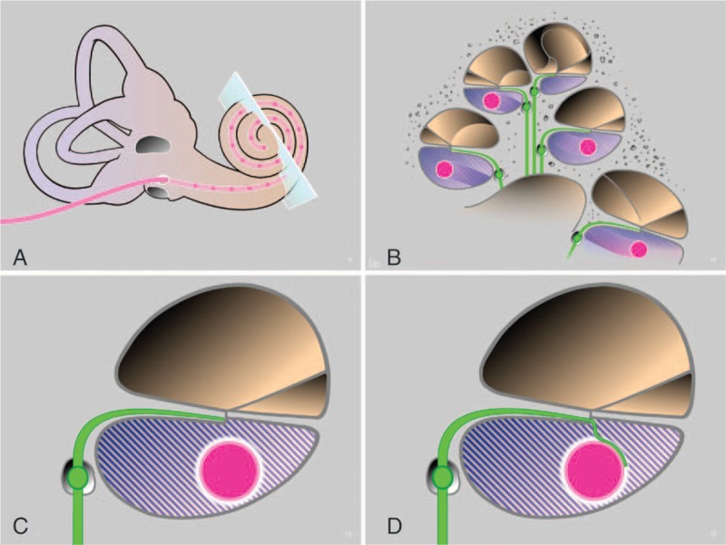
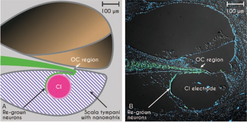
: Cochlear implants (CI) restore functional hearing in the majority of deaf patients. Despite the tremendous success of these devices, some limitations remain. The bottleneck for optimal electrical stimulation with CI is caused by the anatomical gap between the electrode array and the auditory neurons in the inner ear. As a consequence, current devices are limited through 1) low frequency resolution, hence sub-optimal sound quality and 2), large stimulation currents, hence high energy consumption (responsible for significant battery costs and for impeding the development of fully implantable systems). A recently completed, multinational and interdisciplinary project called NANOCI aimed at overcoming current limitations by creating a gapless interface between auditory nerve fibers and the cochlear implant electrode array. This ambitious goal was achieved in vivo by neurotrophin-induced attraction of neurites through an intracochlear gel-nanomatrix onto a modified nanoCI electrode array located in the scala tympani of deafened guinea pigs. Functionally, the gapless interface led to lower stimulation thresholds and a larger dynamic range in vivo, and to reduced stimulation energy requirement (up to fivefold) in an in vitro model using auditory neurons cultured on multi-electrode arrays. In conclusion, the NANOCI project yielded proof of concept that a gapless interface between auditory neurons and cochlear implant electrode arrays is feasible. These findings may be of relevance for the development of future CI systems with better sound quality and performance and lower energy consumption. The present overview/review paper summarizes the NANOCI project history and highlights achievements of the individual work packages.
Conflict of interest statement
The authors disclose no conflicts of interest.
Figures
References
-
- Shannon RF, Fu QJ, Galvin J., 3rd The number of spectral channels required for speech recognition depends on the difficulty of the listening situation. Acta Otolaryngol Suppl 2004; 50–54. - PubMed
-
- Friesen LS, Shannon RV, Baskent D, Wang X. Speech recognition in noise as a function of the number of spectral channels: comparison of acoustic hearing and cochlear implants. J Acoust Soc Am 2001; 110:1150–1163. - PubMed
-
- Wilson BS, Dorman MF. Cochlear implants: current designs and future possibilities. J Rehab Res Dev 2008; 45:695–730. - PubMed
-
- Fritzsch B, Tessarollo L, Coppola E, Reichardt LF. Neurotrophins in the ear: their roles in sensory neuron survival and fiber guidance. Prog Brain Res 2004; 146:265–278. - PubMed
Publication types
MeSH terms
LinkOut - more resources
Full Text Sources
Other Literature Sources
Medical
Research Materials
Miscellaneous
