. 2017 Aug 1;58(10):4318–4331.
doi: 10.1167/iovs.16-21075.

Sema3A Reduces Sprouting of Adult Rod Photoreceptors In Vitro

Affiliations

Sema3A Reduces Sprouting of Adult Rod Photoreceptors In Vitro

Frank Kung et al. Invest Ophthalmol Vis Sci. .

Erratum in

  • Erratum.
    [No authors listed] [No authors listed] Invest Ophthalmol Vis Sci. 2017 Sep 1;58(11):4799. doi: 10.1167/iovs.17-22948a. Invest Ophthalmol Vis Sci. 2017. PMID: 28973336 Free PMC article. No abstract available.

Abstract

Purpose: Rod photoreceptor terminals respond to retinal injury with retraction and sprouting. Since the guidance cue Semaphorin3A (Sema3A) is observed in the retina after injury, we asked whether Sema3A contributes to structural plasticity in rod photoreceptors.

Methods: We used Western blots and alkaline phosphatase (AP)-tagged neuropilin-1 (NPN-1) to detect the expression of Sema3A in an organotypic model of porcine retinal detachment. We then examined Sema3A binding to cultured salamander rod photoreceptors using AP-tagged Sema3A. For functional analysis, we used a microspritzer to apply a gradient of Sema3A-Fc to isolated salamander rod photoreceptors over 24 hours.

Results: Sema3A protein was biochemically detected in porcine retinal explants in the retina 7, 24, and 72 hours after detachment. In sections, NPN-1 receptor was bound to the inner and outer retina. For isolated rod photoreceptors, Sema3A localized to synaptic terminals and to neuritic processes after 1 week in vitro. In microspritzed rod photoreceptors, process initiation occurred away from high concentrations of Sema3A. Sema3A significantly decreased the number of processes formed by rod photoreceptors although the average length of processes was not affected. The cellular orientation of rod photoreceptors relative to the microspritzer also significantly changed over time; this effect was reduced with the Sema3A inhibitor, xanthofulvin.

Conclusion: Sema3A is expressed in the retina after detachment, binds to rod photoreceptors, affects cell orientation, and reduces photoreceptor process initiation in vitro. Our results suggest that Sema3A contributes to axonal retraction in retinal injury, whereas rod neuritic sprouting and regenerative synaptogenesis may require a reduction in semaphorin signaling.


Figures

Figure 1
Figure 1
(A) Experimental setup. Photoreceptor with a retracted but still identifiable axon terminal. Rod photoreceptors have a distinct ellipsoid, which is an accumulation of mitochondria. For microspritzing, a pipette was set 45° relative to the cell's axis. Pipette not to scale. (B) Fluorescent image of microspritzer immediately after spritzing is initiated. Pipette is filled with dextran tretramethyl-rhodamine; highest fluorescence is around the tip. (C) Fluorescent image of microspritzer 24 hours after spritzing was initiated. A gradient still is present, with the highest concentration of dye at the tip of the micropipette. Scale bars: 10 μm.
Figure 2
Figure 2
(A) Western blots for Sema3A. Retinal explants from the same eyes were incubated for 0, 7, 24, and 72 hours and compared. Antibody against Sema3A labeled a 95-kDa band in 7-, 24-, and 72-hour groups, but not in the 0-hour group. GAPDH, from the same SDS-PAGE gel, served as an internal loading control. n = 5 explants per time point, from five eyes from three animals. (B) Quantification of 95 kDA band in Western blots of 0-, 7-, 24-, and 72-hour retinal explant lysates. *P < 0.05, as determined by repeated measures 1-way ANOVA and Bonferroni post hoc analysis. Additional post hoc analysis showed a significant (P < 0.05) linear trend in the increases in expression, from 0, 7, 24, and 72 hours. (C) Representative sections from porcine retinal explants demonstrate binding of NPN1-AP to semaphorin. DIV 0 section treated with 9 nM AP shows no binding (AP only). DIV 0 section treated with 9 nM NPN1-AP shows low levels of binding (DIV 0). Sections treated with 9 nM NPN1-AP after 3 days in vitro show stronger but variable binding in the inner and outer retina (DIV 3). ONL, outer nuclear layer; OPL, outer plexiform layer; INL, inner nuclear layer; IPL, inner plexiform layer; GCL, ganglion cell layer. n = 3 retinal explants per time point, from three eyes from three animals. Scale bar: 50 μm.
Figure 3
Figure 3
Isolated salamander retinal cells bind Sema3A-AP chimera. Rod cells: (A) freshly isolated rod cells from a control culture reacted for endogenous phosphatase activity. No staining is present in the cell body and inner segment that contains the ellipsoid (arrow) and axon terminal (red arrow). (B) Freshly isolated rod photoreceptor shows binding of Sema3A-AP to its surface most prominently over the inner segment (arrow) and at the axon terminal (red arrow). (C) After 3 days in vitro, Sema3A-AP binds along the cell body, inner segment and neuritic extensions. Arrows indicate stained varicosities. Cone cells: (D) freshly isolated cone photoreceptor demonstrates little endogenous phosphatase activity. (E) Freshly isolated cone photoreceptor treated with Sema3A-AP has binding primarily to the area of the ellipsoid (arrow). (F) After 3 days in vitro, cone cell shows staining of Sema3A-AP along the cell body, inner segment, and neuritic extensions. Examples of other freshly isolated cells: (G) a bipolar cell binding Sema3A-AP at the primary dendrite, the Landolt club (arrow), as well as the axon (red arrow). (H) A ganglion cell with binding of Sema3A-AP to cell body and processes. (I) A Müller cell with binding of Sema3A-AP along its entire surface. Apical villi and endfeet are indicated with black and red arrow, respectively. Controls for nonrod cell types had no staining (data not shown). Representative images from n = 3 animals, four cultures per animal and at least three cells per cell type per time point. Scale bar: 10 μm.
Figure 4
Figure 4
Change in direction of process growth and in cell orientation. (A) Typical rod cells after 24 hours of spritzing. 55 μm diameter circles surrounding cells indicate the line from which number of neurites was calculated. Red dashed line indicates original cell axis while the red arrow indicates the final axis. Blue arrow demonstrates amount of change in orientation. Spritzer pipette included in diagram for reference, not to scale. (B) Rose plots indicating cumulative percentages of the direction of growth of all neurites on the basal (axonal) side of the photoreceptor. Blue arrow indicates the mean angle of growth of all of neurites. Neurites grew away from Sema3A-Fc compared to the heat-treated Sema3A-Fc and PBS controls. n = 298 neurites from 63 cells, from 29 animals, at least nine cells per group. *P < 0.05.
Figure 5
Figure 5
Analysis of neurites before spritzing. (A) There was no statistical difference between the average angle of processes before spritzing. (B) There was no statistical difference in the number of processes present in rod photoreceptors before spritzing. Scale bars: Standard deviation. n = 63 cells, 29 animals, nine cells per group.
Figure 6
Figure 6
Significantly fewer processes were present on cells treated with Sema3A-Fc. (A) Number of processes per cell was defined as number of crossings by processes of the 55 μm circle (see Fig. 4). Number of processes was evaluated after spritzing with Sema3A-Fc or controls, PBS, heat-treated-Sema3A-Fc, or Fc alone for 24 hours. (B) Rod photoreceptor neuritic length is not affected by Sema3A application. The average length of all the neurites that grew beyond the 55 μm circle was the same in all conditions. Scale bars: standard deviation. n = 59 cells, 29 animals, at least 10 cells per group. *P < 0.05.
Figure 7
Figure 7
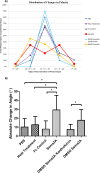
Cell polarity changed with Sema3A-Fc. Cell axis was determined by drawing a line from the center of the nucleus through the center of the ellipsoid. Change in polarity was defined as the degrees from the initial orientation of the photoreceptor to a new orientation after 24 hours of spritzing. A positive change was defined as moving toward the microspritzer, or clockwise, while a negative change was defined as moving away from the spritzer, or counterclockwise. (A) Distribution of changes in polarity after spritzing. Degree change in polarity was binned according to their value. X = Angle in degrees. (B) Average absolute change in polarity after spritzing. The absolute value of each cell's change in polarity for each experimental group was averaged together and compared between groups. Changes in polarity were observed in cells spritzed with Sema3A but not in any of the controls. Scale bars: standard deviation. n = 83 cells, 29 animals, at least 10 cells per group. *P < 0.05.
Figure 8
Figure 8
Rod cells grew more processes in the direction consistent with the change in polarity. Process analysis was done on cells grouped based on the orientation of the rod photoreceptor after 24 hours of spritzing. (Cells with less than 20° of turning were excluded from analysis.) (A) (−) Neurites: processes from cells where the orientation of the rod photoreceptor changed by turning at least 20° away from the spritzer. This included cells spritzed with Sema3A-Fc or Sema3A/DMSO solutions. Average angle of processes was 100.8°. (B) (+) Neurites: processes from cells where the orientation of the rod photoreceptor changed by turning at least 20° toward the spritzer. This included cells spritzed with Sema3A-Fc or Sema3A/DMSO solutions. Average angle of processes was 82.4°. Angle of processes from positively turning cells was significantly less than angle of processes from negatively turning cells. n = 18 cells, 14 animals. P < 0.05.

Similar articles

Cited by

References

    1. Marc RE, Jones BW, Watt CB, Strettoi E. . Neural remodeling in retinal degeneration. Prog Retin Eye Res. 2003; 22: 607– 655. - PubMed
    1. Li ZY, Kljavin IJ, Milam AH. . Rod photoreceptor neurite sprouting in retinitis pigmentosa. J Neurosci. 1995; 15: 5429– 5438. - PMC - PubMed
    1. Fariss RN, Li Z-Y, Milam AH. . Abnormalities in rod photoreceptors, amacrine cells, and horizontal cells in human retinas with retinitis pigmentosa. Am J Ophthalmol. 2000; 129: 215– 223. - PubMed
    1. Clarke RJ, Hognason K, Brimacombe M, Townes-Anderson E. . Cone and rod cells have different target preferences in vitro as revealed by optical tweezers. Mol Vis. 2008; 14: 706– 720. - PMC - PubMed
    1. Sherry DM, St Jules RS, Townes-Anderson E. . Morphologic and neurochemical target selectivity of regenerating adult photoreceptors in vitro. J Comp Neurol. 1996; 376: 476– 488. - PubMed

Publication types