. 2017 Nov:511:40-48.
doi: 10.1016/j.virol.2017.08.011. Epub 2017 Aug 12.

Parvovirus B19 integration into human CD36+ erythroid progenitor cells

Affiliations

Parvovirus B19 integration into human CD36+ erythroid progenitor cells

Tyler Janovitz et al. Virology. 2017 Nov.

Abstract

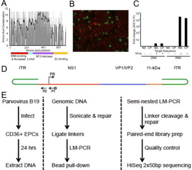
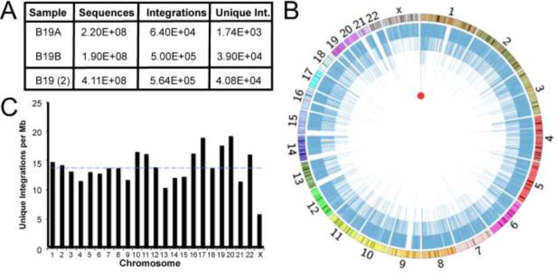
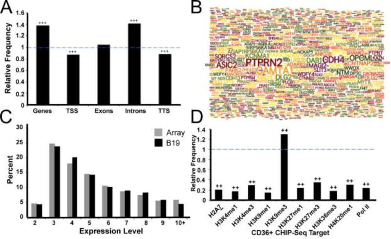
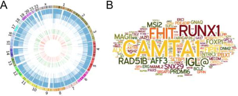
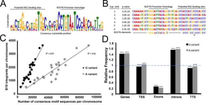
The pathogenic autonomous human parvovirus B19 (B19V) productively infects erythroid progenitor cells (EPCs). Functional similarities between B19V nonstructural protein (NS1), a DNA binding endonuclease, and the Rep proteins of Adeno-Associated Virus (AAV) led us to hypothesize that NS1 may facilitate targeted nicking of the human genome and B19 vDNA integration. We adapted an integration capture sequencing protocol (IC-Seq) to screen B19V infected human CD36+ EPCs for viral integrants, and discovered 40,000 unique B19V integration events distributed throughout the human genome. Computational analysis of integration patterns revealed strong correlations with gene intronic regions, H3K9me3 sites, and the identification of 41 base pair consensus sequence with an octanucleotide core motif. The octanucleotide core has homology to a single region of B19V, adjacent to the P6 promoter TATA box. We present the first direct evidence that B19V infection of erythroid progenitor cells disrupts the human genome and facilitates viral DNA integration.

Keywords: B19; High throughput sequencing; Human erythroid progenitor cell; Integration; Latency; Parvovirus.


Figures

Figure 1
Figure 1. Parvovirus B19 infection of human primary CD36+ erythroid progenitor cells and integration-capture sequencing schematic
(A) Amino acid sequence homology between the major replication proteins of parvovirus B19V, AAV-2, and AAV-5. Black line on graph indicates 10-bp moving average of conservation (11 corresponds to 100% conserved). Colored line indicates functionally active domains established for parvovirus replication proteins. (B) Day 12 CD36+EPCs infected with 2000 ge B19V/cell and harvested 24 hours post inoculation, B19V capsid proteins were detected by fluorescein immunofluorescence with Evans blue counterstaining. (C) Total DNA or RNA harvested at 0 and 24 hours post inoculation with 2000 ge B19V/cell quantified by qPCR or RT-qPCR. Fold change (24/0hr) in B19V NS1 and Capsid region CT values; normalization to cellular actin. (D) Schematic representation of B19V genome organization. Arrows indicate the location of primers used for nested PCR. Biotinylated primer is indicated by dot on arrow end. B19V P6 promoter, arrow indicates direction of transcription. (E) IC-Seq protocol flow diagram (protocol details in materials and methods).
Figure 2
Figure 2. Genome-wide distribution of Parvovirus B19 integration in human erythroid precursors
(A) Summary of integrant-capture sequencing data acquired for B19A and B19B samples with the merge of both data sets. (B) Circos plot presenting genome-wide display of all unique B19V integration events. Chromosomal size and banding phenotype is presented in the external multi-colored ring. The chromosomal position of each unique B19V integration event is indicated as a blue line, where the 40,000 unique integration sites create the internal blue ring. The height of each blue bar corresponds to the read frequency for each site. The red circle indicates the single computationally determined integration hotspot located in CAMTA1 on chromosome 1. (C) Unique B19V integrations per mappable megabase for each chromosome. Dashed blue line represents the mean number of integrants/Mb for all chromosomes.
Figure 3
Figure 3. Parvovirus B19 integration is associated with specific gene regions and histone markers
(A) Relative frequency of B19V integration in genes and specific gene regions; fold enhancement of observed data compared to a random distribution model. The dashed blue line indicates the relative frequency expected in a random model. Presence of +++ represents p < 0.001 using a permutation test. (B) Word cloud of the top 1,000 genes which attracted the highest numbers of B19V integration events; the size of the gene name represents the number of unique integrations received. (C) Percent of B19V integration events occurring within CD36+ erythroid progenitor expression level gene groups (black), compared to the distribution expected based on the gene group array frequency (grey). (D) Enrichment, displayed as relative frequency, of B19V integrations in several histone marker subtypes and Pol II using CD36+ erythroid precursor CHIP-Seq data. Dashed blue line represents expected frequency based on a random model. Presence of ++ indicates p < 0.001 using a permutation test.
Figure 4
Figure 4. Oncogenes targeted by Parvovirus B19 integration
(A) Circos plot presenting total B19V integration events and those located in cancer associated genes. Genome-wide view of unique B19V integration events (blue bars), known genes relating to cancer induction (green), and those cancer-related genes targeted by B19V integration (red). Chromosomal size and banding phenotype is displayed in the external ring. (B) Word cloud diagram of the 287 genes involved with cancer induction that attracted B19V integration events; the size of the gene name correlates to the number of unique integrations.
Figure 5
Figure 5. A large consensus DNA motif was discovered proximal to Parvovirus B19 genomic integration events and resembles the viral promoter
(A) Sequence LOGO diagram of the forty-one base pair consensus motif computationally identified in association with B19V integration (p-value = 1.7E-100). Total column stack height indicates information content, in bits, while individual letter size denotes the probability of the given nucleotide occurring in that position. Labeled bars display possible B19V P6 promoter functional correlation. (B) The sequences of the top six supported B19V consensus motifs associated with integration events. Sites are ranked by p-value, as determined by the probability of a random sequence generating an equal or greater match score. Labeled bars indicate possible B19V P6 promoter correlation. (C) Integrations per chromosome as a function of the highly conserved eight base pair motif core sequences per chromosome. The term variant refers to the least fixed nucleotide position in the core, most commonly the “G variant” (CTGTAGTC) or the “A variant” (CTGTAATC) shown in grey. Simple linear regression and R2 value is presented. (D) Enrichment of “G variant” (black) and “A variant” (grey) eight base pair motif core sequences in genes and specific gene regions, displayed as relative frequency. Dashed blue line denotes expected frequency based on a random model. *** indicates p < 0.001 using a permutation test.

Similar articles

Cited by

References

    1. Adamson-Small LA, Ignatovich IV, Laemmerhirt MG, Hobbs JA. Persistent parvovirus B19 infection in non-erythroid tissues: possible role in the inflammatory and disease process. Virus Res. 2014;190:8–16. - PubMed
    1. Bihari C, Rastogi A, Saxena P, Rangegowda D, Chowdhury A, Gupta N, Sarin SK. Parvovirus b19 associated hepatitis. Hepat Res Treat. 2013;2013:472027. - PMC - PubMed
    1. Bonvicini F, Filippone C, Manaresi E, Zerbini M, Musiani M, Gallinella G. HepG2 hepatocellular carcinoma cells are a non-permissive system for B19 virus infection. J Gen Virol. 2008;89:3034–3038. - PubMed
    1. Brown KE, Anderson SM, Young NS. Erythrocyte P antigen: cellular receptor for B19 parvovirus. Science. 1993;262:114–117. - PubMed
    1. Bua G, Manaresi E, Bonvicini F, Gallinella G. Parvovirus B19 Replication and Expression in Differentiating Erythroid Progenitor Cells. PLoS One. 2016;11:e0148547. - PMC - PubMed

Publication types

MeSH terms