

Design and fabrication of a realistic anthropomorphic heterogeneous head phantom for MR purposes
- PMID: 28806768
- PMCID: PMC5555696
- DOI: 10.1371/journal.pone.0183168
Design and fabrication of a realistic anthropomorphic heterogeneous head phantom for MR purposes
Erratum in
-
Correction: Design and fabrication of a realistic anthropomorphic heterogeneous head phantom for MR purposes.PLoS One. 2018 Feb 7;13(2):e0192794. doi: 10.1371/journal.pone.0192794. eCollection 2018. PLoS One. 2018. PMID: 29415085 Free PMC article.
Abstract
Objective: The purpose of this study is to design an anthropomorphic heterogeneous head phantom that can be used for MRI and other electromagnetic applications.
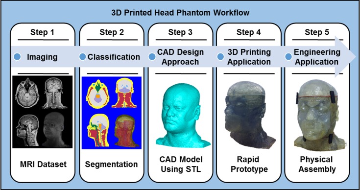
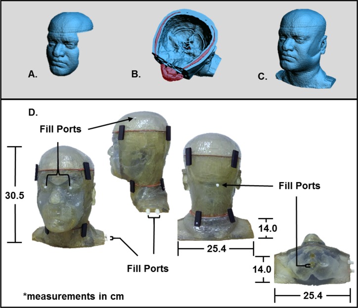
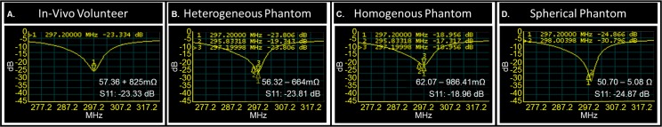
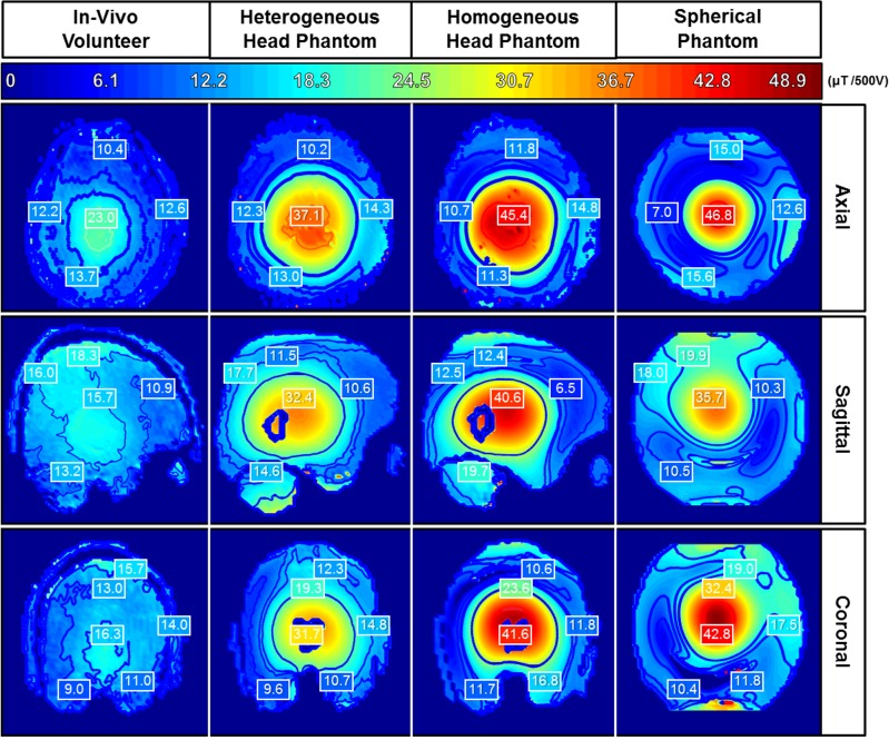
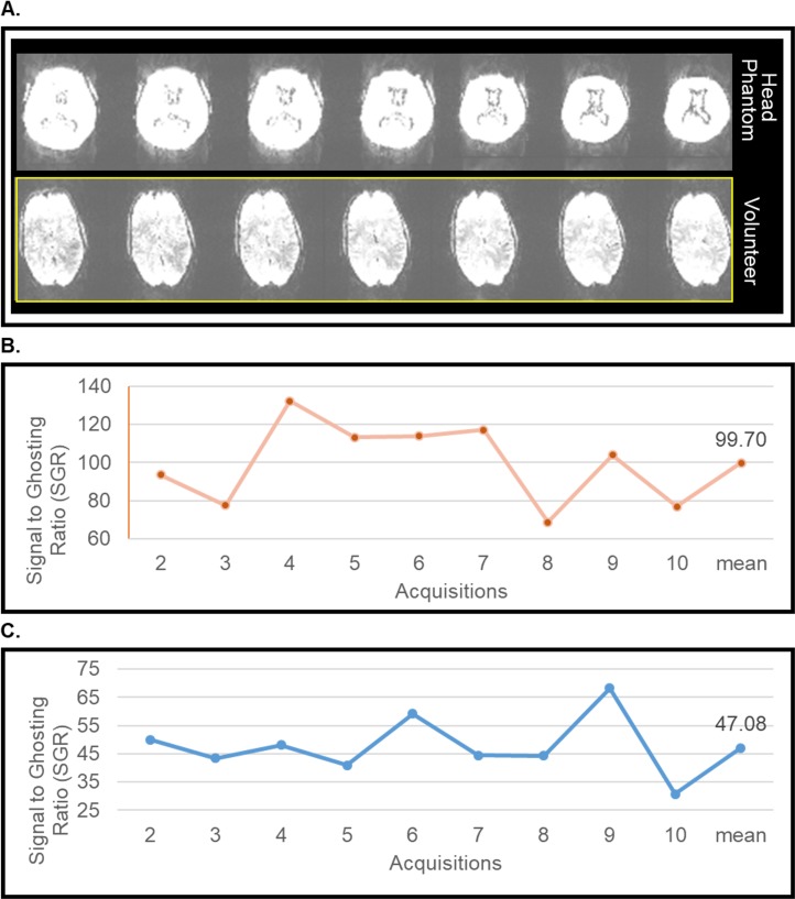
Materials and methods: An eight compartment, physical anthropomorphic head phantom was developed from a 3T MRI dataset of a healthy male. The designed phantom was successfully built and preliminarily evaluated through an application that involves electromagnetic-tissue interactions: MRI (due to it being an available resource). The developed phantom was filled with media possessing electromagnetic constitutive parameters that correspond to biological tissues at ~297 MHz. A preliminary comparison between an in-vivo human volunteer (based on whom the anthropomorphic head phantom was created) and various phantoms types, one being the anthropomorphic heterogeneous head phantom, were performed using a 7 Tesla human MRI scanner.
Results: Echo planar imaging was performed and minimal ghosting and fluctuations were observed using the proposed anthropomorphic phantom. The magnetic field distributions (during MRI experiments at 7 Tesla) and the scattering parameter (measured using a network analyzer) were most comparable between the anthropomorphic heterogeneous head phantom and an in-vivo human volunteer.
Conclusion: The developed anthropomorphic heterogeneous head phantom can be used as a resource to various researchers in applications that involve electromagnetic-biological tissue interactions such as MRI.
Conflict of interest statement
Figures

Similar articles
-
A comprehensive electromagnetic evaluation of an MRI anthropomorphic head phantom.NMR Biomed. 2021 Mar;34(3):e4441. doi: 10.1002/nbm.4441. Epub 2020 Dec 22. NMR Biomed. 2021. PMID: 33354828 Free PMC article.
-
Design, construction and evaluation of an anthropomorphic head phantom with realistic susceptibility artifacts.J Magn Reson Imaging. 2007 Jul;26(1):202-7. doi: 10.1002/jmri.20993. J Magn Reson Imaging. 2007. PMID: 17659546
-
The ADAM-pelvis phantom-an anthropomorphic, deformable and multimodal phantom for MRgRT.Phys Med Biol. 2019 Feb 11;64(4):04NT05. doi: 10.1088/1361-6560/aafd5f. Phys Med Biol. 2019. PMID: 30630152
-
Minimizing magnetic resonance image geometric distortion at 7 Tesla for frameless presurgical planning using skin-adhered fiducials.Med Phys. 2023 Feb;50(2):694-701. doi: 10.1002/mp.16035. Epub 2022 Nov 12. Med Phys. 2023. PMID: 36301228 Review.
-
Anthropomorphic Head MRI Phantoms: Technical Development, Brain Imaging Applications, and Future Prospects.J Magn Reson Imaging. 2025 May 14. doi: 10.1002/jmri.29818. Online ahead of print. J Magn Reson Imaging. 2025. PMID: 40365871 Review.
Cited by
-
Biomimetic phantom with anatomical accuracy for evaluating brain volumetric measurements with magnetic resonance imaging.J Med Imaging (Bellingham). 2021 Jan;8(1):013503. doi: 10.1117/1.JMI.8.1.013503. Epub 2021 Jan 29. J Med Imaging (Bellingham). 2021. PMID: 33532513 Free PMC article.
-
Anthropomorphic brain phantoms for use in MRI systems: a systematic review.MAGMA. 2022 Apr;35(2):277-289. doi: 10.1007/s10334-021-00953-w. Epub 2021 Aug 31. MAGMA. 2022. PMID: 34463866
-
How to design and construct a 3D-printed human head phantom.J 3D Print Med. 2019 Aug;3(3):119-125. doi: 10.2217/3dp-2019-0016. Epub 2019 Aug 21. J 3D Print Med. 2019. PMID: 31929893 Free PMC article.
-
Recent advances on the development of phantoms using 3D printing for imaging with CT, MRI, PET, SPECT, and ultrasound.Med Phys. 2018 Jun 22;45(9):e740-60. doi: 10.1002/mp.13058. Online ahead of print. Med Phys. 2018. PMID: 29933508 Free PMC article. Review.
-
Open-source, customizable phantom for low-field magnetic resonance imaging.MAGMA. 2025 Jun 25. doi: 10.1007/s10334-025-01270-2. Online ahead of print. MAGMA. 2025. PMID: 40560478
References
-
- Caon M. Voxel-based computational models of real human anatomy: a review. Radiat Environ Biophys. 2004;42(4):229–35. doi: 10.1007/s00411-003-0221-8 . - DOI - PubMed
-
- Chen CC, Wan YL, Wai YY, Liu HL. Quality assurance of clinical MRI scanners using ACR MRI phantom: preliminary results. J Digit Imaging. 2004;17(4):279–84. doi: 10.1007/s10278-004-1023-5 ; PubMed Central PMCID: PMCPMC3047180. - DOI - PMC - PubMed
-
- Tofts PS. QA: Quality Assurance, Accuracy, Precision and Phantoms. Quantitative MRI of the Brain. 2003:55–81. doi: 10.1002/0470869526.ch3 - DOI
-
- Radiology ACo. ACR White Paper on Magnetic Resonance (MR) Safety. Radiology. 2004:1–24.
-
- Christ A, Chavannes N, Nikoloski N, Gerber HU, Pokovic K, Kuster N. A numerical and experimental comparison of human head phantoms for compliance testing of mobile telephone equipment. Bioelectromagnetics. 2005;26(2):125–37. doi: 10.1002/bem.20088 . - DOI - PubMed
MeSH terms
Grants and funding
LinkOut - more resources
Full Text Sources
Other Literature Sources
Medical
