

Generation of Xeno-Free, cGMP-Compliant Patient-Specific iPSCs from Skin Biopsy
- PMID: 28806854
- PMCID: PMC5609845
- DOI: 10.1002/cpsc.30
Generation of Xeno-Free, cGMP-Compliant Patient-Specific iPSCs from Skin Biopsy
Abstract
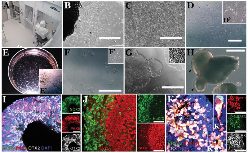
This unit describes protocols for the generation of clinical-grade patient-specific induced pluripotent stem cell (iPSC)-derived retinal cells from patients with inherited retinal degenerative blindness. Specifically, we describe how, using xeno-free reagents in an ISO class 5 environment, one can isolate and culture dermal fibroblasts, generate iPSCs, and derive autologous retinal cells via 3-D differentiation. The universal methods described herein for the isolation of dermal fibroblasts and generation of iPSCs can be employed regardless of disease, tissue, or cell type of interest. © 2017 by John Wiley & Sons, Inc.
Keywords: cGMP; current Good Manufacturing Practice; fibroblasts; induced pluripotent stem cells; photoreceptor precursor cells; retina; xeno-free.
Copyright © 2017 John Wiley & Sons, Inc.
Figures
References
MeSH terms
Grants and funding
LinkOut - more resources
Full Text Sources
Other Literature Sources
