. 2017 Aug 14;14(1):22.
doi: 10.1186/s12987-017-0070-5.

Profiling of metalloprotease activities in cerebrospinal fluids of patients with neoplastic meningitis

Affiliations

Profiling of metalloprotease activities in cerebrospinal fluids of patients with neoplastic meningitis

Catharina Conrad et al. Fluids Barriers CNS. .

Abstract

Background: Neoplastic invasion into leptomeninges and subarachnoid space, resulting in neoplastic meningitis (NM) is a fatal complication of advanced solid and hematological neoplasms. Identification of malignant involvement of the cerebrospinal fluid (CSF) early in the disease course has crucial prognostic and therapeutic implications, but remains challenging. As indicators of extracellular matrix (ECM) degradation and breakdown of the blood-brain-barrier, Matrix Metalloproteases (MMPs) and A Disintegrin and Metalloproteases (ADAMs) are potential analytes for cerebral pathophysiology and metastatic dissemination of tumor cells into the CSF.

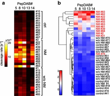
Methods: We compared protease activities in CSF samples from patients with NM and control individuals using FRET-based metalloprotease substrates with distinct enzyme selectivity profiles in a real-time, multiplex approach termed "proteolytic activity matrix assay" (PrAMA). Protease activity dynamics can be tracked by fluorescence changes over time. By simultaneously monitoring a panel of 5 FRET-substrate cleavages, a proteolytic signature can be identified and analyzed to infer the activities of multiple specific proteases. Distinct patterns of substrate cleavage comparing disease vs. control samples allow rapid, reproducible and sensitive discrimination even in small volumes of CSF.

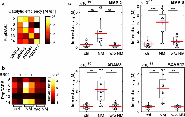
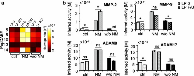
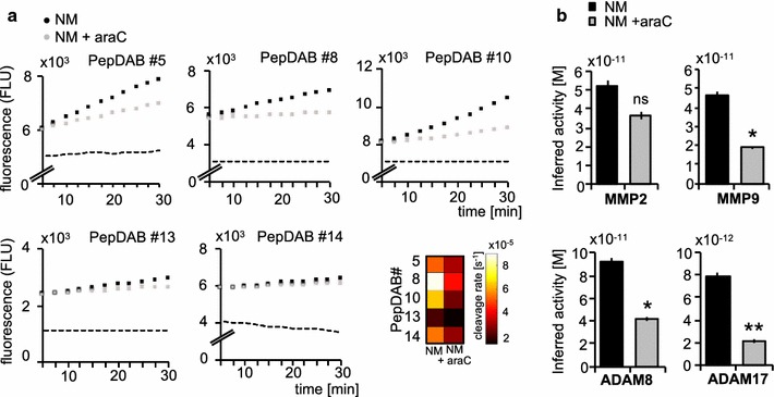
Results: Individual substrate cleavage rates were linked to distinct proteases, and PrAMA computational inference implied increased activities of MMP-9, ADAM8 and ADAM17 (4-5-fold on average) in CSF samples from NM patients that were inhibitable by the metalloprotease inhibitor batimastat (BB-94). The activities of these proteases correlated with blood-brain barrier impairment. Notably, CSF cell counts were not found to directly reflect the protease activities observed in CSF samples from NM patients; this may explain the frequent clinical observation of negative cytology in NM patients.

Conclusion: PrAMA analysis of CSF samples is a potential diagnostic method for sensitive detection of NM and may be suitable for the clinical routine.

Keywords: CSF; FRET-substrates; Metalloproteases; Neoplastic meningitis; Real-time protease activities.


Figures

Fig. 1
Fig. 1
Analysis of protease activities in the CSF of three patient cohorts. Cleavage signatures in CSF samples were analyzed from 12 control individuals (crtl), 12 patients with neoplastic meningitis (NM) and 12 patients with brain metastases of different primary tumors, but without neoplastic meningitis (w/o NM). Protease activities in real-time mode were determined using PEPDab substrates 5, 8, 10, 13 and 14 at final concentrations of 10 µM for 4 h. Heat map summarizing mean cleavage rates of 3 technical replicates for each clinical samples calculated from time-lapse fluorimetry a. Hierarchical bi-clustering result corresponding to (a), with data mean-centered and variance-normalized by row. CSF cleavage patterns of patients with NM (labeled red) cluster closely together, control individuals and patients with brain metastases w/o NM group together without forming main clusters for each condition (b)
Fig. 2
Fig. 2
Inferred protease activitity of MMP-9, ADAM8 and ADAM17 in CSF is increased in neoplastic meningitis. Catalytic efficiencies for FRET-based peptide substrates have been determined previously across a panel of purified recombinant enzymes [13]. Observed cleavage efficiencies for MMP-2, MMP-9, ADAM8 and ADAM17 for PEPDab Substrates 5, 8, 10, 13 and 14 (modified from [13] are shown a. Effects of broad-spectrum MMP-inhibitor batimastat (BB-94+) treatment on observed substrate cleavage in representative CSF samples. Combination of specific substrates and inhibitors allow to infer protease activities accurately (b). PrAMA inferred the cleavage signatures from (Fig. 1a), using parameters from (a). PrAMA inference results in CSF in patients with NM were compared to control individuals and patients with brain metastases w/o NM. The box indicates the interquartile range, blue bar indicates the median, red bar denote the mean values and whiskers indicate the 95% confidence interval. Open dots represent the individual samples (o), extreme values are marked with asterisks. Inferred differences were statistically significant for MMP-9, ADAM8 and ADAM17, stars indicate * p <0.01; ** p <0.005, *** p <0.001 (c)
Fig. 3
Fig. 3
PrAMA inference results are reproducible in follow-up lumbar punctures (LP). Substrate cleavage patterns in CSF samples obtained from same patients with unchanged clinical diagnosis within a time period of 2 months, baseline substrate cleavage in CSF is denoted as LP 0, cleavage rates in CSF of 2-month follow-up (F/U) lumbar punctures as LP F/U. Substrate cleavage is shown for controls (ctrl), patients with neoplastic meningitis (NM) and patients with brain metastases w/o NM in a heatmap (a). PrAMA inferred the cleavage signatures from (Fig. 2a), using parameters from (a). Calculated protease activities are presented as bar graph (light grey, control individuals; grey, NM; black, w/o NM), error bars indicate standard deviation of three technical replicates, dotted columns denote 2-months follow-up lumbar punctures, respectively (b). Note that both, cleavage patterns and PrAMA inference results, remain similiar for controls, NM and the patient with brain metastases w/o NM. Two-tailed, one-sample t test was used to compare initial protease activities with 2-month follow-up measurements, values were not significant (ns, p ≥ 0.05) or significant * (p ≤ 0.05)
Fig. 4
Fig. 4
Protease activity decreases following intrathecal treatment with liposomal cytarabin. In a 62-year old breast cancer patient with leptomeningeal metastasis, protease activities were analyzed prior and after intrathecal treatment with liposomal cytarabin. Time-lapse fluorimetry output of the same patient with neoplastic meningitis arising from breast cancer previous (NM, black dots) and following treatment of NM with Cytarabin (araC) (NM + AraC, grey dots) for 3 months. Substrate tracking (PEPDab 5, 8, 10, 13 and 14) for 30 min averaged over 3 technical replicates is shown, dashed line indicates the negative control. Cleavage rates were calculated as described previously using a non-linear kinetic model to fit the time-lapse data (a). PrAMA inference results for samples analysed in A, using significance threshold σT = 1.4. Error bars indicate standard deviation of technical triplicates (b). Two-tailed, one-sample t test was used to compare cleavage rates pre and post treatment with araC. Values were not significant (ns, p ≥ 0.05), significant * (p ≤ 0.05) or highly significant ** (p ≤ 0.01)

Similar articles

Cited by

References

    1. Gleissner B, Chamberlain MC. Neoplastic meningitis. Lancet Neurol. 2006;5:443–452. doi: 10.1016/S1474-4422(06)70443-4. - DOI - PubMed
    1. Strik H, Prömmel P. Diagnosis and individualized therapy of neoplastic meningitis. Expert Rev Anticancer Ther. 2010;10:1137–1148. doi: 10.1586/era.10.86. - DOI - PubMed
    1. Chamberlain MC, Glantz M, Groves MD, Wilson WH. Diagnostic tools for neoplastic meningitis: detecting disease, identifying patient risk, and determining benefit of treatment. Semin Oncol. 2009;36:S35–S45. doi: 10.1053/j.seminoncol.2009.05.005. - DOI - PubMed
    1. Prömmel P, Pilgram-Pastor S, Sitter H, Buhk JH, Strik H. Neoplastic meningitis: How MRI and CSF cytology are influenced by CSF cell count and tumor type. Sci World J. 2013;2013:248072. doi: 10.1155/2013/248072. - DOI - PMC - PubMed
    1. Grossman SA, Krabak MJ. Leptomeningeal carcinomatosis. Cancer Treat Rev. 1999;25:103–119. doi: 10.1053/ctrv.1999.0119. - DOI - PubMed