. 2017 Oct:125:376-385.
doi: 10.1016/j.neuropharm.2017.08.011. Epub 2017 Aug 12.

Phosphodiesterase 4b expression plays a major role in alcohol-induced neuro-inflammation

Affiliations

Phosphodiesterase 4b expression plays a major role in alcohol-induced neuro-inflammation

Diana V Avila et al. Neuropharmacology. 2017 Oct.

Abstract

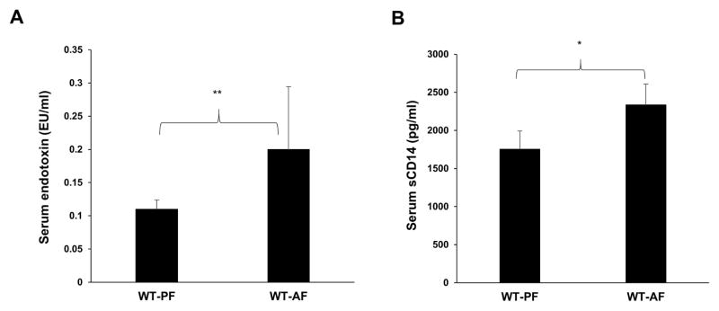
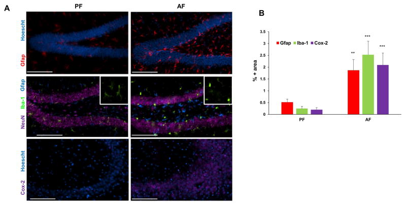
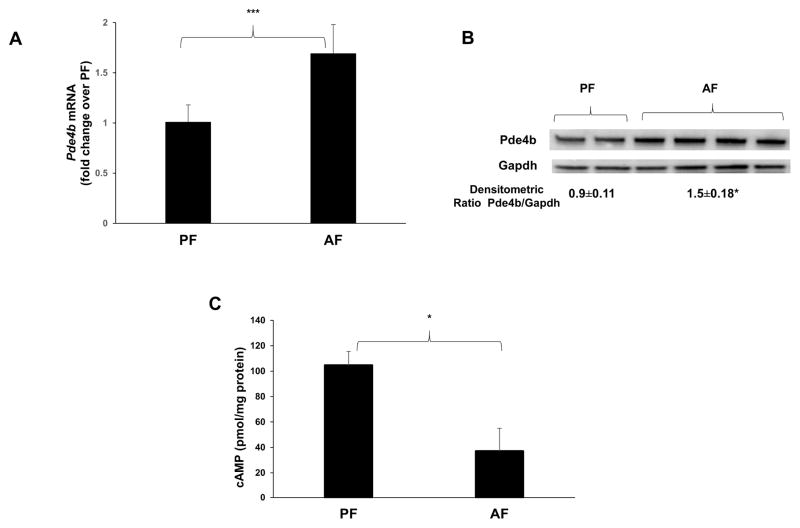
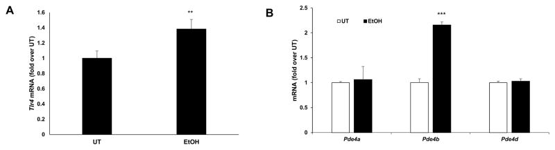
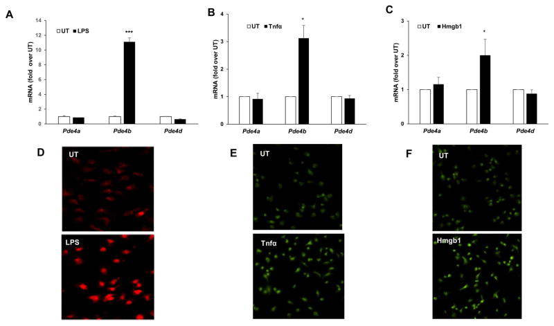
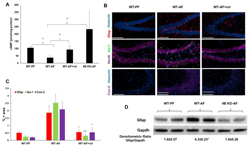
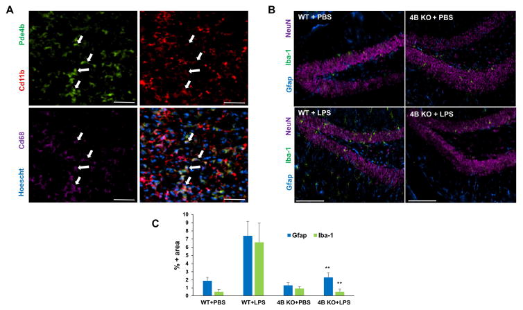
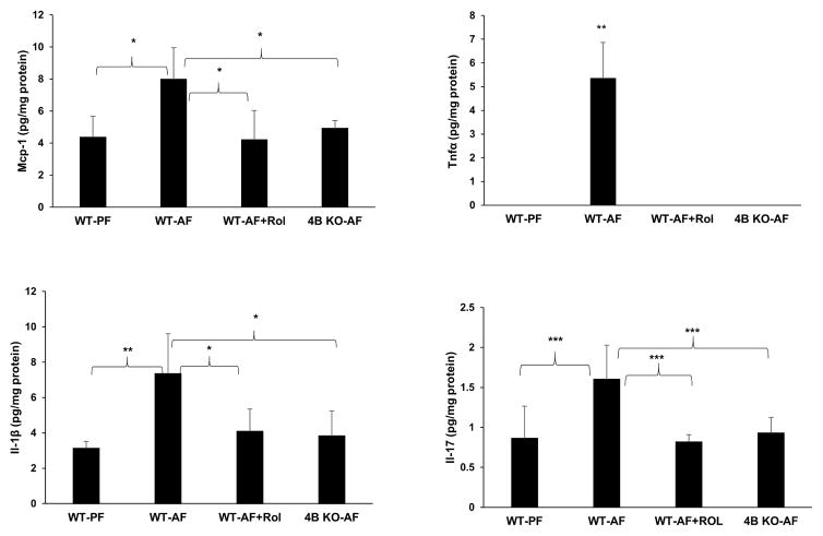
It is increasingly evident that alcohol-induced, gut-mediated peripheral endotoxemia plays a significant role in glial cell activation and neuro-inflammation. Using a mouse model of chronic alcohol feeding, we examined the causal role of endotoxin- and cytokine-responsive Pde4 subfamily b (Pde4b) expression in alcohol-induced neuro-inflammation. Both pharmacologic and genetic approaches were used to determine the regulatory role of Pde4b. In C57Bl/6 wild type (WT) alcohol fed (WT-AF) animals, alcohol significantly induced peripheral endotoxemia and Pde4b expression in brain tissue, accompanied by a decrease in cAMP levels. Further, along with Pde4b, there was a robust activation of astrocytes and microglia accompanied by significant increases in the inflammatory cytokines (Tnfα, Il-1β, Mcp-1 and Il-17) and the generalized inflammatory marker Cox-2. At the cellular level, alcohol and inflammatory mediators, particularly LPS, Tnfα and Hmgb1 significantly activated microglial cells (Iba-1 expression) and selectively induced Pde4b expression with a minimal to no change in Pde4a and d isoforms. In comparison, the alcohol-induced decrease in brain cAMP levels was completely inhibited in WT mice treated with the Pde4 specific pharmacologic inhibitor rolipram and in Pde4b-/- mice. Moreover, all the observed markers of alcohol-induced brain inflammation were markedly attenuated. Importantly, glial cell activation induced by systemic endotoxemia (LPS administration) was also markedly decreased in Pde4b-/- mice. Taken together, these findings strongly support the notion that Pde4b plays a critical role in coordinating alcohol-induced, peripheral endotoxemia mediated neuro-inflammation and could serve as a significant therapeutic target.

Keywords: Alcohol; Microglial activation; Neuroinflammation; Pde4b; Rolipram (PubChem CID: 5092); cAMP.


Conflict of interest statement

Conflict of Interest: The authors declare no competing financial interests

Figures

Figure 1
Figure 1
Alcohol increases systemic endotoxin and sCD14 levels in mice. A, Serum endotoxin levels were measured using LAL assay 1 week after starting 5% alcohol and B, Serum sCD14 levels were measured using sCD14 ELISA kit after 2 weeks of starting 5% alcohol. Data are presented as means and S.D. (n=5–7 in each group). *P < 0.05, **P<0.01.
Figure 2
Figure 2
Alcohol induces glial activation and neuro-inflammation. A, Representative images of the inflammatory state of brains of their respective treatment groups: astrocytic activation marker Gfap (red, hippocampus-dentate gyrus), microglial marker Iba-1 (green, hippocampus—dentate gyrus) and inflammatory marker Cox-2 (purple, hippocampus-CA3 region), in the brains of PF and AF mice after two weeks of feeding. Hoescht (blue) and NeuN (purple) staining are used as nuclear and neuronal markers, respectively. Bar = 100 μm. B, Quantitation of IHC staining. The percentage of image area positive for Gfap, Iba-1, and Cox-2 are significantly higher (**p<0.01, ***p<0.001) in alcohol-fed mice (AF) relative to control pair-fed animals (PF). Data are means ± SD (n = 5–7 in each group).
Figure 3
Figure 3
Alcohol increases Pde4b expression and decreases cAMP levels in the brain. A, mRNA levels of brain Pde4b quantified by real time qPCR and B, Immunoblot analysis of brain homogenates from WT mice that were PF and AF for 1 week. (n = 5–7 in each group). C, brain cAMP levels measured by using cAMP ELISA kit were normalized by protein content. Data are presented as means ± SD (n = 5–7 in each group). *P < 0.05.
Figure 4
Figure 4
Ethanol-mediated increase of Tlr4 and Pde4b mRNA expression in primary microglial cells. mRNA levels of Tlr4, Pde4a, b and d were quantified by real time qPCR in primary microglia cells treated with 50 mM ethanol (EtOH) for 24 hours. Data are presented as mean ± SD from 3 independent experiments, ** P<0.01 and *** P<0.001 compared UT.
Figure 5
Figure 5
Increase of Pde4b expression in primary microglial cells in response to LPS, Tnfα and Hmgb1. mRNA levels of Pde4a, b and d were quantified by real time qPCR in A, cells treated with LPS, 100 ng/ml for 90 minutes. B, cells treated with recombinant Tnfα, 20 ng/ml for 90 minutes. C, cells treated with recombinant Hmgb1, 10 μg/ml for 90 minutes. Data are presented as mean ± SD from 3 independent experiments, *P<0.05, and *** P<0.001 compared UT. Representative images of IHC for Pde4b protein in D, cells treated with LPS, 100 ng/ml for 3 hours, E, in cells treated with Tnfα, 20 ng/ml for 3 hours, F, in cells treated with Hmgb1, 10 μg/ml for 3 hours.
Figure 6
Figure 6
Pde4 inhibition prevents alcohol-induced effects on cAMP and inflammation in the brain. A, Brains from PF and AF mice were lysed, and cellular cAMP levels were measured. Obtained cAMP values were normalized by protein content. Data are presented as means ± SD (n=5–7 in each group). *P < 0.05. B, Cox-2, Gfap and Iba-1 expression in mouse brains. Images are representative of the inflammatory state of the whole brain of their respective treatment groups. (n = 5–7 in each group). C, Quantitation of IHC staining. The percentage of image area positive for Gfap, Iba-1, and Cox-2 are significantly lower (*p<0.05, ***p<0.001) in rolipram-treated alcohol-fed mice (WT-AF+rol) relative to alcohol-fed animals (WT-AF). (n = 5–7 in each group). Bar = 100 μm. Data are means ± S.D. D, Immunoblot analysis of brain Gfap of WT and Pde4b/ mice fed alcohol for 2 weeks. *p<0.05 compared to WT-PF and 4B KO-AF.
Figure 7
Figure 7
Pde4b/ mice are protected from endotoxin-induced glial activation. A, IHC staining for Pde4b (green), Cd11b (red) and Cd68 (violet) demonstrating the localization of Pde4b in macrophage/microglia of WT mice 6 h after LPS administration. Bar = 50 μm. B, Expression of CD11b/c, Gfap, and NeuN in the dentate gyrus. C, Iba-1 immunostaining is up-regulated within 6 hours of LPS exposure. Images are representative of the inflammatory state of the whole brains of their respective treatment groups. C, Quantitation of IHC staining. The percentage of image area positive for Iba-1 and Gfap are significantly lower (**p<0.01) in LPS-injected Pde4b/ mice (4B KO+LPS, n=4) relative to LPS-injected WT mice (WT+LPS, n=4). Data are means ± S.D.
Figure 8
Figure 8
Pde4 inhibition prevents alcohol-induced increases in neuro-inflammatory cytokines/chemokines. Mcp-1, Tnfα, Il-1β and Il-17 were measured in brain homogenates of wild type PF, AF, AF with rolipram treatment and Pde4b/- mice. Obtained values were normalized by protein content. Data are presented as mean ± S.D (n = 5–7 in each group). *P<0.05, **P<0.01, ***P<0.01.

Similar articles

Cited by

References

    1. Alfonso-Loeches S, Pascual-Lucas M, Blanco AM, Sanchez-Vera I, Guerri C. Pivotal role of TLR4 receptors in alcohol-induced neuroinflammation and brain damage. J Neurosci. 2010;30:8285–8295. - PMC - PubMed
    1. Avila DV, Barker DF, Zhang J, McClain CJ, Barve S, Gobejishvili L. Dysregulation of hepatic cAMP levels via altered Pde4b expression plays a critical role in alcohol-induced steatosis. J Pathol. 2016;240:96–107. - PMC - PubMed
    1. Banks WA, Robinson SM. Minimal penetration of lipopolysaccharide across the murine blood-brain barrier. Brain Behav Immun. 2010;24:102–109. - PMC - PubMed
    1. Bazil V, Strominger JL. Shedding as a mechanism of down-modulation of CD14 on stimulated human monocytes. J Immunol. 1991;147:1567–1574. - PubMed
    1. Bianchi ME. HMGB1 loves company. J Leukoc Biol. 2009;86:573–576. - PubMed

MeSH terms

LinkOut - more resources