. 2017 Nov:174:120-127.
doi: 10.1016/j.jsbmb.2017.08.005. Epub 2017 Aug 12.

Identification and characterization of 5α-cyprinol-sulfating cytosolic sulfotransferases (Sults) in the zebrafish (Danio rerio)

Affiliations

Identification and characterization of 5α-cyprinol-sulfating cytosolic sulfotransferases (Sults) in the zebrafish (Danio rerio)

Katsuhisa Kurogi et al. J Steroid Biochem Mol Biol. 2017 Nov.

Abstract

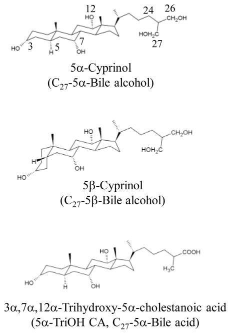
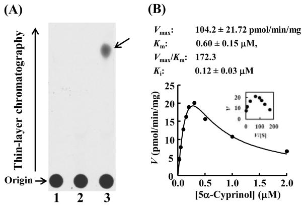
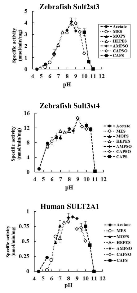
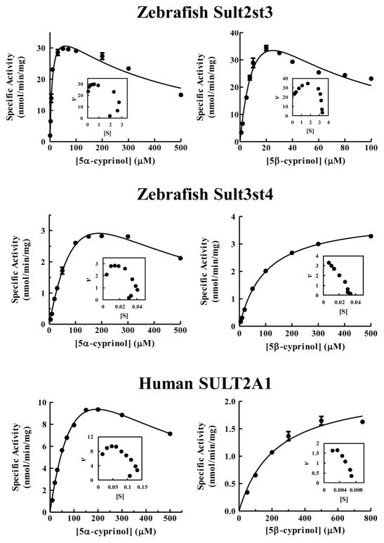
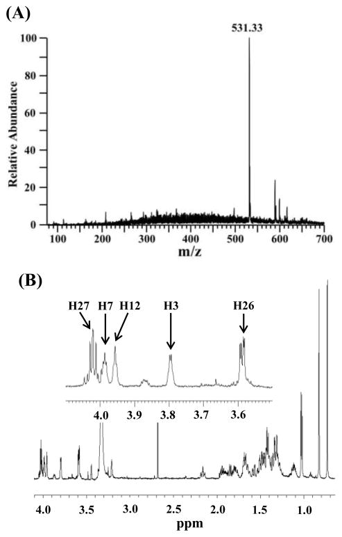
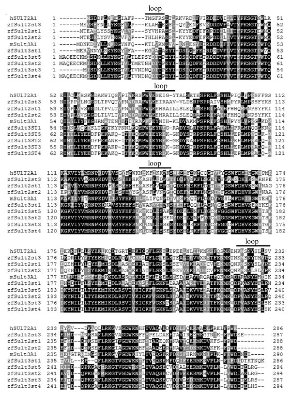
5α-Cyprinol 27-sulfate is the major biliary bile salt present in cypriniform fish including the zebrafish (Danio rerio). The current study was designed to identify the zebrafish cytosolic sulfotransferase (Sult) enzyme(s) capable of sulfating 5α-cyprinol and to characterize the zebrafish 5α-cyprinol-sulfating Sults in comparison with human SULT2A1. Enzymatic assays using zebrafish homogenates showed 5α-cyprinol-sulfating activity. A systematic analysis, using a panel of recombinant zebrafish Sults, revealed two Sult2 subfamily members, Sult2st2 and Sult2st3, as major 5α-cyprinol-sulfating Sults. Both enzymes showed higher activities using 5α-cyprinol as the substrate, compared to their activity with DHEA, a representative substrate for mammalian SULT2 family members, particularly SULT2A1. pH-Dependence and kinetics experiments indicated that the catalytic properties of zebrafish Sult2 family members in mediating the sulfation of 5α-cyprinol were different from those of either zebrafish Sult3st4 or human SULT2A1. Collectively, these results imply that both Sult2st2 and Sult2st3 have evolved to sulfate specifically C27-bile alcohol, 5α-cyprinol, in Cypriniform fish, whereas the enzymatic characteristics of zebrafish Sult3 members, particularly Sult3st4, correlated with those of human SULT2A1.

Keywords: 5α-cyprinol; Cytosolic sulfotransferase; SULT; Sulfation; Zebrafish.


Figures

Figure 1
Figure 1
Stereochemical structures of 5α- and 5β-cyprinol and 3α,7α,12α-Trihydroxy-5α-cholestanoic acid (5α-TriOH CA). 5α-cyprinol and 5α-TriOH CA possess a planar structure due to the 5α-configuration at A/B ring junction, in contrast to 5β-cyprinol which has a bent orientation at the C5 position.
Figure 2
Figure 2
Zebrafish homogenate exhibits a significant 5α-cyprinol-sulfating activity. (A) Enzymatic sulfation of cyprinol was performed with 7-dpf zebrafish homogenate under assay condition as described in Materials and Methods. The figure shows the autoradiograph of the TLC analysis for the reaction product without substrate (lane 1), without homogenate (lane 2), or with substrate and homogenate (lane 3). (B) Kinetic analysis for the sulfation of 5α-Cyprinol by zebrafish homogenate. The fitted curve was generated using substrate inhibition kinetics. An Eadie-Hofstee plot is inserted in the fitting curve. Kinetic parameters compiled in this figure were calculated based on the substrate inhibition kinetics. The data are calculated mean ± SD derived from three experiments.
Figure 3
Figure 3
Zebrafish Sult2st3, zebrafish Sult3st4, and human SULT2A1 exhibit differential pH profiles in the sulfation of 5α-cyprinol. The enzymatic assays were performed using different pH-buffer systems as indicated under standard assay conditions as described in Materials and Methods. The data are calculated mean ± SD derived from three experiments.
Figure 4
Figure 4
Zebrafish Sult2st3, but not zebrafish Sult3st4 and human SULT2A1, displays similar kinetics in the sulfation of 5α- and 5β-cyprinol. Figures in the left panels show the kinetic assay with 5α-cyprinol and in the right panels the kinetics with 5β-cyprinol. The fitting curves were generated using Michaelis-Menten or substrate inhibition kinetics. Eadie-Hofstee plots are inserted under each fitting curve. The data are calculated mean ± SD derived from three experiments.
Figure 5
Figure 5
Sulfation of 5α-cyprinol by zebrafish Sult2st3 occurs specifically at the 27-hydroxyl group. (A) Negative mode ESI-Mass spectrometry profile of sulfated 5α-cyprinol. (B) 1H-NMR spectrum of the sulfated 5α-cyprinol. Assigned protons, H3, H7, H12, H26, H27 are referred to the 5α-cyprinol in Figure 1.
Figure 6
Figure 6
Substrate recognition sites of zebrafish Sult2 and Sult3 family members differ significantly from those of human SULT2A1 and mouse Sult3A1. Alignment of amino acid sequences of members of SULT2 and SULT3 family were performed with the Clustal Omega tool. Identical and similar residues conserved among the enzymes analyzed are drawn in black and gray. Four loop structures involved in the substrate recognition are shown in the figure.

Similar articles

References

    1. Alnouti Y. Bile Acid sulfation: a pathway of bile acid elimination and detoxification. Toxicol Sci. 2009;108:225–246. - PubMed
    1. Hofmann AF. Detoxification of lithocholic acid, a toxic bile acid: relevance to drug hepatotoxicity. Drug Metab Rev. 2004;36:703–722. - PubMed
    1. Radominska A, Comer KA, Zimniak P, Falany J, Iscan M, Falany CN. Human liver steroid sulphotransferase sulphates bile acids. Biochem J. 1990;272:597–604. - PMC - PubMed
    1. Otterness DM, Wieben ED, Wood TC, Watson RWG, Madden BJ, Mccormick DJ, Weinshilboum RM. Human liver dehydroepiandrosterone sulfotransferase: molecular cloning and expression of cDNA. Mol Pharmacol. 1992;41:865–872. - PubMed
    1. Kitada H, Miyata M, Nakamura T, Tozawa A, Honma W, Shimada M, Nagata K, Sinal CJ, Guo GL, Gonzalez FJ, Yamazoe Y. Protective role of hydroxysteroid sulfotransferase in lithocholic acid-induced liver toxicity. J Biol Chem. 2003;278:17838–17844. - PubMed

Publication types

LinkOut - more resources