Review
. 2018 Jan:84:407-423.
doi: 10.1016/j.neubiorev.2017.08.004. Epub 2017 Aug 12.

A critical evaluation of validity and utility of translational imaging in pain and analgesia: Utilizing functional imaging to enhance the process

Affiliations
Review

A critical evaluation of validity and utility of translational imaging in pain and analgesia: Utilizing functional imaging to enhance the process

Jaymin Upadhyay et al. Neurosci Biobehav Rev. 2018 Jan.

Abstract

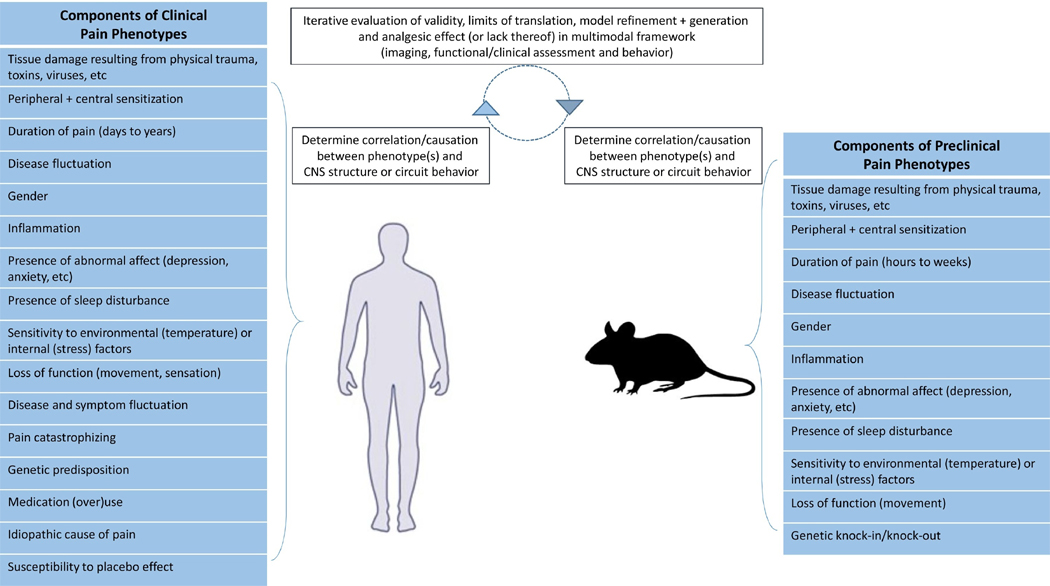
Assessing clinical pain and metrics related to function or quality of life predominantly relies on patient reported subjective measures. These outcome measures are generally not applicable to the preclinical setting where early signs pointing to analgesic value of a therapy are sought, thus introducing difficulties in animal to human translation in pain research. Evaluating brain function in patients and respective animal model(s) has the potential to characterize mechanisms associated with pain or pain-related phenotypes and thereby provide a means of laboratory to clinic translation. This review summarizes the progress made towards understanding of brain function in clinical and preclinical pain states elucidated using an imaging approach as well as the current level of validity of translational pain imaging. We hypothesize that neuroimaging can describe the central representation of pain or pain phenotypes and yields a basis for the development and selection of clinically relevant animal assays. This approach may increase the probability of finding meaningful new analgesics that can help satisfy the significant unmet medical needs of patients.

Keywords: Animal model; Brain imaging; Pain; Translation; Validity.


Figures

Figure 1
Figure 1. Convergence of phenotypes and CNS properties in clinical and pre-clinical settings
A model of clinical and preclinical pain experimentation considers the use of pain-related phenotypes in conjunction CNS function to assess and improve the overall validity of preclinical pain investigations.
Figure 2
Figure 2. Common CNS Circuitry Implicated in Processing Evoked Pain Stimuli in Healthy Human Subjects and Naïve Rats
Noxious thermal stimulation (7/10 pain rating) applied to the dorsum of the foot in healthy human subjects or noxious electrical stimulation administered to the paw of naïve rats commonly elicit BOLD (human fMRI data) or cerebral blood volume (rat fMRI data) changes in multiple CNS structures from the brainstem to cortex. CI – Cingulate cortex, S1 – Primary Somatosensory Cortex, S2 – Secondary Somatosensory Cortex, RSC – Retrospenial Cortex. Data was adapted and reproduced with permission from (Zhao et al., 2012) and (Upadhyay et al., 2015).
Figure 3
Figure 3. Common CNS Functional Responses During Evoked, Sensory Stimulation and CSD in Clinical and Preclinical Migraine States
In 8 migraine patients with extracephalic allodynia, increased thalamic BOLD responses to heat and brush stimuli were observed during migraine attack in comparison to migraine and allodynia free states in the same patients (A.) Thalamic activation to mechanical stimuli following dural administration of an Inflammatory soup (B.). Heightened electrophysiological responses of sensitized thalamic-trigeminovascular neurons to extracephalic skin stimuli following application of inflammatory soup to the respective dural afferents (C.). CSD can be measured and quantified in both migraine patients (D.) and preclinical migraine models (E.). Br – Brush, CL – Centrolateral thalamic nucleus, CM – Centromedian thalamic nucleus, CSD – Cortical spreading depression, LI – Limitans nucleus, MD – Mediodorsal thalamic group, PC – Posterior commissure, PF – Parafascicular thalamic nucleus, Pi – Pinch, Pr – Pressure, Pul – Pulvinar, V1 – Primary visual cortex, VPL – Ventral posterior lateral thalamic nucleus. Data was adapted and reproduced with permission from (Hadjikhani et al., 2001), (Burstein et al., 2010), (Becerra et al., 2016) and (Becerra et al., 2017).
Figure 4
Figure 4. Changes in Putaminal Function Commonly Observed in Knee OA Patients with Pain and the MMT Model of Knee OA
In 26 knee OA patients with pain, the magnitude of cerebral blood flow (CBF) in the putamen correlated with patient reported levels of pain with (A.) and without regression of pain catastrophizing scores (PCS) (B.) Compared with the sham condition, increased functional connectivity in the MMT model was observed when the left caudate-putamen (contralateral to MMT knee) was used as the seed region (C.). Following intravenous celecoxib administration and compared to the saline condition, decreases in caudate-putamen functional connectivity was measured (D.), while BOLD fMRI signals within the bilateral caudate-putamen was present only in MMT animals (E.), but not in sham animals (data not shown). The MMT model possesses not only aberrant functional properties in the CNS, but also, the expected peripheral pathology in the knee joint (synovial lesions (red arrow), subchondral bone lesions (yellow arrow) and cartilage hyper-intensities (blue arrow)) as measured by contrast enhanced MRI (F). Data was adapted and reproduced with permission from (Upadhyay et al., 2013) and (Cottam et al., 2016).

References

    1. Abaei M, Sagar DR, Stockley EG, Spicer CH, Prior M, Chapman V, Auer DP. Neural correlates of hyperalgesia in the monosodium iodoacetate model of osteoarthritis pain. Mol Pain. 2016;12 - PMC - PubMed
    1. Albrecht DS, MacKie PJ, Kareken DA, Hutchins GD, Chumin EJ, Christian BT, Yoder KK. Differential dopamine function in fibromyalgia. Brain Imaging Behav. 2016;10:829–839. - PMC - PubMed
    1. Amirmohseni S, Segelcke D, Reichl S, Wachsmuth L, Gorlich D, Faber C, Pogatzki-Zahn E. Characterization of incisional and inflammatory pain in rats using functional tools of MRI. Neuroimage. 2016;127:110–122. - PubMed
    1. Andrews N, Harper S, Issop Y, Rice AS. Novel, nonreflex tests detect analgesic action in rodents at clinically relevant concentrations. Ann N Y Acad Sci. 2011;1245:11–13. - PubMed
    1. Asad AB, Seah S, Baumgartner R, Feng D, Jensen A, Manigbas E, Henry B, Houghton A, Evelhoch JL, Derbyshire SW, Chin CL. Distinct BOLD fMRI Responses of Capsaicin-Induced Thermal Sensation Reveal Pain-Related Brain Activation in Nonhuman Primates. PLoS One. 2016;11:e0156805. - PMC - PubMed