. 2017 Oct 24;61(11):e01202-17.
doi: 10.1128/AAC.01202-17. Print 2017 Nov.

Impact of Type III Secretion Effectors and of Phenoxyacetamide Inhibitors of Type III Secretion on Abscess Formation in a Mouse Model of Pseudomonas aeruginosa Infection

Affiliations

Impact of Type III Secretion Effectors and of Phenoxyacetamide Inhibitors of Type III Secretion on Abscess Formation in a Mouse Model of Pseudomonas aeruginosa Infection

Bryan J Berube et al. Antimicrob Agents Chemother. .

Abstract

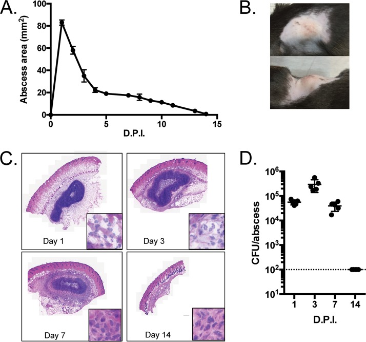
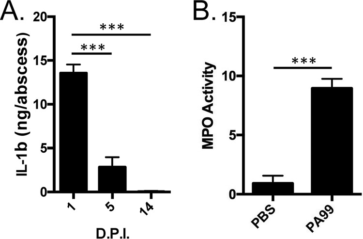
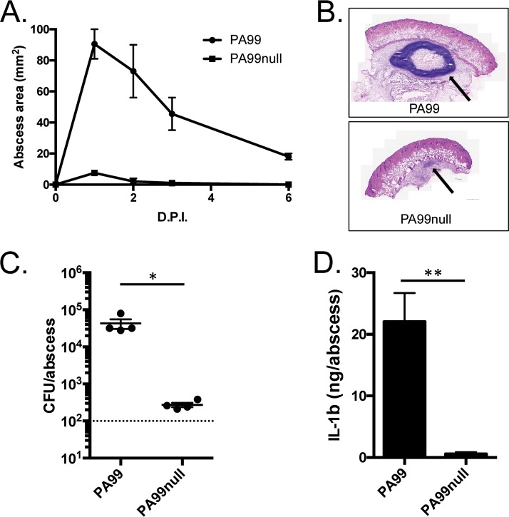
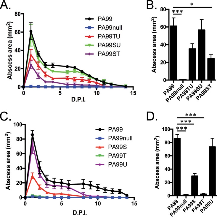
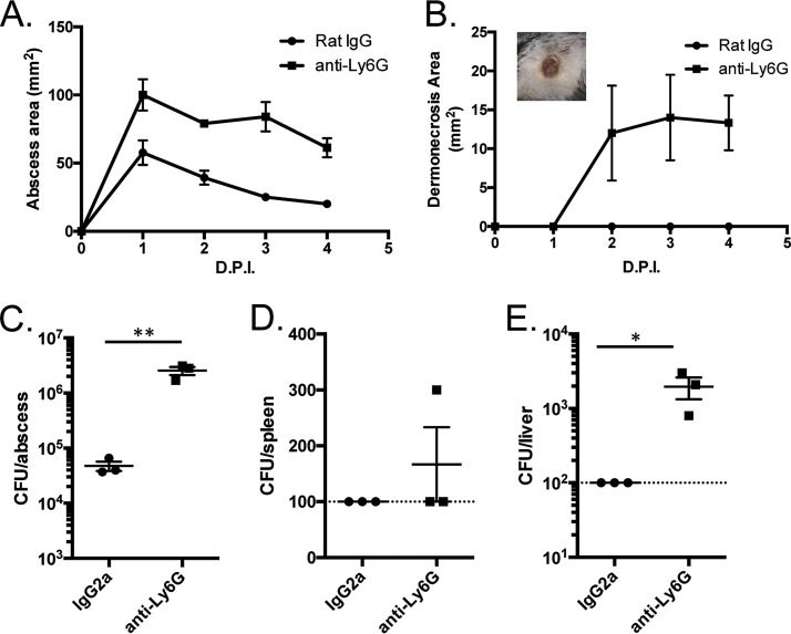
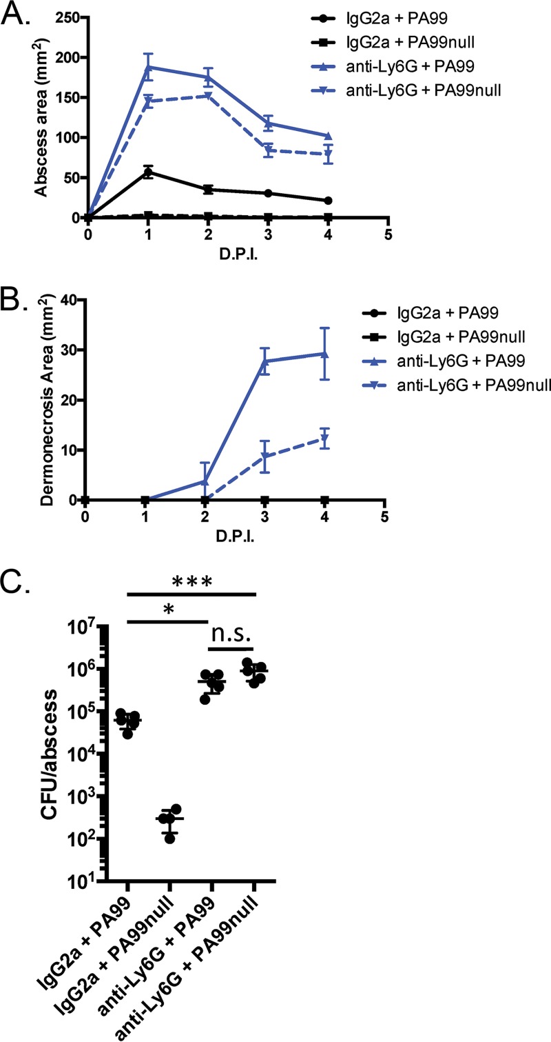
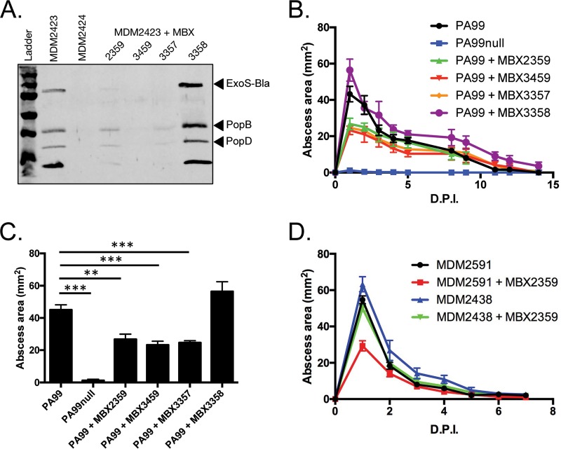
Pseudomonas aeruginosa is a leading cause of intra-abdominal infections, wound infections, and community-acquired folliculitis, each of which may involve macro- or microabscess formation. The rising incidence of multidrug resistance among P. aeruginosa isolates has increased both the economic burden and the morbidity and mortality associated with P. aeruginosa disease and necessitates a search for novel therapeutics. Previous work from our group detailed novel phenoxyacetamide inhibitors that block type III secretion and injection into host cells in vitro In this study, we used a mouse model of P. aeruginosa abscess formation to test the in vivo efficacy of these compounds against the P. aeruginosa type III secretion system (T3SS). Bacteria used the T3SS to intoxicate infiltrating neutrophils to establish abscesses. Despite this antagonism, sufficient numbers of functioning neutrophils remained for proper containment of the abscesses, as neutrophil depletion resulted in an increased abscess size, the formation of dermonecrotic lesions on the skin, and the dissemination of P. aeruginosa to internal organs. Consistent with the specificity of the T3SS-neutrophil interaction, P. aeruginosa bacteria lacking a functional T3SS were fully capable of causing abscesses in a neutropenic host. Phenoxyacetamide inhibitors attenuated abscess formation and aided in the immune clearance of the bacteria. Finally, a P. aeruginosa strain resistant to the phenoxyacetamide compound was fully capable of causing abscess formation even in the presence of the T3SS inhibitors. Together, our results further define the role of type III secretion in murine abscess formation and demonstrate the in vivo efficacy of phenoxyacetamide inhibitors in P. aeruginosa infection.

Keywords: Pseudomonas aeruginosa; abscesses; neutrophils; phenoxyacetamide inhibitors; type III secretion.


Figures

FIG 1
FIG 1
Characteristics of subcutaneous abscesses caused by P. aeruginosa strain PA99 in mice. (A to D) C57BL/6J mice were injected in the subcutaneous space with 1.2 × 10⁶ CFU of P. aeruginosa strain PA99. (A) The abscess size was determined daily by measuring the length and width of the abscess. Five mice were used per group. Symbols represent means, and error bars represent standard errors of the means. (B) Photographs of representative abscesses. (C) Abscess tissues were harvested at the indicated times postinfection via punch biopsy and were subjected to H&E staining. For each panel, the inset shows a portion of the micrograph at enhanced magnification. (D) Abscess tissues were harvested at the indicated times postinfection, homogenized, and spot plated to assess the number of CFU. Each symbol represents a single mouse; five mice were used per group. The dotted line represents the limit of detection. For all experiments, the data shown are representative of those from at least three independent experiments. D.P.I., days postinfection.
FIG 2
FIG 2
Inflammatory responses in nascent abscesses. C57BL/6J mice were infected in the subcutaneous space with strain PA99. (A) Abscess tissues were harvested and homogenized at the indicated times postinfection, and IL-1β levels were determined by ELISA. Data are means plus standard errors of the means. Experiments were done on three mice per group and were performed at least twice. Statistical analyses were performed by a one-way ANOVA and Dunnett's multiple-comparison test. ***, P < 0.001 compared to day 1. (B) Abscess tissues were harvested and homogenized at 24 h postinfection, and myeloperoxidase activity was measured as a surrogate for neutrophil activity. Experiments were done on five mice per group. Data are means plus standard deviations. Data were analyzed by an unpaired t test. ***, P < 0.001. D.P.I., days postinfection; PBS, phosphate-buffered saline; MPO, myeloperoxidase.
FIG 3
FIG 3
The role of type III secretion in abscess formation. C57BL/6J mice were infected in the subcutaneous space with either PA99 or PA99null. (A) The abscess size was measured daily over a 2-week period. Data are the mean abscess sizes for 5 mice per group, and error bars represent standard errors of the means. (B) Abscess tissues were harvested at 24 h postinfection, and cross sections of the abscesses were stained with H&E. Arrows, the abscess formed under the skin. (C) Abscess tissues were harvested by punch biopsy, and bacterial loads were determined by plating serial dilutions of tissue homogenates. Each symbol represents the abscess from a single mouse. Data are means plus standard deviations and were analyzed using an unpaired t test. Experiments used four mice per group and were performed at least three times. *, P < 0.05. (D) Abscess tissues were harvested at 24 h postinfection and homogenized. IL-1β levels were determined by ELISA. Data are means plus standard errors of the means and were analyzed using unpaired t tests. *, P < 0.05; **, P < 0.01. Experiments were done on three mice per group and were performed at least twice. D.P.I., days postinfection.
FIG 4
FIG 4
Roles of type III secretion effector proteins in abscess formation. C57BL/6J mice were infected in the subcutaneous space with P. aeruginosa strains lacking either a single type III effector (A and B) or two type III effectors (C and D). (A, C) Abscess sizes were measured over a 2-week period, and the peak abscess sizes at 24 h postinfection (from panels A and C) were plotted in bar graphs (B and D). The data in panels A and C represent mean abscess areas at each time point, and error bars represent standard errors of the means. The data in panels B and D represent means plus standard errors of the means and were analyzed by one-way ANOVA and Dunnett's multiple-comparison test. *, P < 0.05; ***, P < 0.001. Experiments were done on five mice per group. D.P.I., days postinfection.
FIG 5
FIG 5
Characteristics of abscesses in neutropenic mice. C57BL/6J mice were injected in the peritoneum with IgG2a control antibodies or anti-Ly6G antibodies to deplete the neutrophils. The mice were then infected in the subcutaneous space with P. aeruginosa strain PA99. (A, B) Abscess areas (A) and dermonecrotic areas (B) were measured daily following infection. (B, inset) Picture of a representative dermonecrotic skin lesion from an anti-Ly6G antibody-treated mouse. (C to E) Abscess (C), spleen (D), and liver (E) tissues were homogenized at 24 h postinfection and plated for CFU enumeration. The dashed lines represent the limit of detection. Data are means plus standard errors of the means. Experiments were done on three mice per group and were performed at least twice. Data were analyzed by unpaired t tests. *, P < 0.05; **, P < 0.01. D.P.I., days postinfection.
FIG 6
FIG 6
Role of type III secretion in abscess formation in a neutropenic mouse. C57BL/6J mice were injected in the peritoneum with IgG2a antibodies or anti-Ly6G antibodies to deplete the neutrophils. The mice were then infected in the subcutaneous space with P. aeruginosa strains PA99 or PA99null. (A, B) Abscess areas (A) and dermonecrotic areas (B) were measured daily following infection. (C) Mice were sacrificed at 24 h postinfection. Abscess tissues were harvested, homogenized, and plated for CFU enumeration. Data are means plus standard errors of the means. Statistics were calculated by a one-way ANOVA and Tukey's multiple-comparison test. *, P < 0.05, ***, P < 0.001; n.s., no significant difference. Four mice were used in the group treated with IgG2a and infected with PA99null; 5 mice per group were used for all other conditions. D.P.I., days postinfection.
FIG 7
FIG 7
Structures of novel phenoxyacetamide inhibitors. The chemical structures of MBX 3357, MBX 3358, and MBX 3459 are shown. MBX 3357 and MBX 3459 have inhibitory activity; MBX 3358 is an inactive stereoisomer of MBX 3357.
FIG 8
FIG 8
P. aeruginosa abscess formation in the presence of T3SS inhibitors. (A) P. aeruginosa strains were grown under secretion-inducing conditions in the presence or absence of T3SS inhibitors. The secretion of type III effectors into the supernatant was examined by Western blotting. (B, C) C57BL/6J mice were infected in the subcutaneous space with P. aeruginosa PA99 strains in the presence or absence of various phenoxyacetamide T3SS inhibitors (the final concentration of 100 μM was delivered in the inoculum), as indicated. Five mice were infected per group, and the experiment was repeated twice. (B) The abscess size was measured over the subsequent 2 weeks postinfection. (C) The peak abscess size (at day 1 postinfection) was plotted in a bar graph. (D) P. aeruginosa strains susceptible or resistant to phenoxyacetamide compounds were injected into the subcutaneous space of C57BL/6J mice in the presence or absence of the inhibitor MBX 2359. Experiments were performed on two different days, and the results were pooled. A total of 10 mice per group were used. The abscess size was measured over the subsequent 2 weeks. Data are means plus standard errors of the means. Statistics were calculated by a one-way ANOVA and Dunnett's multiple-comparison test. **, P < 0.01; ***, P < 0.001. D.P.I., days postinfection.

Similar articles

Cited by

References

    1. Stryjewski M, Sexton DJ. 2003. Pseudomonas aeruginosa infections in specific types of patients and clinical settings, p 1–15. In Hauser A, Rello J (ed), Severe infections caused by Pseudomonas aeruginosa. Kluwer Academic Publishers, Boston, MA.
    1. Fiorillo L, Zucker M, Sawyer D, Lin AN. 2001. The Pseudomonas hot-foot syndrome. N Engl J Med 345:335–338. doi:10.1056/NEJM200108023450504. - DOI - PubMed
    1. Silverman AR, Nieland ML. 1983. Hot tub dermatitis: a familial outbreak of Pseudomonas folliculitis. J Am Acad Dermatol 8:153–156. doi:10.1016/S0190-9622(83)70017-4. - DOI - PubMed
    1. Solomkin JS, Mazuski JE, Bradley JS, Rodvold KA, Goldstein EJ, Baron EJ, O'Neill PJ, Chow AW, Dellinger EP, Eachempati SR, Gorbach S, Hilfiker M, May AK, Nathens AB, Sawyer RG, Bartlett JG. 2010. Diagnosis and management of complicated intra-abdominal infection in adults and children: guidelines by the Surgical Infection Society and the Infectious Diseases Society of America. Clin Infect Dis 50:133–164. doi:10.1086/649554. - DOI - PubMed
    1. Sievert DM, Ricks P, Edwards JR, Schneider A, Patel J, Srinivasan A, Kallen A, Limbago B, Fridkin S, National Healthcare Safety Network (NHSN) Team and Participating NHSN Facilities. 2013. Antimicrobial-resistant pathogens associated with healthcare-associated infections: summary of data reported to the National Healthcare Safety Network at the Centers for Disease Control and Prevention, 2009-2010. Infect Control Hosp Epidemiol 34:1–14. doi:10.1086/668770. - DOI - PubMed

MeSH terms

LinkOut - more resources