. 2017 Nov;102(5):1261-1269.
doi: 10.1189/jlb.4A0317-116R. Epub 2017 Aug 14.

Human mucosal-associated invariant T (MAIT) cells possess capacity for B cell help

Affiliations

Human mucosal-associated invariant T (MAIT) cells possess capacity for B cell help

Michael S Bennett et al. J Leukoc Biol. 2017 Nov.

Abstract

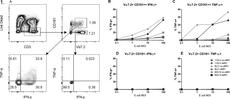
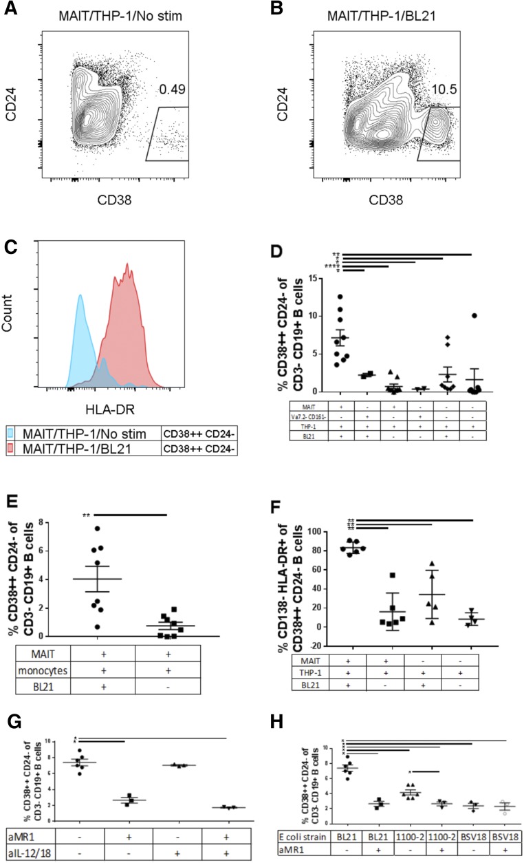
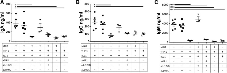
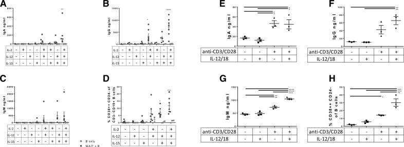
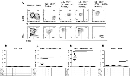
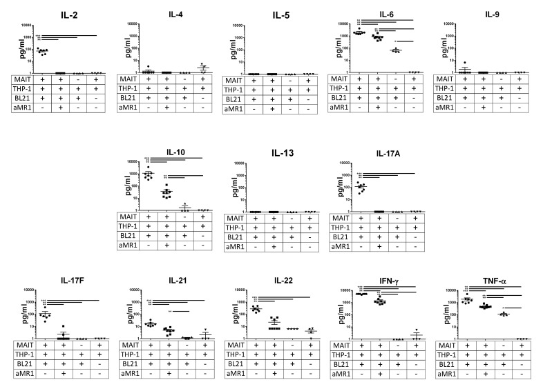
Mucosal-associated invariant T (MAIT) cells are an innate-like T cell subset, restricted by the nonclassic MHC class I-related protein MR1 and enriched at mucosal sites. Human studies have shown an association between MAIT cells and pathogen-specific antibody responses. In this study, we investigate the effect of human MAIT cells on B cells ex vivo. We found that supernatants from microbe- or cytokine-stimulated MAIT cells, when added to purified autologous B cells, increase frequencies of plasmablasts and promote IgA, IgG, and IgM production. We found effects to be mostly MR1-dependent and that the increases in plasmablasts are likely a result of increased differentiation from memory B cells. Furthermore, microbe-activated MAIT cell supernatant contains multiple cytokines known to stimulate B cells, including IL-6, -10, and -21. This study thus provides the first direct evidence of a newly identified role of MAIT cells in providing help to B cells.

Keywords: B cell help; MR-1; antibody; plasmablast.


Figures

Figure 1.
Figure 1.. MR1-dependent activation of MAIT cells by E. coli.
(A) Gating strategy of PBMCs stimulated overnight with formaldehyde-fixed BL21, 1100-2, or BSV18 E. coli, with or without anti-MR1 antibody (10 µg/ml) at 0.1, 1, 10, and 100 MOI, with 1.25 µg/ml anti-CD28. Levels of TNF-α and IFN-γ in (B and C) MAIT (Vα7.2+CD161++) and (D and E) non-MAIT (Vα7.2+CD161) T cell populations were measured by flow cytometry. Data are from a single donor and are representative of 2 independent experiments.
Figure 2.
Figure 2.. B cell phenotypic changes after addition of E. coli-stimulated MAIT cell supernatant.
We cultured B cells from different subjects in the presence of supernatant from subject-matched MAIT cells and THP-1 cells (A) or MAIT/THP-1/BL21 (B). Of CD38++CD24 cells, we measured HLA-DR (C) in B cells stimulated with MAIT/THP-1 (blue) or MAIT/THP-1/BL21 supernatant. Frequency of CD38++CD24 cells after addition of MAIT supernatant with THP-1 (D) and primary monocytes (E) as APCs, frequency of CD138HLA-DR+ of CD38++CD24 B cells (F), the effect of addition of anti-MR1 (10 µg/ml) and/or anti-IL-12 (5 µg/ml) and anti-IL-18 (5 µg/ml) during MAIT/THP-1/BL21 culture on the ability of resulting supernatant to differentiate B cells (G), the effect of different E. coli strains, with or without added anti-MR1 in MAIT/THP-1/E. coli supernatant (H). Data are means ± sem, and significance was determined by Mann-Whitney U tests. Data are representative of 3 independent experiments. *P ≤ 0.05; **P ≤ 0.01; ****P ≤ 0.00001.
Figure 3.
Figure 3.. MR1 is essential for induction of B cell antibody production by MAIT/THP-1/BL21 supernatant.
We cultured B cells from different subjects for 7 d in supernatant from subject-matched MAIT cells and THP-1 cells incubated overnight with BL21. (A–C) IgA, IgG, and IgM levels for the different conditions, with anti-MR1 (10 µg/ml), anti-IL-12/18, or both at 5 µg/ml each during initial MAIT/THP-1/BL21 stimulation or anti-CD40L (20 µg/ml) added to supernatant after MAIT culture and before addition to B cells. Data are means ± sem, and significance was determined by 1-way ANOVA using Tukey’s multiple-comparisons test. Data are representative of 2 independent experiments. *P ≤ 0.05; **P ≤ 0.01; ****P ≤ 0.00001.
Figure 4.
Figure 4.. Induction of antibody production and plasmablast differentiation by TCR- and cytokine-stimulated MAIT cells.
(A–D) We cocultured Vα7.2+CD161++ MAIT cells and B cells from different subjects in the presence of several combinations of IL-2, -12, and -15 for 7 d and measured antibody levels and B cell phenotype. Data are means ± sem, and were analyzed by 2-way ANOVA, with Fisher’s LSD test, comparing MAIT+B cells with B cells only. We also cultured Vα7.2+CD161++ T cells for 24 h with plate-bound anti-CD3/CD28 stimulation and/or recombinant IL-12/18, and supernatant with IL-12/18 blocking antibodies was applied to B cells for 7 d, followed by ELISA (E–G) and flow cytometry (H) for B cell phenotype. Data shown are mean ± sem, and significance was determined by 1-way ANOVA using Tukey’s multiple comparisons test. Data are representative of 2 independent experiments. *P ≤ 0.05; **P ≤ 0.01; ***P ≤ 0.001; ****P ≤ 0.00001.
Figure 5.
Figure 5.. Activated MAIT supernatant induces plasmablast differentiation from memory B cells.
(A) B cells from multiple subjects were sorted and then pooled into CD3CD19+ B cell populations based on IgD, CD27, CD38, and CD24 expression. (B–E) Supernatant from cocultures of different combinations of MAIT cells from multiple subjects, THP-1 cells, and BL21 (100 MOI per THP-1 cell) were added to different populations of sort-purified B cells, each population containing 150,000 IgD+CD27 cells per well plus either no extra cells (B), 150,000 added IgD+CD27+CD38 cells (C), 150,000 added IgD-CD27+CD38 cells (D), or 150,000 IgDCD27+CD38++ cells (E), and cultured for 7 d. After 7 d, B cells were assayed for the frequency of CD38++CD24 plasmablasts. Data are s ± sem, and representative of 2 independent experiments. Significance was determined by 1-way ANOVA using Tukey’s multiple comparisons test. ****P ≤ 0.00001.
Figure 6.
Figure 6.. Cytokine profile of ex vivo-stimulated MAIT cells.
Vα7.2+ primary T cells from multiple subjects were cultured overnight with THP-1 cells in the presence or absence of fixed E. coli (strain BL21) at an MOI of 100 per THP-1 cell and with or without anti-MR1 (10 µg/ml) and cytokine levels were measured with a T-helper-specific multiplex cytokine array. Data shown are means ± sem, significance was determined by Mann-Whitney U tests and are representative of 2 independent experiments. **P ≤ 0.01; ***P ≤ 0.001.

References

    1. Crotty S. (2015) A brief history of T cell help to B cells. Nat. Rev. Immunol. 15, 185–189. - PMC - PubMed
    1. Dellabona P., Abrignani S., Casorati G. (2014) iNKT-cell help to B cells: a cooperative job between innate and adaptive immune responses. Eur. J. Immunol. 44, 2230–2237. - PubMed
    1. Kjer-Nielsen L., Patel O., Corbett A. J., Le Nours J., Meehan B., Liu L., Bhati M., Chen Z., Kostenko L., Reantragoon R., Williamson N. A., Purcell A. W., Dudek N. L., McConville M. J., O’Hair R. A., Khairallah G. N., Godfrey D. I., Fairlie D. P., Rossjohn J., McCluskey J. (2012) MR1 presents microbial vitamin B metabolites to MAIT cells. Nature 491, 717–723. - PubMed
    1. Walker L. J., Kang Y. H., Smith M. O., Tharmalingham H., Ramamurthy N., Fleming V. M., Sahgal N., Leslie A., Oo Y., Geremia A., Scriba T. J., Hanekom W. A., Lauer G. M., Lantz O., Adams D. H., Powrie F., Barnes E., Klenerman P. (2012) Human MAIT and CD8αα cells develop from a pool of type-17 precommitted CD8+ T cells. Blood 119, 422–433. - PMC - PubMed
    1. Eckle S. B., Birkinshaw R. W., Kostenko L., Corbett A. J., McWilliam H. E., Reantragoon R., Chen Z., Gherardin N. A., Beddoe T., Liu L., Patel O., Meehan B., Fairlie D. P., Villadangos J. A., Godfrey D. I., Kjer-Nielsen L., McCluskey J., Rossjohn J. (2014) A molecular basis underpinning the T cell receptor heterogeneity of mucosal-associated invariant T cells. J. Exp. Med. 211, 1585–1600. - PMC - PubMed

MeSH terms

LinkOut - more resources