. 2017 Oct 1;77(19):5301-5312.
doi: 10.1158/0008-5472.CAN-17-0418. Epub 2017 Aug 14.

Nuclear FAK and Runx1 Cooperate to Regulate IGFBP3, Cell-Cycle Progression, and Tumor Growth

Affiliations

Nuclear FAK and Runx1 Cooperate to Regulate IGFBP3, Cell-Cycle Progression, and Tumor Growth

Marta Canel et al. Cancer Res. .

Abstract

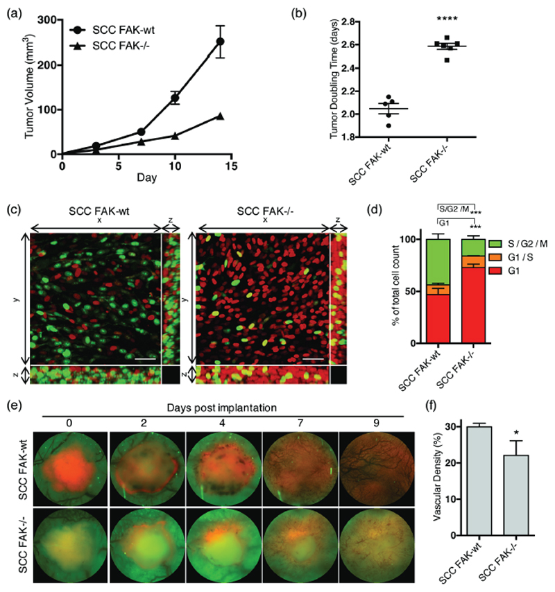
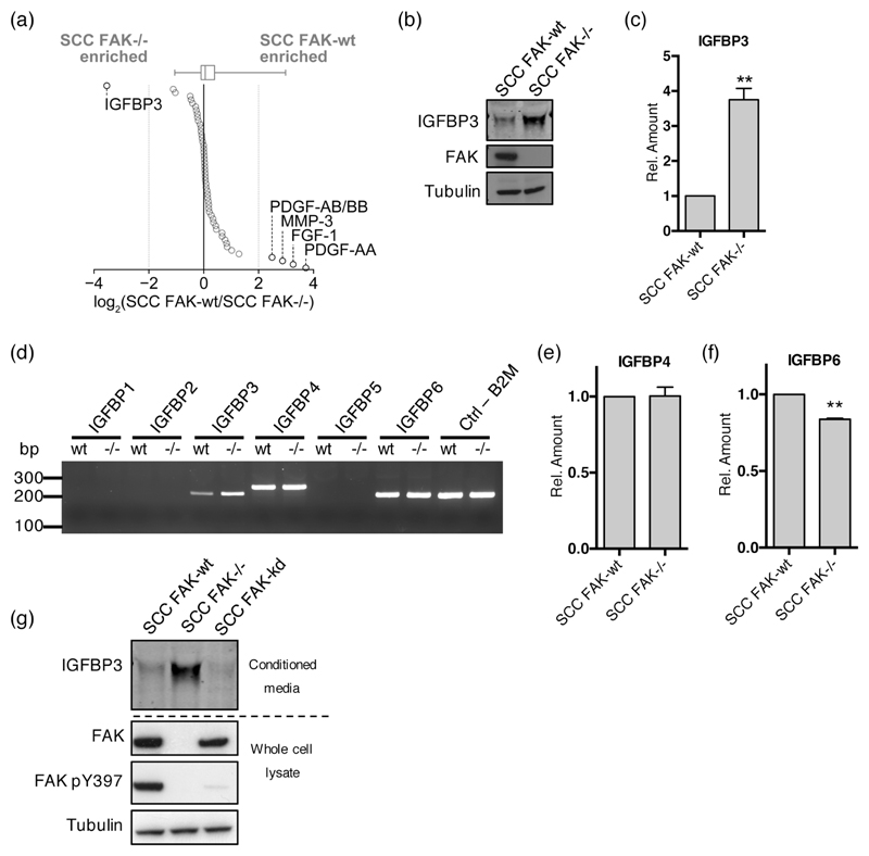
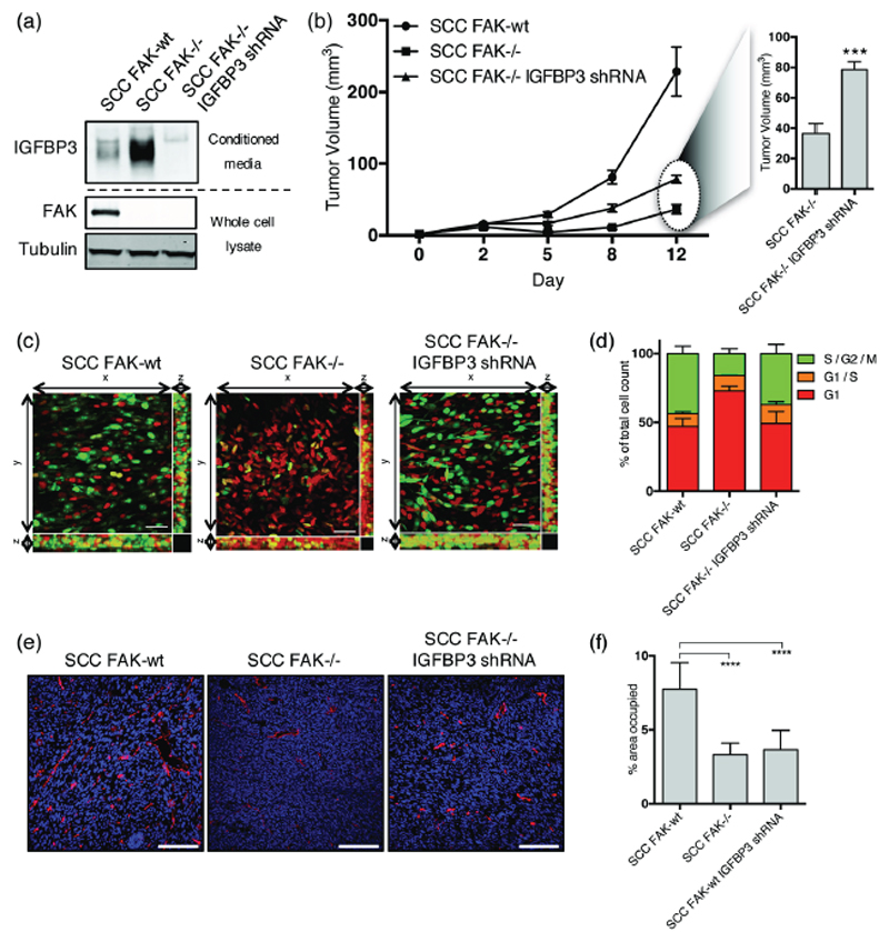
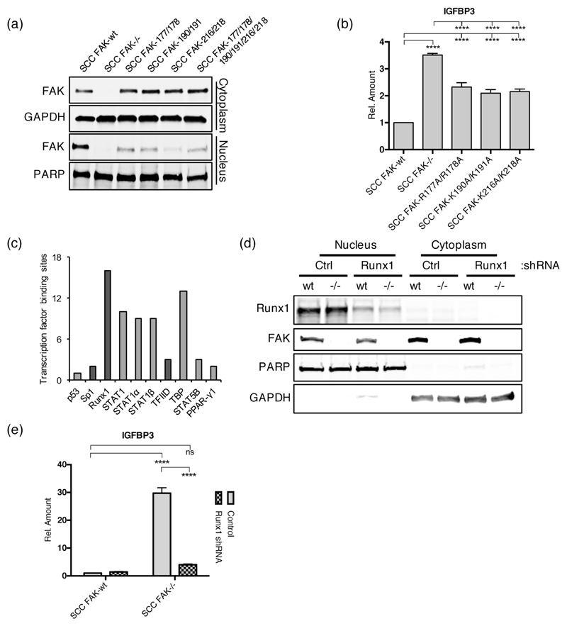
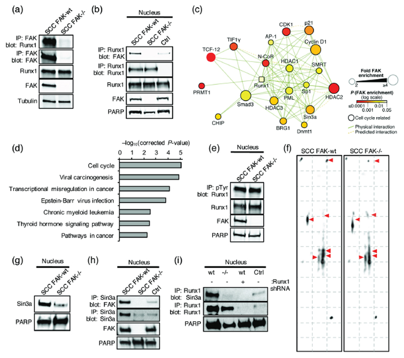
Nuclear focal adhesion kinase (FAK) is a potentially important regulator of gene expression in cancer, impacting both cellular function and the composition of the surrounding tumor microenvironment. Here, we report in a murine model of skin squamous cell carcinoma (SCC) that nuclear FAK regulates Runx1-dependent transcription of insulin-like growth factor binding protein 3 (IGFBP3), and that this regulates SCC cell-cycle progression and tumor growth in vivo Furthermore, we identified a novel molecular complex between FAK and Runx1 in the nucleus of SCC cells and showed that FAK interacted with a number of Runx1-regulatory proteins, including Sin3a and other epigenetic modifiers known to alter Runx1 transcriptional function through posttranslational modification. These findings provide important new insights into the role of FAK as a scaffolding protein in molecular complexes that regulate gene transcription. Cancer Res; 77(19); 5301-12. ©2017 AACR.


Conflict of interest statement

Conflict of interest: The authors declare no potential conflicts of interest.

Figures

Figure 1
Figure 1. FAK regulates SCC tumor growth, cell cycle and angiogenesis in vivo.
(a) Growth of SCC FAK-wt and SCC FAK-/- tumor xenografts in CD-1 nude mice. n = 5 - 6 tumors per group. (b) SCC FAK-wt and SCC FAK-/- tumor doubling time. Unpaired T-test, ****p < 0.0001. (c) Intra-vital imaging of FUCCI expressing SCC FAK-wt and SCC FAK-/- cells 24 hours post-implantation under dorsal skinfold windows. (d) Quantitation of FUCCI cell cycle distribution from 3-dimentional image stacks shown in panel c. Sidak’s corrected 2way ANOVA, ***p < 0.001. n = 4 tumors per group. (e) Longitudinal imaging of tumour angiogenesis following implantation of tumour fragments under dorsal skinfold windows. Red – tagRFP labelled SCC tumor, Green – tissue autofluorescence. (f) Quantitation of blood vessel density at day 9. Unpaired T-test, *p = 0.0306. Data in all graphs represented as mean +/- s.e.m. n = 3 tumors per group.
Figure 2
Figure 2. FAK negatively regulates the expression of IGFBP3 but not other IGFBP family members.
(a) Relative secreted levels of 53 angiogenesis-related proteins measured by antibody capture array from media conditioned by SCC FAK-wt and FAK-/- cells. Proteins are ordered by fold change. Dotted gray lines indicate four-fold enrichment; proteins changed by at least four fold are indicated. Box-and-whisker plot summarizes the median (line), 25th and 75th percentiles (box) and 5th and 95th percentiles (whiskers). (b) Representative anti-IGFBP3 western blot from concentrated conditioned media. (c) (q)RT-PCR analysis of IGFBP3 transcript levels in SCC FAK-wt and FAK-/- cells. n = 3. (d) Representative PCR analysis of IGFBP family transcript levels in SCC FAK-wt and FAK-/- cells. (e) (q)RT-PCR analysis of IGFBP4 transcript levels in SCC FAK-wt and FAK-/- cells. n = 3. (f) (q)RT-PCR analysis of IGFBP6 transcript levels in SCC FAK-wt and FAK-/- cells. n = 3. (g) Representative anti-IGFBP3 western blot showing secreted IGFBP3 protein levels from SCC FAK-wt, SCC FAK-/-, and SCC FAK-kd cells. Data in all graphs represented as mean +/- s.e.m. Unpaired T-test, **p < 0.01.
Figure 3
Figure 3. IGFBP3 regulates cell cycle progression but not tumor angiogenesis.
(a) Representative anti-IGFBP3 western blot from concentrated media conditioned by either SCC FAK-wt, SCC FAK-/-, or SCC FAK-/- IGFBP3 shRNA cells. Anti-FAK western blot shows FAK expression status and Anti-tubulin western blot was used a loading control. (b) Left - Growth of SCC FAK-wt, SCC FAK-/-, and SCC FAK-/- IGFBP3 shRNA tumor xenografts in CD-1 nude mice. Right – Average volume of SCC FAK-/- and SCC FAK-/- IGFBP3 shRNA tumors at day 12. Unpaired T-test, ***p < 0.001. n = 6 tumors per group. (c) Intra-vital imaging of FUCCI expressing SCC FAK-wt, SCC FAK-/-, and SCC FAK-/- IGFBP3 shRNA cells 24 hours post-implantation under dorsal skinfold windows. (d) Quantitation of FUCCI cell cycle distribution from 3-dimentional image stacks shown in panel c. Values shown for SCC FAK-wt and SCC FAK-/- cells are repeated from Fig. 1d. n = 4 tumors per group. (e) Fluorescent staining of frozen tissue sections using anti-CD31 antibody (red). Nuclei labelled using DAPI (blue). (f) Quantitation of the % area occupied by CD31+ cells. Tukey’s corrected one-way ANOVA, ****p < 0.0001. Data in all graphs represented as mean +/- s.e.m. n = 3 tumors per group with an average of 3 fields measured per tumor.
Figure 4
Figure 4. Nuclear FAK and RUNX1 regulate IGFBP3 expression.
(a) Representative anti-FAK western blot of cytoplasmic and nuclear fractions prepared from a series of SCC cells expressing FAK nuclear localization signal (NLS) mutants. (b) (q)RT-PCR analysis of IGFBP3 expression in SCC cells expressing FAK NLS mutants. Tukey’s corrected 1way ANOVA, ****p < 0.0001. Data in all graphs represented as mean +/- s.e.m. n = 3. (c) Predicted transcription factor binding sites in the promoter of Igfbp3. Transcription factors that interact with nuclear FAK in SCC cells are displayed in dark grey. (d) Representative western blot showing Runx1 depletion using shRNA. (e) (q)RT-PCR analysis of IGFBP3 expression in control and Runx1 depleted SCC FAK-wt and SCC FAK-/- cells. Sidak’s corrected 2way ANOVA, ****p < 0.0001. ns = not significant. n = 3.
Figure 5
Figure 5. Nuclear FAK interacts with Runx1.
(a) Representative western blot of anti-FAK immunoprecipitation probed with anti-Runx1 antibody. (b) Representative western blot of anti-Runx1 immunoprecipitation probed with anti-FAK antibody. IgG control (Ctrl). (c) Interaction network analysis of physical or predicted direct binders of Runx1 that interact with FAK in the nucleus of SCC cells (see Supplementary Table 2). Runx1 is shown as a square node. Protein node size is proportional to fold enrichment in nuclear FAK immunoprecipitations. Node color indicates significance of enrichment in nuclear FAK immunoprecipitations. (d) Pathway enrichment analysis of KEGG terms in the nuclear FAK interactome of Runx1 binders (Q < 0.01) (see Supplementary Fig. 5). (e) Representative western blot of anti-phospho-tyrosine (pTyr) immunoprecipitation probed with anti-Runx1 antibody. (f) 2D gel electrophoresis probed with anti-Runx1 antibody. (g) Representative western blot of SCC FAK-wt and FAK-/- nuclear lysates probed with anti-Sin3a antibody. (h) Western blot of anti-Sin3a immunoprecipitation from SCC FAK-wt and FAK-/- nuclear lysates probed with anti-FAK antibody. IgG control (Ctrl) was done using SCC FAK-wt nuclear lysates. (i) Western blot of anti-Runx1 immunoprecipitation from SCC FAK-wt, SCC FAK-/-, and SCC FAK-wt Runx1 shRNA nuclear lysates probed with anti-Sin3a antibody. IgG control (Ctrl) was done using SCC FAK-wt nuclear lysates.

References

    1. McLean GW, Carragher NO, Avizienyte E, Evans J, Brunton VG, Frame MC. The role of focal-adhesion kinase in cancer - a new therapeutic opportunity. Nature reviews Cancer. 2005;5:505–15. - PubMed
    1. Sulzmaier FJ, Jean C, Schlaepfer DD. FAK in cancer: mechanistic findings and clinical applications. Nature reviews Cancer. 2014;14:598–610. - PMC - PubMed
    1. Ashton GH, Morton JP, Myant K, Phesse TJ, Ridgway RA, Marsh V, et al. Focal adhesion kinase is required for intestinal regeneration and tumorigenesis downstream of Wnt/c-Myc signaling. Developmental cell. 2010;19:259–69. - PMC - PubMed
    1. Lahlou H, Sanguin-Gendreau V, Zuo D, Cardiff RD, McLean GW, Frame MC, et al. Mammary epithelial-specific disruption of the focal adhesion kinase blocks mammary tumor progression. Proceedings of the National Academy of Sciences of the United States of America. 2007;104:20302–7. - PMC - PubMed
    1. Luo M, Fan H, Nagy T, Wei H, Wang C, Liu S, et al. Mammary epithelial-specific ablation of the focal adhesion kinase suppresses mammary tumorigenesis by affecting mammary cancer stem/progenitor cells. Cancer research. 2009;69:466–74. - PMC - PubMed

Publication types

MeSH terms

Substances