. 2017 Aug 29;114(35):9469-9474.
doi: 10.1073/pnas.1702441114. Epub 2017 Aug 14.

BDNF-TrkB controls cocaine-induced dendritic spines in rodent nucleus accumbens dissociated from increases in addictive behaviors

Affiliations

BDNF-TrkB controls cocaine-induced dendritic spines in rodent nucleus accumbens dissociated from increases in addictive behaviors

Ethan M Anderson et al. Proc Natl Acad Sci U S A. .

Abstract

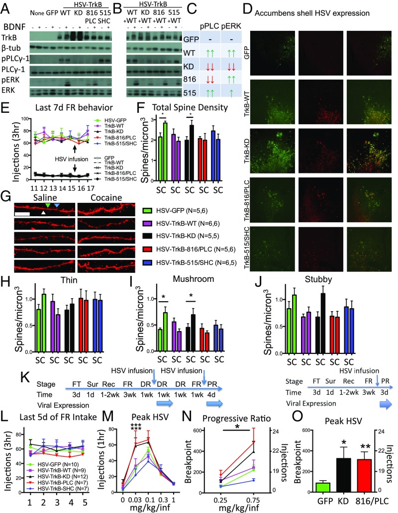
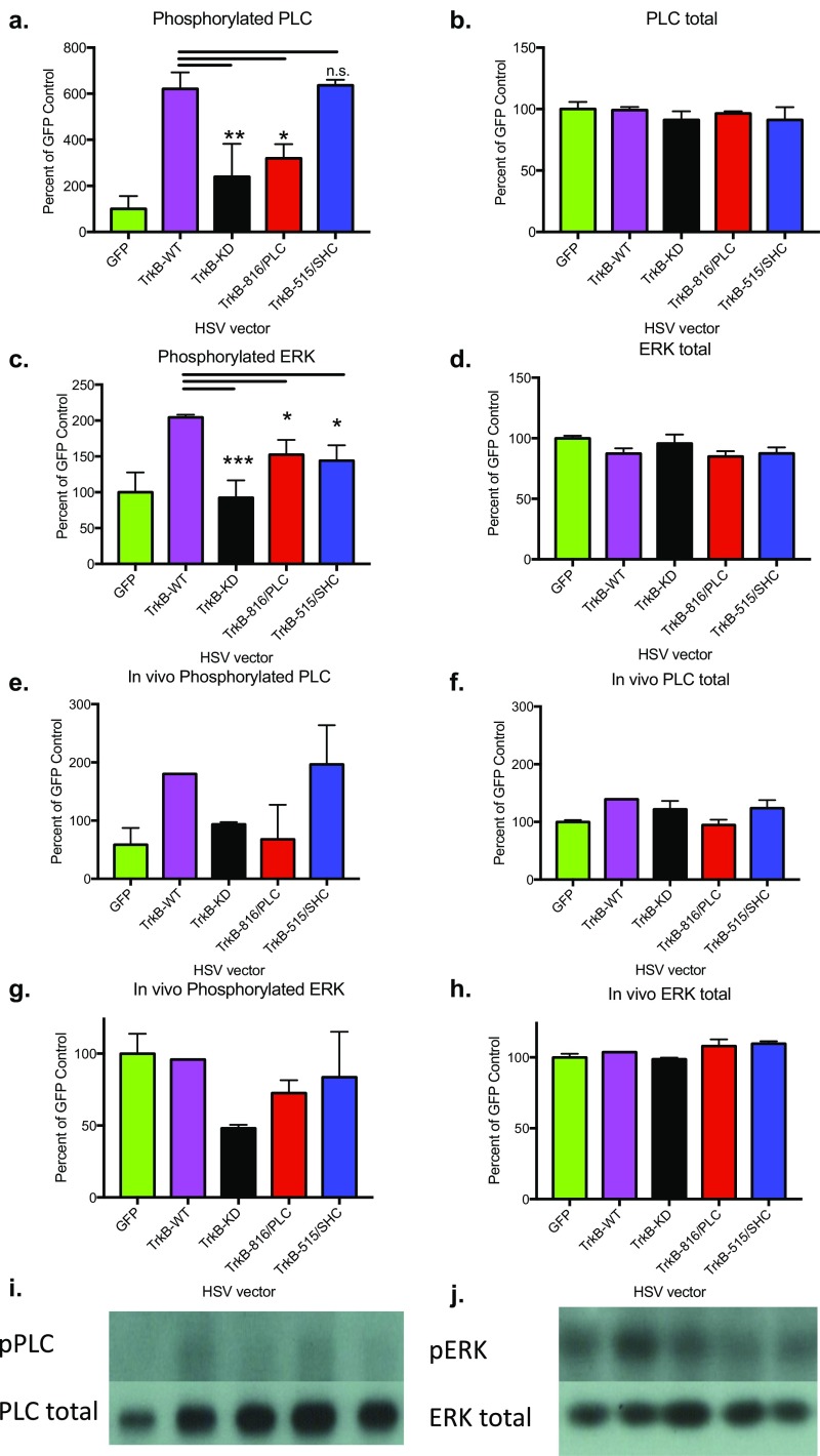
Chronic cocaine use is associated with prominent morphological changes in nucleus accumbens shell (NACsh) neurons, including increases in dendritic spine density along with enhanced motivation for cocaine, but a functional relationship between these morphological and behavioral phenomena has not been shown. Here we show that brain-derived neurotrophic factor (BDNF) signaling through tyrosine kinase B (TrkB) receptors in NACsh neurons is necessary for cocaine-induced dendritic spine formation by using either localized TrkB knockout or viral-mediated expression of a dominant negative, kinase-dead TrkB mutant. Interestingly, augmenting wild-type TrkB expression after chronic cocaine self-administration reverses the sustained increase in dendritic spine density, an effect mediated by TrkB signaling pathways that converge on extracellular regulated kinase. Loss of TrkB function after cocaine self-administration, however, leaves spine density intact but markedly enhances the motivation for cocaine, an effect mediated by specific loss of TrkB signaling through phospholipase Cgamma1 (PLCγ1). Conversely, overexpression of PLCγ1 both reduces the motivation for cocaine and reverses dendritic spine density, suggesting a potential target for the treatment of addiction in chronic users. Together, these findings indicate that BDNF-TrkB signaling both mediates and reverses cocaine-induced increases in dendritic spine density in NACsh neurons, and these morphological changes are entirely dissociable from changes in addictive behavior.

Keywords: BDNF-TrkB; PLC; accumbens shell; cocaine addiction; dendritic spines.


Conflict of interest statement

The authors declare no conflict of interest.

Figures

Fig. 1.
Fig. 1.
Cocaine induction of spines is blocked by TrkB knockdown. (A) Localized expression of GFP (green) and TrkB (blue) after intra-NACsh infusions of HSV-GFP, HSV-Cre, or HSV-TrkB-KD (with bicistronic GFP) in floxed TrkB mice. (Magnification, 10×.) (B) Total spine density of saline- and cocaine-treated (5 × 20 mg/kg, i.p.) mice during HSV expression. (C) Representative images of saline- and cocaine-treated dendrites expressing GFP only, Cre recombinase, or TrkB-KD. Representative spines are labeled white (thin), blue (mushroom), and green (stubby) to illustrate their categorization. (Scale bar, 10 µm.) (D) Thin, (E) mushroom, and (F) stubby spine density after binge cocaine-dosing paradigm. Data are expressed as mean ± SEM. **P < 0.01 compared with saline.
Fig. S1.
Fig. S1.
Lack of evidence for retrograde infection with NACsh infusions of HSV-Cre. GFP from the HSV-Cre virus was detected via IHC in the NAC, but not in areas projecting to the NAC including ventral hippocampus (vHIPP), prelimbic prefrontal cortex (PL), and ventral tegmental area (VTA). Representative images are shown. (Magnification, 4×.)
Fig. 2.
Fig. 2.
BDNF-TrkB signaling reverses dendritic spines induced by chronic CSA and is dissociated from increased CSA behaviors caused by the selective loss of BDNF-TrkB-PLC activity in NACsh neurons. (A) Protein levels of TrkB, β-tubulin, pPLCγ, PLCγ, pERK, and ERK either with or without the addition of BDNF. Results from HEK293 cells infected with no vector, HSV-GFP, HSV-TrkB-WT, HSV-TrkB-KD, HSV-TrkB-816/PLC, or HSV-TrkB-515/SHC alone. (B) Coinfection with HSV-TrkB-WT reveals pathway-specific dominant negative profiles of TrkB signaling mutants. (C) Summary of TrkB-WT and dominant negative TrkBs HSV on pPLC and pERK. (D) Localized expression of GFP (green) and TrkB (red) 3 d after intra-NACsh infusions of HSV-GFP and HSV-TrkB vectors. (Magnification, 10×.) (E) Cocaine and saline SA rates before and after infusions of HSV vectors. (F) Quantification of total spine density in saline and CSA expressing TrkB viral vectors. (G) Representative images of dendritic segments expressing HSV vectors from saline and CSA animals. Representative spines are labeled white (thin), blue (mushroom), and green (stubby) to illustrate their categorization. (Scale bar, 10 µm.) (H) Quantification of thin, (I) mushroom, and (J) stubby spine density after SA. (K, Left) Experimental time course depicting operant training with food pellets (FT), surgery (Sur), recovery (Rec), and fixed ratio (FR) CSA training, followed by dose–response (DR) before, during, and after HSV infusion. A second HSV infusion is given before CSA testing on a PR reinforcement schedule. (K, Right) A modified experimental time course depicting 1 HSV infusion before PR. (L) Average CSA (FR5) in study groups before (M) dose–response testing during peak HSV-mediated expression. (N) CSA on a PR schedule during HSV expression after a second HSV infusion; the asterisk above a line represents a significant main effect of vector after a mixed factorial ANOVA. (O) CSA (0.75 mg/kg/injection) on a PR schedule during HSV expression after a single HSV infusion. Data are expressed as mean ± SEM. *P < 0.05; **P < 0.01; ***P < 0.001 compared with HSV-GFP. C, cocaine; S, saline.
Fig. S2.
Fig. S2.
Mutant TrkB receptors selectively alter BDNF-induced PLC and ERK phosphorylation events. HEK293 cells were infected with HSV-GFP (n = 3), HSV-TrkB-WT (n = 6), HSV-TrkB-KD (n = 2), HSV-TrkB-816/PLC (n = 3), or HSV-TrkB-515/SHC (n = 3), and then 100 ng/mL BDNF or PBS was added 1 d later and cells were harvested after 30 min. Results from Western blots are shown here, and BDNF-treated samples were analyzed with a one-way ANOVA. (A) TrkB vectors had significant effects on phosphorylated PLC (F4,12 = 9.81; P = 0.0009), but not (B) PLC total protein levels. (C) TrkB vectors also had significant effects on phosphorylated ERK (F4,12 = 8.342; P = 0.0019), but not (D) total ERK protein levels. Similar effects were also observed in vivo. Rats received cannulation surgery and, after recovery, were infused with HSV-GFP (n = 5), HSV-TrkB-WT (n = 1), HSV-TrkB-KD (n = 3), HSV-TrkB-816/PLC (n = 2), or HSV-TrkB-515/SHC (n = 2). After 3 d, during peak expression HSV times, rats were infused with 1 μL BDNF (0.5 mg/μL) and sacked 30 min later. Compared with in vitro cell culture data, similar profiles were observed for (E) phosphorylated PLC, (F) PLC total protein, (G) phosphorylated ERK, and (H) ERK total, suggesting these TrkB mutants work the same in vivo. Qualitative representative images are shown for (I) pPLC/PLC total and (J) pERK/ERK total. Data are expressed as mean ± SEM. *P < 0.05; **P < 0.01; ***P < 0.001 compared with WT control. n.s., not significant.
Fig. S3.
Fig. S3.
Cocaine-induced spines are reversed by TrkB-WT, but not TrkB-KD, overexpression after 17 d of daily 20 mg/kg i.p. cocaine injections in mice. Mice were infused with HSV-GFP, HSV-TrkB-WT, or HSV-TrkB-KD before the 2 additional i.p. injections and perfused 1 d later to mimic the 17-d CSA treatment conditions shown in Fig. 2 E–J. (A) Cocaine i.p. injections increased total spine density in HSV-GFP mice compared with saline-injected controls. This increase was reversed in mice infused with HSV-TrkB-WT, but not HSV-TrkB-KD (effect of drug, F1,17 = 6.799 [P = 0.0184]; interaction, F2,17 = 5.496 [P = 0.0144\). (B) The increases in total spine density is reflected by an increase in mushroom spine density (effect of drug, F1,17 = 17.09 [P = 0.0007]; interaction, F2,17 = 4.105 [P = 0.0351]), but not (C) thin nor (D) stubby spines similar to findings in CSA rats. Data are expressed as mean ± SEM. *P < 0.05 compared with saline control. Coc, cocaine; Sal, saline.
Fig. S4.
Fig. S4.
Similar baseline SA for fixed ratio dose–response testing is seen in all HSV groups, and no HSV vector caused long-lasting changes in postexpression CSA behavior. (A) Dose–response testing was not significantly different between groups before GFP, TrkB-WT, TrkB-KD, TrkB-816/PLC, or TrkB-515/SHC expression (effect of dose, F4,152 = 159.2; P < 0.001). (B) Dose–response testing was not significantly different after the second week of HSV expression (effect of dose, F4,152 = 102.7 [P < 0.001]). (C) No significant change in FR5 intake was evident the third week after HSV expression. (D) No significant differences in FR intake was evident for rats before expression of GFP, TrkB-KD, or TrkB-816/PLC before PR testing after a single infusion of HSV vector. (E) Dose–response testing was not significantly different before GFP or PLCγ1 expression (effect of dose, F4,52 = 52.38 [P < 0.001]). (F) Dose–response testing was not significantly different after the second week of GFP or PLCγ1 expression (effect of dose, F4,52 = 46.73 [P < 0.001]). (G) No significant change in FR5 intake was evident the third week after GFP or PLCγ1 expression.
Fig. 3.
Fig. 3.
PLCγ overexpression both reverses dendritic spines induced by chronic CSA and reduces CSA behaviors. (A) Western blot depicting increased phosphorylated and total PLCγ in HEK293 cells infected with HSV-PLCγ compared with HSV-TrkB-WT. (B) Time course of pERK after addition of the PLC activator m-3M3FBS (100 µM) over the course of 30 min. (C) Localized expression of GFP (green) and PLCγ (blue) 3 d after intra-NACsh infusions of HSV-GFP or HSV-PLCγ vectors. (D) Cocaine and saline SA rates before and after HSV-PLCγ or HSV-GFP infusions. (E) Quantification of mushroom spine density in saline and CSA overexpressing PLCγ or GFP. (F) Representative images of dendritic segments expressing HSV- PLCγ from saline and CSA animals. Representative spines are labeled white (thin), blue (mushroom), and green (stubby) to illustrate their categorization. (Scale bar, 10 µm.) (G) Average CSA (FR5) in study groups before (H) dose–response testing during peak HSV-mediated expression of PLCγ. (I) CSA on a PR schedule during GFP or PLCγ expression after a second HSV infusion (Fig. 2K, Left for time line). The asterisk above a line represents a significant main effect of vector after a mixed factorial ANOVA. Data are expressed as mean ± SEM. *P < 0.05 compared with HSV-GFP controls. C, cocaine; n.s., not significant; S, saline.
Fig. S5.
Fig. S5.
The effect of HSV-PLC versus HSV-GFP on total, thin, or stubby spines in the NACsh. Saline- and cocaine-treated rats infused with HSV-GFP or HSV-PLC were analyzed for (A) total (effect of drug, F1,23 = 6.71 [P = 0.0164]), (B) thin, and (C) stubby spines. Data are expressed as mean ± SEM. Coc, cocaine; Sal, saline.

References

    1. Dennis ML, Foss MA, Scott CK. An eight-year perspective on the relationship between the duration of abstinence and other aspects of recovery. Eval Rev. 2007;31:585–612. - PubMed
    1. Graham DL, et al. Tropomyosin-related kinase B in the mesolimbic dopamine system: Region-specific effects on cocaine reward. Biol Psychiatry. 2009;65:696–701. - PMC - PubMed
    1. Graham DL, et al. Dynamic BDNF activity in nucleus accumbens with cocaine use increases self-administration and relapse. Nat Neurosci. 2007;10:1029–1037. - PubMed
    1. Bahi A, Boyer F, Chandrasekar V, Dreyer JL. Role of accumbens BDNF and TrkB in cocaine-induced psychomotor sensitization, conditioned-place preference, and reinstatement in rats. Psychopharmacology (Berl) 2008;199:169–182. - PubMed
    1. Li X, et al. Different roles of BDNF in nucleus accumbens core versus shell during the incubation of cue-induced cocaine craving and its long-term maintenance. J Neurosci. 2013;33:1130–1142. - PMC - PubMed

Publication types

LinkOut - more resources