. 2017 Aug 29;114(35):9439-9444.
doi: 10.1073/pnas.1707032114. Epub 2017 Aug 14.

Immune evasion by a staphylococcal inhibitor of myeloperoxidase

Affiliations

Immune evasion by a staphylococcal inhibitor of myeloperoxidase

Nienke W M de Jong et al. Proc Natl Acad Sci U S A. .

Abstract

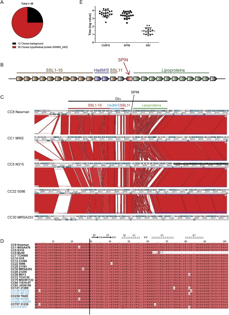
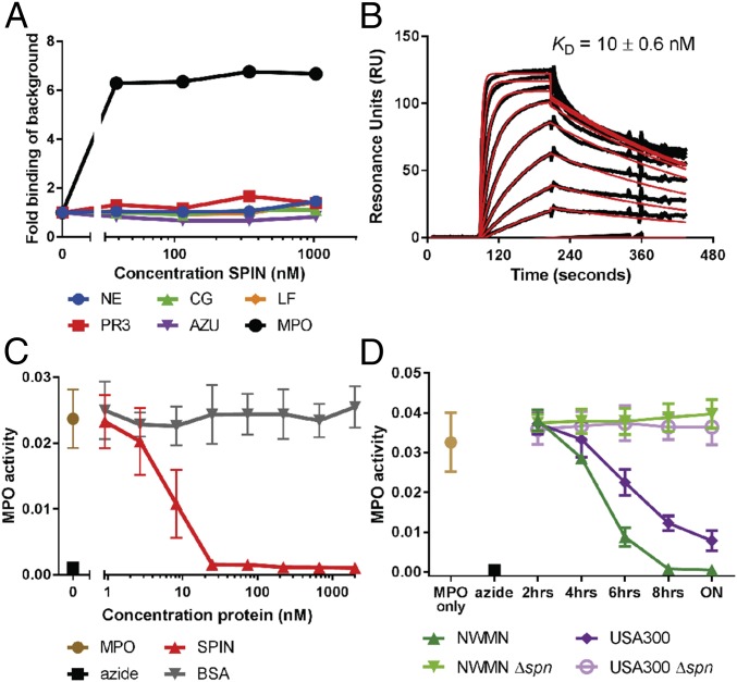
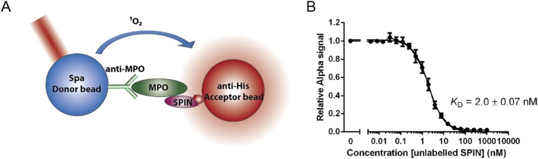
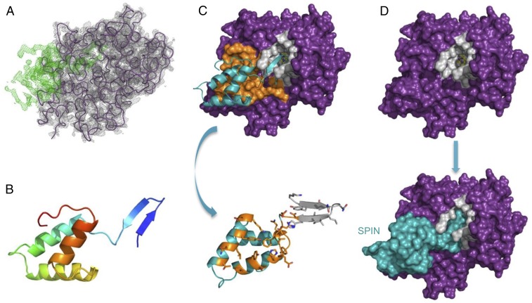
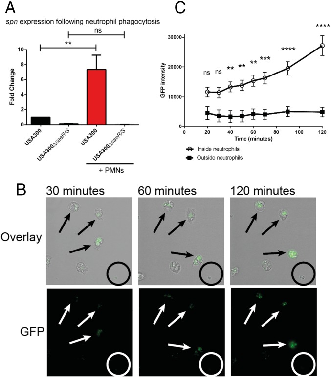
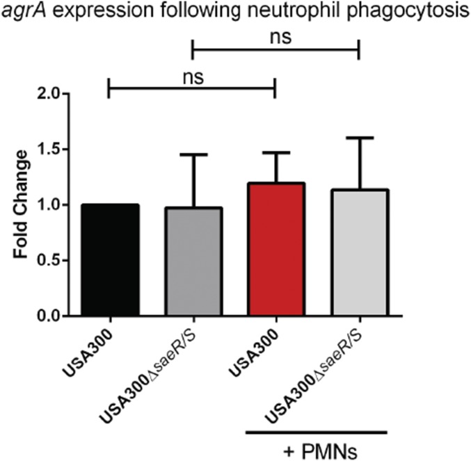
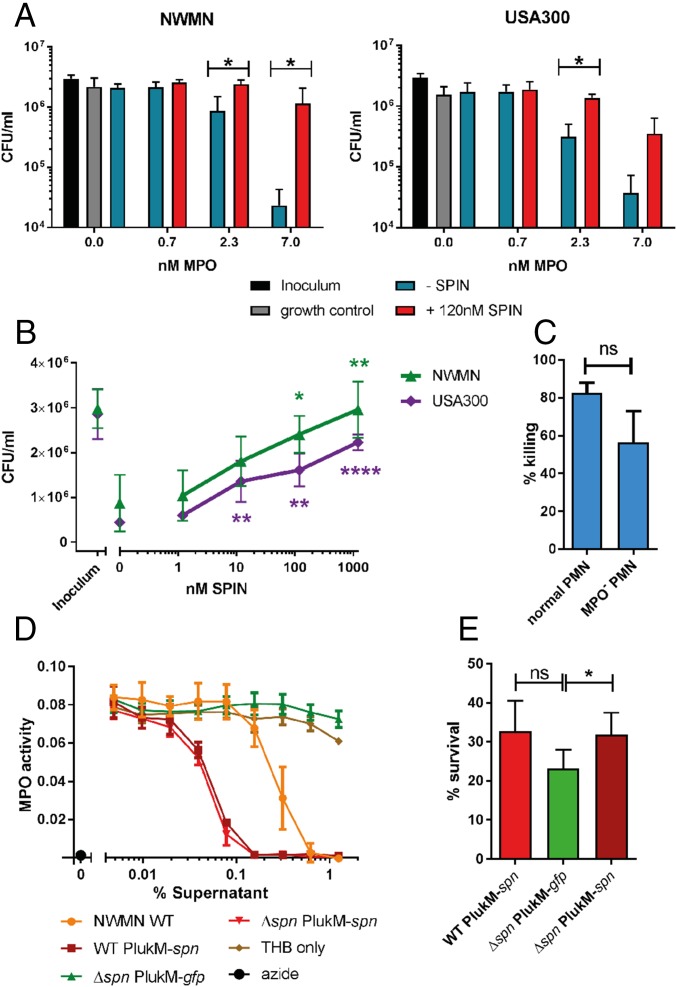
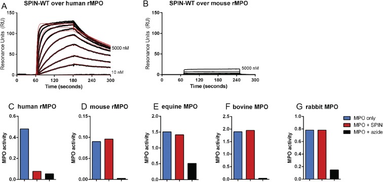
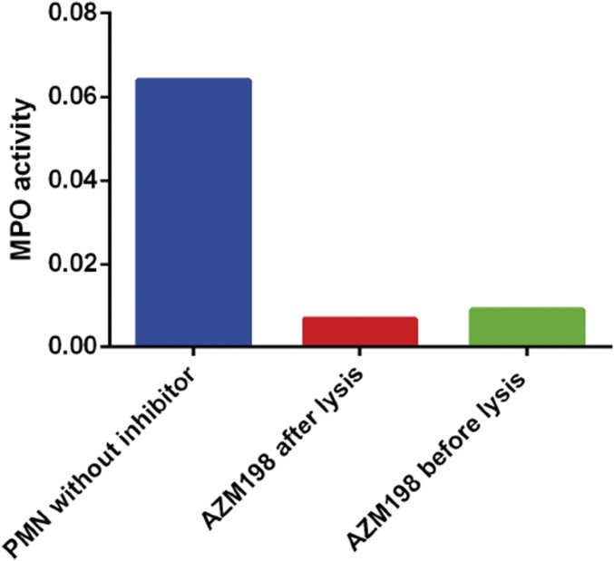
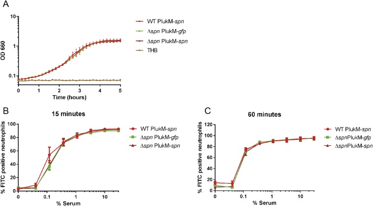
Staphylococcus aureus is highly adapted to its host and has evolved many strategies to resist opsonization and phagocytosis. Even after uptake by neutrophils, S. aureus shows resistance to killing, which suggests the presence of phagosomal immune evasion molecules. With the aid of secretome phage display, we identified a highly conserved protein that specifically binds and inhibits human myeloperoxidase (MPO), a major player in the oxidative defense of neutrophils. We have named this protein "staphylococcal peroxidase inhibitor" (SPIN). To gain insight into inhibition of MPO by SPIN, we solved the cocrystal structure of SPIN bound to a recombinant form of human MPO at 2.4-Å resolution. This structure reveals that SPIN acts as a molecular plug that prevents H2O2 substrate access to the MPO active site. In subsequent experiments, we observed that SPIN expression increases inside the neutrophil phagosome, where MPO is located, compared with outside the neutrophil. Moreover, bacteria with a deleted gene encoding SPIN showed decreased survival compared with WT bacteria after phagocytosis by neutrophils. Taken together, our results demonstrate that S. aureus secretes a unique proteinaceous MPO inhibitor to enhance survival by interfering with MPO-mediated killing.

Keywords: Staphylococcus aureus; immune evasion; myeloperoxidase; neutrophil; phagocytosis.


Conflict of interest statement

The authors declare no conflict of interest.

Figures

Fig. S1.
Fig. S1.
Identification of SPIN as a potential staphylococcal evasion protein. (A) Output from phages of secretome phage display after four rounds of selection against granular proteins of neutrophils showed enrichment of hypothetical ORF NWMN_0402. (B) Gene distribution of S. aureus genomic island α. The SPIN gene is located in the middle, downstream of the ssl cluster with the coding sequence oriented in the reverse direction. Proteins indicated in gray have an unknown function. (C) Genomic region of Giα (also known as νSaα) is compared between S. aureus strains Newman clonal complex (CC)8, MW2 (CC1), N315 (CC5), 5096 (CC22), and MRSA252 (CC30) using the Artemis Comparison Tool. Red blocks are indicative of homology between strains. GIα, SPIN (NWMN_0402), and the proteins encoded by genes in GIα are indicated at the top of the figure; these genes encode SSL proteins, type I restriction-modification system modification (HsdM), and specificity (HsdS) subunits and lipoproteins. (D) Amino acid sequence alignment of SPIN with signal sequence from Newman strain to 88 isolates, clustered by their clonal complex. The clonal complexes are both of human and animal origin (CC130, CC151, CC398, CC425, CC1173 are animal-associated and indicated in blue). The cleavage site that yields the mature protein is located between ADA-KV, and is indicated by a black line. Red indicates homology and the secondary structure of SPIN is indicated on top. (E) Anti-SPIN titers were determined from 20 healthy laboratory workers and compared with anti-CHIPS and anti-SEI titers by ELISA. Five donors for anti-SEI titers were below the detection limit. The values represent the logarithmic of the dilution factor that gave an OD450 of 0.2 after subtraction of the background. Each dot represents one individual in one experiment. The graph is representative of two trials.
Fig. 1.
Fig. 1.
SPIN binds and inhibits MPO. (A) An ELISA-type binding assay of C-His SPIN to all candidate proteins. AZU, azurocidin; CG, cathepsin G; LF, lactoferrin; MPO, myeloperoxidase; NE, neutrophil elastase; PR3, proteinase 3. (B) Characterization of SPIN binding to MPO by SPR, where SPIN was injected over a surface of native MPO. (C and D) Recombinant SPIN and S. aureus supernatant inhibit MPO in a dose-dependent manner. ON is overnight culture. Bars express SD with n = 3 for BD.
Fig. S2.
Fig. S2.
Determining KD of SPIN to MPO by AlphaScreen equilibrium competitive binding assay. An AlphaScreen equilibrium binding signal was observed by capturing MPO via antibodies to protein A donor beads and using C-His SPIN bound to anti-His acceptor beads (A). A concentration series of untagged SPIN was used to compete for MPO binding and a KD of 2 ± 0.07 nM was derived from nonlinear curve-fitting (B).
Fig. S3.
Fig. S3.
MPO activity from overnight culture supernatant of different S. aureus strains. (A and B) Supernatant from USA300 (A) or Newman (B) overnight culture was diluted and preincubated with native MPO. WT strains show inhibition of MPO, whereas the KO strains (Δspn) do not. (C) Truncated SPIN of MU50 doesn’t inhibit MPO. MPO activity from supernatant of MU50 is on the THB control line. The lines of THB, Mu50, and both KOs show background inhibition at high concentration due to the color of the undiluted THB. Sodium azide is used as a positive control for complete MPO inhibition. Bars express SD with n = 3.
Fig. 2.
Fig. 2.
Structural basis for inhibition of MPO by SPIN. (A) Electron density maps (2.4-Å resolution) calculated after initial placement of an MPO model (Rfree = 28%). 2FoFc density contoured at 1.5 σ (gray cage) is shown for the MPO model (purple wire), and FoFc density contoured at 3.0 σ (green cage) is attributable to SPIN. (B) Structure of the SPIN polypeptide depicted as a ribbon diagram. N terminus of the protein is indicated in indigo, the C terminus in red. The orientation of SPIN has been maintained across A and B for clarity. (C) Representation of the final model for the SPIN/MPO complex. (Upper) SPIN is shown as a cyan ribbon while MPO is depicted as a molecular surface. Residues comprising the first SPIN binding interface are colored orange, and residues lining the MPO active site channel are colored gray. (Lower) SPIN is drawn with the residues found at the first interface colored orange, and residues interacting with the MPO active site channel are colored gray. The sidechains of interfacing residues are depicted in ball-and-stick convention. Note the orientation of SPIN in the Lower panel is flipped 180° in the viewing plane relative to the Upper panel. (D) Surface representations provide insight into the physical basis for MPO inhibition by SPIN. (Upper) MPO is shown as a molecular surface with the residues lining the active site channel in gray. (Lower) SPIN is drawn as a cyan molecular surface according to its position in the final model of the SPIN/MPO complex. The location of the reactive site heme from native human MPO (33) is shown as a colored ball-and-stick. Note that the SPIN β-hairpin appears to completely occlude access of small molecules to the reactive site heme.
Fig. 3.
Fig. 3.
Production of SPIN is up-regulated after phagocytosis inside neutrophils. (A) Analysis of spn expression following exposure to human neutrophils. Bars express SD with n = 3. Statistical significance was determined using one-way ANOVA. (B) Time-lapse analysis of SPIN expression shown as GFP fluorescence promoter-reporter USA300 in a fibrin-thrombin matrix gel. An overlay of bright-field and GFP (Upper) and GFP alone (Lower) is shown. (Magnification: 40×.) The time indicates the duration after start of phagocytosis. Arrows indicate bacteria inside neutrophils and the circle indicates a colony of bacteria growing outside neutrophils. One representative experiment is shown from three independent experiments. (C) Quantification of total GFP signal from neutrophil-resident or free bacteria. Three different experiments with 44 neutrophils and 26 colonies growing outside neutrophils were analyzed. Bars express SEM. Significance was determined by two-way ANOVA with Bonferroni posttest correction for multiple comparison. **P ≤ 0.01, ***P ≤ 0.001, ****P ≤ 0.0001; ns, not significant.
Fig. S4.
Fig. S4.
Analysis of agrA expression following exposure to human neutrophils. Bars express SD with n = 3. Statistical significance was determined using one-way ANOVA; ns, not significant.
Fig. S5.
Fig. S5.
Promoter expression of SPIN after 2 h in THB culture. Bacteria were grown overnight in THB where GFP fluorescence and absorbance at OD660 was both measured in 10-min intervals, GFP signal was subtracted from the background of USA300 WT strain. The promoter of SPIN is activated after 2 h, when bacteria are within the late exponential phase. Data are representative of three independent experiments with duplicate measurements.
Fig. 4.
Fig. 4.
SPIN is important for evasion of MPO-dependent neutrophil killing. (A) S. aureus strains Newman (Left) and USA300 (Right) were killed in a dose-dependent manner by adding 0.7–7.0 nM MPO in a coupled glucose oxidase-MPO system. Addition of 120 nM SPIN prevented ROS killing. Statistical significance was determined by two-way ANOVA with Bonferroni posttest correction for multiple comparison. (B) SPIN provides dose-dependent protection from HOCl-mediated killing for strains Newman and USA300 relative to 0 nM SPIN. Statistical significance was determined by one-way ANOVA with Bonferroni posttest correction for multiple comparison. (C) Differential bacterial survival of Newman WT from neutrophils with active or membrane-permeable AZM198-inactivated MPO. (D) Supernatants of overnight culture of strains WT PlukM-spn, Δspn PlukM-spn, Δspn PlukM-gfp were diluted, tested for MPO activity, and compared with Newman WT. Bars express SD with n = 3 (AD). (E) Effect of S. aureus survival from isolated neutrophils after 60 min of phagocytosis. Statistical analysis determined by paired Student t test for C and E. Bars express SEM with n = 6. *P ≤ 0.05, **P ≤ 0.01, ****P ≤ 0.0001; ns, not significant.
Fig. S6.
Fig. S6.
SPIN does not inhibit murine, bovine, equine, or rabbit MPO. (A and B) Direct binding of SPIN to human recombinant MPO (rMPO) (A) and mouse rMPO (B) determined by SPR. SPIN is able to bind to human rMPO, but not to mouse rMPO. (C) SPIN inhibits human rMPO. “MPO only” shows MPO activity; adding 1 µg/mL SPIN inhibits the activity of MPO similar to azide control. (D) SPIN cannot inhibit mouse rMPO. Adding 50 µg/mL SPIN does not change the activity. Azide is able to completely inhibit the activity of the mouse rMPO. (EG) SPIN cannot inhibit bovine, equine, and rabbit MPO. Neutrophils from cow, horse, and rabbit were lysed and incubated with 10 µg/mL SPIN (cow and horse) or 9 µg/mL SPIN (rabbit).
Fig. S7.
Fig. S7.
MPO inhibitor AZM198 is able to cross neutrophil membrane and inhibit intracellular MPO. Polymorphonuclear (PMN) lysate without the inhibitor shows MPO activity. PMNs incubated with 10 µM inhibitor for 1 h at room temperature, washed, and then lysed, are devoid of MPO activity. The activity was comparable to samples which were incubated with the inhibitor after lysis. Thus, the MPO inhibitor AZM198 crosses the neutrophil membrane and is suitable to use for bactericidal assays to test the MPO-dependent killing of bacteria.
Fig. S8.
Fig. S8.
No difference in growth or phagocytosis between mutant strains WT PlukM-spn, Δspn PlukM-gfp, and Δspn PlukM-spn. (A) Newman mutants show growth with similar kinetics in THB. Absorbance at OD660 was measured every 10 min. (B and C) No difference in phagocytosis of WT PlukM-spn, Δspn PlukM-gfp, and Δspn PlukM-spn by neutrophils. The three Newman strains were labeled with FITC, washed, and treated with indicated dilutions of NHS. The percentage FITC-positive neutrophils was measured after 15 or 60 min of phagocytosis by FACS. All strains were phagocytosed by neutrophils at comparable levels. Bars express SD with n = 3 for AC.

References

    1. Lowy FD. Staphylococcus aureus infections. N Engl J Med. 1998;339:520–532. - PubMed
    1. Rigby KM, DeLeo FR. Neutrophils in innate host defense against Staphylococcus aureus infections. Semin Immunopathol. 2012;34:237–259. - PMC - PubMed
    1. Faurschou M, Borregaard N. Neutrophil granules and secretory vesicles in inflammation. Microbes Infect. 2003;5:1317–1327. - PubMed
    1. Babior BM, Lambeth JD, Nauseef W. The neutrophil NADPH oxidase. Arch Biochem Biophys. 2002;397:342–344. - PubMed
    1. Klebanoff SJ, Hamon CB. Role of myeloperoxidase-mediated antimicrobial systems in intact leukocytes. J Reticuloendothel Soc. 1972;12:170–196. - PubMed

Publication types

Associated data

LinkOut - more resources