. 2017 Oct 18;85(11):e00280-17.
doi: 10.1128/IAI.00280-17. Print 2017 Nov.

Cytosolic Phospholipase A2α Promotes Pulmonary Inflammation and Systemic Disease during Streptococcus pneumoniae Infection

Affiliations

Cytosolic Phospholipase A2α Promotes Pulmonary Inflammation and Systemic Disease during Streptococcus pneumoniae Infection

Rudra Bhowmick et al. Infect Immun. .

Abstract

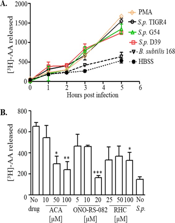
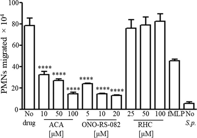
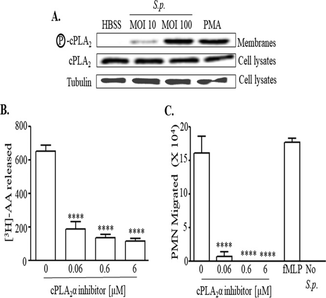
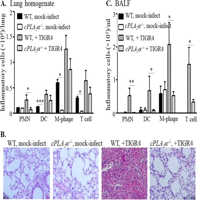
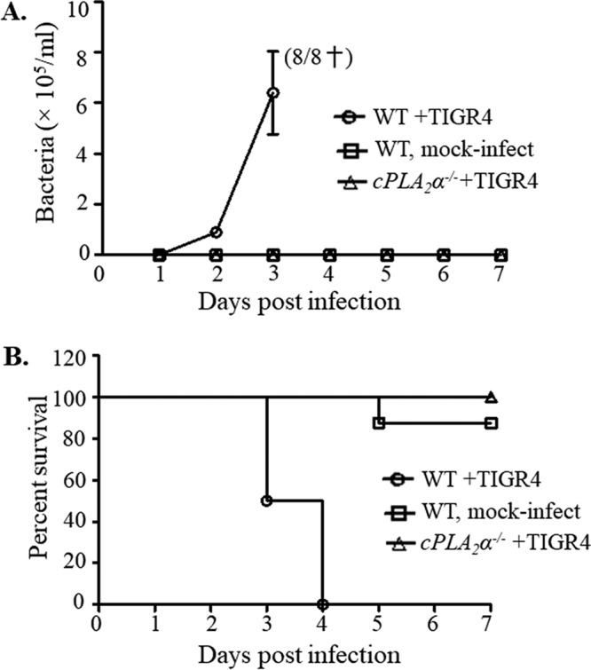
Pulmonary infection by Streptococcus pneumoniae is characterized by a robust alveolar infiltration of neutrophils (polymorphonuclear cells [PMNs]) that can promote systemic spread of the infection if not resolved. We previously showed that 12-lipoxygenase (12-LOX), which is required to generate the PMN chemoattractant hepoxilin A3 (HXA3) from arachidonic acid (AA), promotes acute pulmonary inflammation and systemic infection after lung challenge with S. pneumoniae As phospholipase A2 (PLA2) promotes the release of AA, we investigated the role of PLA2 in local and systemic disease during S. pneumoniae infection. The group IVA cytosolic isoform of PLA2 (cPLA2α) was activated upon S. pneumoniae infection of cultured lung epithelial cells and was critical for AA release from membrane phospholipids. Pharmacological inhibition of this enzyme blocked S. pneumoniae-induced PMN transepithelial migration in vitro Genetic ablation of the cPLA2 isoform cPLA2α dramatically reduced lung inflammation in mice upon high-dose pulmonary challenge with S. pneumoniae The cPLA2α-deficient mice also suffered no bacteremia and survived a pulmonary challenge that was lethal to wild-type mice. Our data suggest that cPLA2α plays a crucial role in eliciting pulmonary inflammation during pneumococcal infection and is required for lethal systemic infection following S. pneumoniae lung challenge.

Keywords: Streptococcus pneumoniae; inflammation; neutrophils; phospholipase.


Figures

FIG 1
FIG 1
S. pneumoniae infection of airway epithelial cells triggers arachidonic acid (AA) release. (A) H292 cell monolayers were labeled with [3H]AA as detailed in Materials and Methods and infected with the indicated S. pneumoniae (S.p.) strains (each of a different capsular serotype) or with B. subtilis strain 168 at a multiplicity of infection (MOI) of 10 (1 ×10⁷ CFU/monolayer). Supernatants were collected at the indicated times postinfection, and radioactive counts released into the supernatants were measured by scintillation counting. Labeled H292 cell monolayers treated with PMA and HBSS+Ca/Mg were used as positive and negative controls, respectively. Shown are results from a representative of two experiments. (B) H292 cell monolayers labeled with [3H]AA were treated with the indicated concentrations of pan-PLA2 inhibitors ACA or ONO-RS-082 or the DAG lipase inhibitor RHC-80267 (labeled as RHC) prior to infection with S. pneumoniae TIGR4, as detailed in Materials and Methods. Labeled H292 cell monolayers treated with HBSS+Ca/Mg were used as negative controls. Radioactive counts released into supernatants were determined by scintillation counting. Shown are results from a representative of three experiments. *, P < 0.05; **, P < 0.01; ***, P < 0.001 (compared with the no-drug control, using one-way ANOVA).
FIG 2
FIG 2
PLA2 is critical for S. pneumoniae-elicited PMN transepithelial migration. H292 cell monolayers were treated with different concentrations of pan-PLA2 inhibitors ACA and ONO-RS-082 or the DAG lipase inhibitor RHC-80267 prior to infection with S. pneumoniae TIGR4, as detailed in Materials and Methods. PMNs were added basolaterally, and the number of PMNs that migrated to the apical side was calculated using the myeloperoxidase (MPO) assay. Monolayers treated with fMLP and HBSS+Ca/Mg were used as positive and negative controls, respectively. Shown are results from a representative of three experiments. ****, P = 0.0001 compared to the no-drug control, using one-way ANOVA.
FIG 3
FIG 3
cPLA2 is activated upon pneumococcal infection of pulmonary epithelial monolayers and is critical for eliciting AA release and PMN transepithelial migration. (A) H292 cell monolayers were infected with S. pneumoniae TIGR4 at an MOI of 10 or 100. Monolayers treated with PMA and HBSS+Ca/Mg were used as positive and negative controls, respectively. Cell membranes were prepared as described in Materials and Methods and were probed with anti-phospho-cPLA2, anti-cPLA2, or antitubulin antibody. Shown are results from a representative of two independent experiments. (B) H292 cell monolayers labeled with [3H]AA were treated with the indicated concentrations of cPLA2α inhibitor prior to infection with S. pneumoniae TIGR4, as detailed in Materials and Methods. Radioactive counts in supernatants were measured by scintillation counting. Shown are results from a representative of three experiments. ****, P = 0.0001 compared to the untreated control, using one-way ANOVA. (C) H292 cell monolayers were treated with the indicated concentrations of the cPLA2α inhibitor prior to infection with S. pneumoniae TIGR4, as detailed in Materials and Methods. PMNs were added basolaterally, and the number of PMNs that migrated to the apical side was calculated using the MPO assay. Monolayers treated with fMLP and HBSS+Ca/Mg were used as positive and negative controls, respectively. Shown are results from a representative of three experiments. ****, P < 0.0001 compared to the respective no-drug controls, using one-way ANOVA.
FIG 4
FIG 4
cPLA2α promotes pulmonary inflammation in S. pneumoniae-infected mice. Mice (cPLA2α−/− or their WT littermates) were mock infected with PBS (n = 4 for each strain, per experiment) or infected intratracheally with S. pneumoniae TIGR4 (n = 5 for each strain, per experiment), as detailed in Materials and Methods. Lungs and bronchoalveolar lavage fluid (BALF) were collected from the mock-infected or TIGR4-infected mice. Cells present in the digested lungs and BALF were stained with relevant MAbs, and the fluorescence intensities of the stained cells were determined by flow cytometry. Collected data were analyzed to determine the numbers of PMNs, dendritic cells (DC), macrophages (M-phage), or T cells in the lungs (A) and in the BALF (C). Statistical significance was analyzed by one-way ANOVA followed by individual Student's t test analyses. *, P < 0.05; **, P < 0.005; ***, P < 0.0005. For histological analyses (B), H&E-stained lung sections were prepared and examined by light microscopy (original magnification, ×20). Shown are results from a representative of two independent experiments.
FIG 5
FIG 5
Ablation of cPLA2α leads to decreased bacteremia and increased survival in an otherwise lethal S. pneumoniae lung challenge. cPLA2α−/− mice or their littermate WT controls were intratracheally inoculated with TIGR4 (see Materials and Methods). A control group of WT mice received PBS. (A) Bacteremia was determined by plating tail vein blood at the specified time points. Shown are results from a combination of two experiments. Death of all infected WT mice is indicated by a dagger. (B) Survival of animals was monitored over a 7-day postinfection period. The log rank (Mantel-Cox) test was performed for survival curve analysis. The experiment was performed twice, with similar results. Shown are results from a combination of two experiments. A total of 8 mice were used per group; panels A and B represent the same cohorts of mice.

References

    1. Gillespie SH, Balakrishnan I. 2000. Pathogenesis of pneumococcal infection. J Med Microbiol 49:1057–1067. doi:10.1099/0022-1317-49-12-1057. - DOI - PubMed
    1. McCullers JA, Tuomanen EI. 2001. Molecular pathogenesis of pneumococcal pneumonia. Front Biosci 6:D877–D889. - PubMed
    1. Lynch JP III, Zhanel GG. 2010. Streptococcus pneumoniae: epidemiology and risk factors, evolution of antimicrobial resistance, and impact of vaccines. Curr Opin Pulm Med 16:217–225. - PubMed
    1. O'Brien KL, Wolfson LJ, Watt JP, Henkle E, Deloria-Knoll M, McCall N, Lee E, Mulholland K, Levine OS, Cherian T. 2009. Hib and Pneumococcal Global Burden of Disease Study Team: burden of disease caused by Streptococcus pneumoniae in children younger than 5 years: global estimates. Lancet 374:893–902. doi:10.1016/S0140-6736(09)61204-6. - DOI - PubMed
    1. Doerschuk CM, Markos J, Coxson HO, English D, Hogg JC. 1994. Quantitation of neutrophil migration in acute bacterial pneumonia in rabbits. J Appl Physiol 77:2593–2599. - PubMed

Publication types

MeSH terms