

Modeling and validating three dimensional human normal cervix and cervical cancer tissues in vitro
- PMID: 28808216
- PMCID: PMC5460612
- DOI: 10.7555/JBR.31.20160150
Modeling and validating three dimensional human normal cervix and cervical cancer tissues in vitro
Abstract
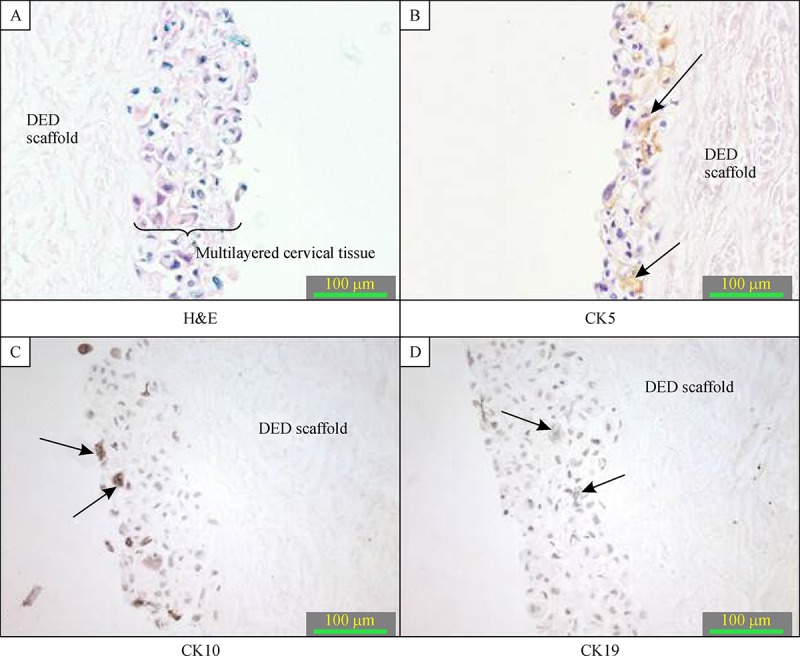
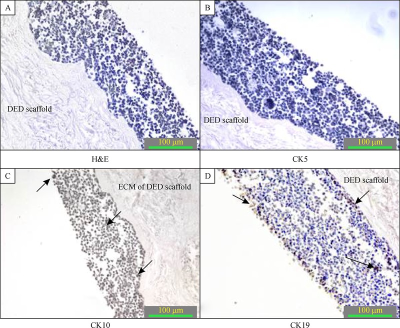
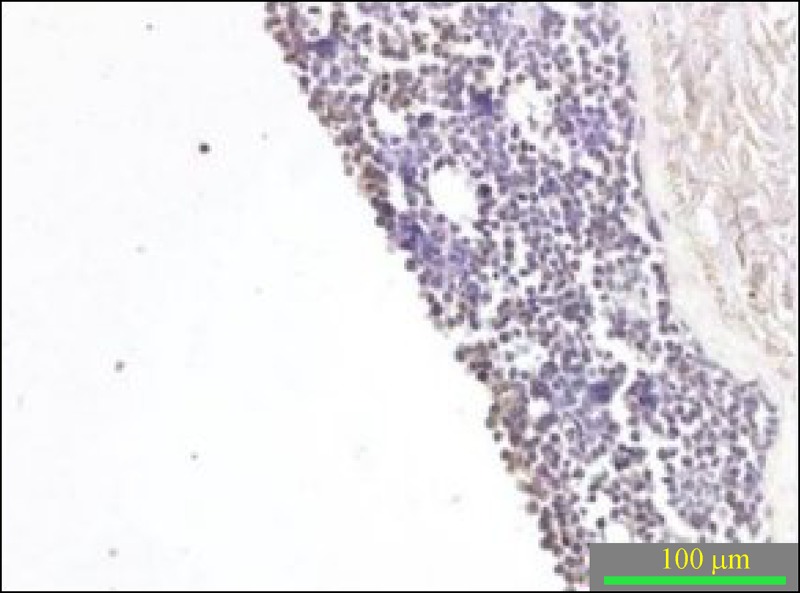
The use of three dimensional in vitro systems in cancer research is a promising path for developing effective anticancer therapies. The aim of this study was to engineer a functional 3-Din vitro model of normal and cancerous cervical tissue.Normal epithelial and immortalized cervical epithelial carcinoma cell lines were used to construct 3-D artificial normal cervical and cervical cancerous tissues. De-epidermised dermis (DED) was used as a scaffold for both models. Morphological analyses were conducted by using hematoxylin and eosin staining and characteristics of the models were studied by analyzing the expression of different structural cytokeratins and differential protein marker Mad1 using immunohistochemical technique.Haematoxylin and eosin staining results showed that normal cervical tissue had multi epithelial layers while cancerous cervical tissue showed dysplastic changes. Immunohistochemistry staining results revealed that for normal cervix model cytokeratin 10 was expressed in the upper stratified layer of epithelium while cytokeratin 5 was expressed mainly in the middle and basal layer. Cytokeratin 19 was weakly expressed in a few basal cells. Cervical cancer model showed cytokeratin 19 expression in different epithelial layers and weak or no expression for cytokeratin 5 and cytokeratin 10. Mad1 expression was detected in some suprabasal cells.The 3-Din vitro models showed stratified epithelial layers and expressed the same types and patterns of differentiation marker proteins as seen in correspondingin vivo tissue in either normal cervical or cervical cancerous tissue. Findings imply that they can serve as functional normal and cervical cancer models.
Conflict of interest statement
The authors reported no conflict of interests.
Figures
Similar articles
-
Keratin subtypes in carcinomas of the uterine cervix: implications for histogenesis and differential diagnosis.Cancer Res. 1990 Aug 15;50(16):5143-52. Cancer Res. 1990. PMID: 1696167
-
Expression of simple epithelial type cytokeratins in stratified epithelia as detected by immunolocalization and hybridization in situ.J Cell Biol. 1988 May;106(5):1635-48. doi: 10.1083/jcb.106.5.1635. J Cell Biol. 1988. PMID: 2453518 Free PMC article.
-
Cytokeratins 8, 18 and 19 in endometrial epithelial cells during the normal menstrual cycle and in women receiving Norplant.Contraception. 1993 Nov;48(5):481-93. doi: 10.1016/0010-7824(93)90137-v. Contraception. 1993. PMID: 7506133
-
Expression of cytokeratins in early neoplastic epithelial lesions of the uterine cervix.Int J Gynecol Pathol. 1985;4(4):300-13. doi: 10.1097/00004347-198512000-00003. Int J Gynecol Pathol. 1985. PMID: 2417968 Review.
-
[Cytokeratin detection as a diagnostic tool in oncology].Pol Merkur Lekarski. 2007 Sep;23(135):209-11. Pol Merkur Lekarski. 2007. PMID: 18080697 Review. Polish.
Cited by
-
Bioengineering models of female reproduction.Biodes Manuf. 2020 Sep;3(3):237-251. doi: 10.1007/s42242-020-00082-8. Epub 2020 Jun 16. Biodes Manuf. 2020. PMID: 32774987 Free PMC article.
-
Multi-cellular engineered living systems to assess reproductive toxicology.Reprod Toxicol. 2024 Aug;127:108609. doi: 10.1016/j.reprotox.2024.108609. Epub 2024 May 16. Reprod Toxicol. 2024. PMID: 38759876 Free PMC article. Review.
-
Three-dimensional models of the cervicovaginal epithelia to study host-microbiome interactions and sexually transmitted infections.Pathog Dis. 2022 Aug 27;80(1):ftac026. doi: 10.1093/femspd/ftac026. Pathog Dis. 2022. PMID: 35927516 Free PMC article.
-
Bioengineering Approaches to Improve Gynecological Cancer Outcomes.Curr Opin Biomed Eng. 2022 Jun;22:100384. doi: 10.1016/j.cobme.2022.100384. Epub 2022 Mar 30. Curr Opin Biomed Eng. 2022. PMID: 35874185 Free PMC article.
-
Engineered reproductive tissues.Nat Biomed Eng. 2020 Apr;4(4):381-393. doi: 10.1038/s41551-020-0525-x. Epub 2020 Apr 6. Nat Biomed Eng. 2020. PMID: 32251392 Free PMC article. Review.
References
-
- Kim JB. Three-dimensional tissue culture models in cancer biology[J]. Semin Cancer Biol, 2005, 15(5): 365–377. - PubMed
-
- Ellingsen C, Natvig I, Gaustad JV, et al. Human cervical carcinoma xenograft models for studies of the physiological microenvironment of tumors[J]. J Cancer Res ClinOncol, 2009, 135(9): 1177–1184. - PubMed
-
- Padrón JM, van der Wilt CL, Smid K, et al. The multilayered postconfluent cell culture as a model for drug screening[J]. Crit Rev Oncol Hematol, 2000, 36(2-3): 141–157. - PubMed
-
- Sun T, Jackson S, Haycock JW, et al. Culture of skin cells in 3D rather than 2D improves their ability to survive exposure to cytotoxic agents[J]. J Biotechnol, 2006, 122(3): 372–381. - PubMed
-
- Benam KH, Dauth S, Hassell B, et al. Engineered in vitro disease models[J]. Annu Rev Pathol, 2015, 10(10): 195–262. - PubMed
LinkOut - more resources
Full Text Sources
Research Materials