Gaining access to the periphery of the lung: Bronchoscopic and transthoracic approaches
- PMID: 28808487
- PMCID: PMC5541963
- DOI: 10.4103/atm.ATM_416_16
Gaining access to the periphery of the lung: Bronchoscopic and transthoracic approaches
Abstract
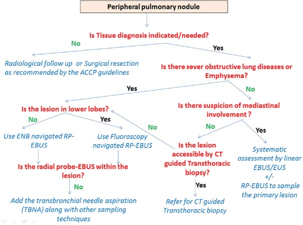
Globally, lung cancer remains the leading cause of cancer-related death. Annual low-dose computed tomography has been recommended as a screening test for early detection of lung cancers. Implementing this screening strategy is expected to challenge pulmonologist to confirm the nature of the increasing number of detected pulmonary nodules. Clinicians are obliged to use the less invasive and most efficient and safe means to set diagnoses. Hence, the field of diagnostic modalities, especially the advanced diagnostic bronchoscopy is witnessing rapid evolution to fulfill these unmet needs. This review highlights the available diagnostic modalities, describes their advantages and discusses the limitations of each technique. It also suggests an integrated diagnostic algorithm based on the best available evidence. A search of the PubMed database was conducted using relevant terms described at methodology; only articles in English were reviewed by November 2016.
Keywords: Bronchoscopic modalities; image-guided transthoracic needle aspiration; interventional pulmonology; lung cancer; navigation bronchoscopy; peripheral lung nodule.
Conflict of interest statement
There are no conflicts of interest.
Figures
References
-
- American Cancer Society. Cancer facts and figures 2016. Atlanta: American Cancer Society; 2016.
-
- World Health Organization. Cancer. Fact Sheet Number 297. [Last updated on 2017 Feb]. Available from: http://www.who.int/mediacentre/factsheets/fs297/en/
-
- Groome PA, Bolejack V, Crowley JJ, Kennedy C, Krasnik M, Sobin LH, et al. The IASLC Lung Cancer Staging Project: Validation of the proposals for revision of the T, N, and M descriptors and consequent stage groupings in the forthcoming (seventh) edition of the TNM classification of malignant tumours. J Thorac Oncol. 2007;2:694–705. - PubMed
Publication types
LinkOut - more resources
Full Text Sources
Other Literature Sources