Case Reports
. 2017:2017:9460958.
doi: 10.1155/2017/9460958. Epub 2017 Jul 20.

A Case of Superficial Femoral Arteriovenous Fistula and Severe Venous Stasis Ulceration, Managed with an Iliac Extender Prosthesis

Affiliations
Case Reports

A Case of Superficial Femoral Arteriovenous Fistula and Severe Venous Stasis Ulceration, Managed with an Iliac Extender Prosthesis

Nicole Ilonzo et al. Case Rep Vasc Med. 2017.

Abstract

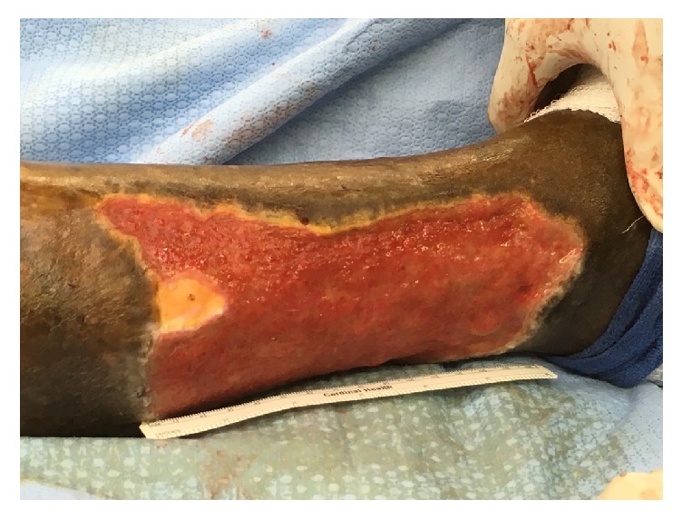
Most femoral artery arteriovenous fistulas occur as a result of percutaneous interventions. However, arteriovenous fistulas can occur in the setting of trauma, with resultant consequences such as heart failure, steal syndrome, or venous insufficiency. Indications for endovascular repair in this setting are limited to patients who are at too high risk for anesthesia, have a hostile groin, or would not survive significant bleeding. We report the case of a traumatic femoral arteriovenous fistula, causing severe venous insufficiency and arteriomegaly, in a 58-year-old male, with history of traumatic gunshot wound complicated by popliteal DVT. Surgical options for arteriovenous fistula include open and endovascular repair but this patient's fistula was more suitable for endovascular repair for reasons that will be discussed.


Figures

Figure 1
Figure 1
Right lateral leg venous stasis ulcer.
Figure 2
Figure 2
CT angiogram showing a superficial femoral arteriovenous fistula with arteriomegaly of the right external iliac artery.
Figure 3
Figure 3
Right lower extremity angiogram showing a patent and enlarged right superficial femoral artery with a distal AV fistula connecting the superficial femoral artery to the femoral vein with venous flow upwards.
Figure 4
Figure 4
Right lower extremity angiogram showing possibility of a venous malformation, which was not seen on previous imaging.
Figure 5
Figure 5
A superficial femoral artery cutdown was performed revealing an enlarged SFA.
Figure 6
Figure 6
Superficial femoral arteriovenous fistula occluded after placement of iliac extender stent.
Figure 7
Figure 7
Wound healed within 3 months.

Similar articles

Cited by

References

    1. Beebe-Dimmer J. L., Pfeifer J. R., Engle J. S., Schottenfeld D. The epidemiology of chronic venous insufficiency and varicose veins. Annals of Epidemiology. 2005;15(3):175–184. doi: 10.1016/j.annepidem.2004.05.015. - DOI - PubMed
    1. Krishnan S., Nicholls S. C. Chronic venous insufficiency: clinical assessment and patient selection. Seminars in Interventional Radiology. 2005;22(3):169–177. doi: 10.1055/s-2005-921961. - DOI - PMC - PubMed
    1. Vasudevan B. Venous leg ulcers: Pathophysiology and Classification. Indian Dermatology Online Journal. 2014;5(3):p. 366. doi: 10.4103/2229-5178.137819. - DOI - PMC - PubMed
    1. Chiesa R., Marone E. M., Limoni C., Volontè M., Petrini O. Chronic venous disorders: correlation between visible signs, symptoms, and presence of functional disease. Journal of Vascular Surgery. 2007;46(2):322–330. doi: 10.1016/j.jvs.2007.04.030. - DOI - PubMed
    1. Aslim E. Traumatic arteriovenous fistulae 25 years after gunshot injury. European Journal of Vascular and Endovascular Surgery. 2016;52(3):p. 316. doi: 10.1016/j.ejvs.2016.06.012. - DOI - PubMed

Publication types

LinkOut - more resources