

Possibilities for arthroscopic treatment of the ageing sternoclavicular joint
- PMID: 28808624
- PMCID: PMC5534402
- DOI: 10.5312/wjo.v8.i7.536
Possibilities for arthroscopic treatment of the ageing sternoclavicular joint
Abstract
Aim: To investigate if there are typical degenerative changes in the ageing sternoclavicular joint (SCJ), potentially accessible for arthroscopic intervention.
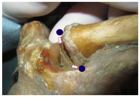
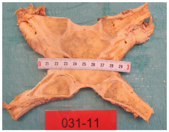
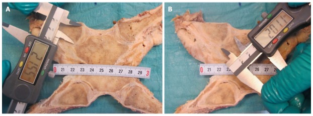
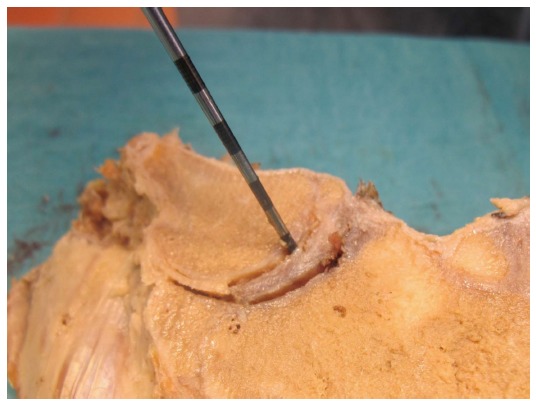
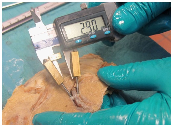
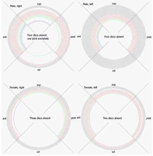
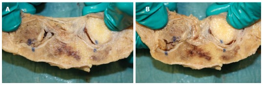
Methods: Both SCJs were obtained from 39 human cadavers (mean age: 79 years, range: 59-96, 13 F/26 M). Each frozen specimen was divided frontally with a band saw, so that both SCJs were opened in the same section through the center of the discs. After thawing of the specimens, the condition of the discs was evaluated by probing and visual inspection. The articular cartilages were graded according to Outerbridge, and disc attachments were probed. Cranio-caudal heights of the joint cartilages were measured. Superior motion of the clavicle with inferior movement of the lateral clavicle was measured.
Results: Degenerative changes of the discs were common. Only 22 discs (28%) were fully attached and the discs were thickest superiorly. We found a typical pattern: Detachment of the disc inferiorly in connection with thinning, fraying and fragmentation of the inferior part of the disc, and detachment from the anterior and/or posterior capsule. Severe joint cartilage degeneration ≥ grade 3 was more common on the clavicular side (73%) than on the sternal side (54%) of the joint. In cadavers < 70 years 75% had ≤ grade 2 changes while this was the case for only 19% aged 90 years or more. There was no difference in cartilage changes when right and left sides were compared, and no difference between sexes. Only one cadaver - a woman aged 60 years - had normal cartilages.
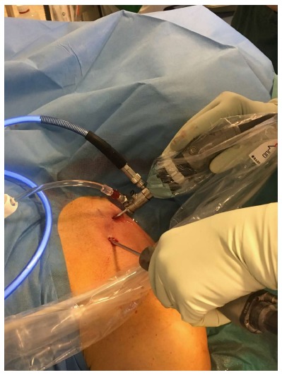
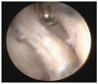
Conclusion: Changes in the disc and cartilages can be treated by resection of disc, cartilage, intraarticular osteophytes or medial clavicle end. Reattachment of a degenerated disc is not possible.
Keywords: Arthroscopy; Cartilage; Degenerative; Disc; Sternoclavicular.
Conflict of interest statement
Conflict-of-interest statement: The study was approved by the head of the body donation program at Department of Cellular and Molecular Medicine (ICMM) at the University of Copenhagen.
Figures
Similar articles
-
A cadaveric study of the structural anatomy of the sternoclavicular joint.Clin Anat. 2012 Oct;25(7):903-10. doi: 10.1002/ca.22021. Epub 2012 Jan 3. Clin Anat. 2012. PMID: 22991168
-
Macroscopic and histological observations on the human sternoclavicular joint disc.Anat Sci Int. 2009 Sep;84(3):182-8. doi: 10.1007/s12565-009-0014-5. Epub 2009 Feb 17. Anat Sci Int. 2009. PMID: 19221859
-
Surgical anatomy of the sternoclavicular joint: a qualitative and quantitative anatomical study.J Bone Joint Surg Am. 2014 Oct 1;96(19):e166. doi: 10.2106/JBJS.M.01451. J Bone Joint Surg Am. 2014. PMID: 25274794
-
Sternoclavicular Joint Instability: Symptoms, Diagnosis And Management.Orthop Res Rev. 2020 Jul 28;12:75-87. doi: 10.2147/ORR.S170964. eCollection 2020. Orthop Res Rev. 2020. PMID: 32801951 Free PMC article. Review.
-
Instability of the sternoclavicular joint: current concepts in classification, treatment and outcomes.Bone Joint J. 2013 Jun;95-B(6):721-31. doi: 10.1302/0301-620X.95B6.31064. Bone Joint J. 2013. PMID: 23723264 Review.
References
-
- Negri JH, Malavolta EA, Assunção JH, Gracitelli ME, Pereira CA, Bolliger Neto R, Croci AT, Ferreira Neto AA. Assessment of the function and resistance of sternoclavicular ligaments: A biomechanical study in cadavers. Orthop Traumatol Surg Res. 2014;100:727–731. - PubMed
-
- Tytherleigh-Strong GM, Getgood AJ, Griffiths DE. Arthroscopic intra-articular disk excision of the sternoclavicular joint. Am J Sports Med. 2012;40:1172–1175. - PubMed
-
- Tytherleigh-Strong G, Griffith D. Arthroscopic excision of the sternoclavicular joint for the treatment of sternoclavicular osteoarthritis. Arthroscopy. 2013;29:1487–1491. - PubMed
LinkOut - more resources
Full Text Sources
Other Literature Sources
