. 2017 Aug 4;3(8):eaao0027.
doi: 10.1126/sciadv.aao0027. eCollection 2017 Aug.

A conformational checkpoint between DNA binding and cleavage by CRISPR-Cas9

Affiliations

A conformational checkpoint between DNA binding and cleavage by CRISPR-Cas9

Yavuz S Dagdas et al. Sci Adv. .

Abstract

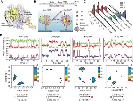
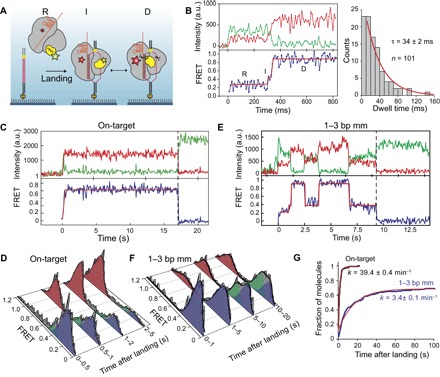
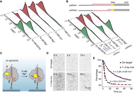
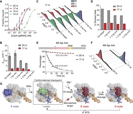
The Cas9 endonuclease is widely used for genome engineering applications by programming its single-guide RNA, and ongoing work is aimed at improving the accuracy and efficiency of DNA targeting. DNA cleavage of Cas9 is controlled by the conformational state of the HNH nuclease domain, but the mechanism that governs HNH activation at on-target DNA while reducing cleavage activity at off-target sites remains poorly understood. Using single-molecule Förster resonance energy transfer, we identified an intermediate state of Streptococcus pyogenes Cas9, representing a conformational checkpoint between DNA binding and cleavage. Upon DNA binding, the HNH domain transitions between multiple conformations before docking into its active state. HNH docking requires divalent cations, but not strand scission, and this docked conformation persists following DNA cleavage. Sequence mismatches between the DNA target and guide RNA prevent transitions from the checkpoint intermediate to the active conformation, providing selective avoidance of DNA cleavage at stably bound off-target sites.


Figures

Fig. 1
Fig. 1. HNH conformational dynamics reveal a distinct I state as a function of PAM-distal mismatches.
(A) Model shows HNH labeling sites under different conformations of Cas9, using sgRNA-bound (4ZT0) and dsDNA-bound (5F9R) structures. The cysteine-light Cas9 construct is labeled with Cy3 and Cy5 at S867C and S355C positions. (B) Top: Cas9 was incubated with 55-bp-long dsDNA substrates that include PAM and target sequences. Mismatches were introduced at the PAM-distal site. Bottom: DNA binding to Cas9 results in HNH interconversion, determined by a transition from a low to high FRET state. Scissors show the DNA cleavage sites. (C) Steady-state smFRET histograms for Cas9 in the absence and presence of 200 nM DNA targets. A multi-Gaussian fitting (black curve) reveals D, I, and R states of HNH. (D) Representative time traces (top), transition density plots (TDPs; middle), and rates of the major transitions in TDPs (bottom) for various DNA substrates. a.u., arbitrary units.
Fig. 2
Fig. 2. Real-time kinetics of HNH activation immediately after DNA binding.
(A) Schematic for observation of Cas9 conformational dynamics upon landing onto surface-immobilized DNA. (B) Left: A representative smFRET trajectory recorded at 100 Hz shows a brief visit to the I state between initial on-target binding and transitioning into the D state. Right: Single exponential fit (red curve) to the I state dwell time histogram reveals its lifetime (τ, ±95% confidence interval). (C and E) A representative smFRET trajectory at 10 Hz of Cas9 after landing to an on-target and 1–3 bp mm DNA. t = 0 s and dashed vertical lines represent time of landing and acceptor photobleaching, respectively. (D and F) Time-dependent changes in the conformational distribution of Cas9 after DNA landing. (G) Cumulative distribution of first transition to the D state after DNA landing. Red curves show fit to a single exponential function (±95% confidence interval).
Fig. 3
Fig. 3. HNH activation requires divalent cation but is independent of nuclease activity.
(A) smFRET histograms of Cas9 bound to on-target dsDNA in the absence and presence of a divalent cation. (B) Top: The on-target DNA was truncated at the 5′ end of the NTS one base after the target sequence (pdDNA1) and four bases after PAM (pdDNA2). Bottom: smFRET histograms of Cas9 bound to pdDNAs in the absence and presence of a divalent cation. (C) Cleavage of pdDNA1 was initiated by replacing EDTA with 5 mM Mg2+ and monitored by dissociation of the Cy5-labeled NTS 5′ end from Cas9. (D) Still images of Cy5-pdDNA1 bound to surface-immobilized Cas9 after Mg2+ addition (t = 0 s). (E) Percentage of Cy5 spots remain at the surface after Mg2+ flow. Red curves represent fit to single exponential decay (mean ± 95% confidence interval).
Fig. 4
Fig. 4. Truncation of the gRNA traps the HNH domain in the checkpoint intermediate with fewer mismatches on the DNA.
(A) On-target DNA binding assay with 20-nt and truncated gRNAs. (B) Bulk cleavage rates of the DNA substrates by Cas9 assembled with 20- and 17-nt gRNAs. (C) Steady-state smFRET histograms of Cas9 guided with a 17-nt gRNA. (D) D population of Cas9 guided with 20- and 17-nt gRNAs. (E) Top: A single mismatch was introduced at the 4th bp after the PAM-distal end (blue arrowhead). Bottom: Real-time cleavage of 4th bp mm by Cas9 guided with 20- and 17-nt gRNAs. Black curves represent fit to a single exponential decay (mean ± 95% confidence interval). (F) Steady-state smFRET histograms of Cas9 with 20- and 17-nt gRNAs bound to 4th bp mm. (G) Model for substrate-dependent HNH activation. DNA binding triggers transition from R (blue) to I (green) conformation, which serves as a conformational checkpoint between DNA binding and cleavage. In Mg2+, recognition of an on-target locks HNH in the catalytically active D conformation (red), which is destabilized after NTS release. HNH activation is prohibited when the RNA-DNA complementarity drops below a threshold (red cross).

References

    1. van der Oost J., Westra E. R., Jackson R. N., Wiedenheft B., Unravelling the structural and mechanistic basis of CRISPR–Cas systems. Nat. Rev. Microbiol. 12, 479–492 (2014). - PMC - PubMed
    1. Barrangou R., Marraffini L. A., CRISPR-Cas systems: Prokaryotes upgrade to adaptive immunity. Mol. Cell 54, 234–244 (2014). - PMC - PubMed
    1. Mohanraju P., Makarova K. S., Zetsche B., Zhang F., Koonin E. V., van der Oost J., Diverse evolutionary roots and mechanistic variations of the CRISPR-Cas systems. Science 353, aad5147 (2016). - PubMed
    1. Wright A. V., Nuñez J. K., Doudna J. A., Biology and Applications of CRISPR Systems: Harnessing nature’s toolbox for genome engineering. Cell 164, 29–44 (2016). - PubMed
    1. Sternberg S. H., Redding S., Jinek M., Greene E. C., Doudna J. A., DNA interrogation by the CRISPR RNA-guided endonuclease Cas9. Nature 507, 62–67 (2014). - PMC - PubMed

Publication types