. 2013 Jan 18;6(1):255-264.
doi: 10.3390/ma6010255.

Nanomagnetic Gene Transfection for Non-Viral Gene Delivery in NIH 3T3 Mouse Embryonic Fibroblasts

Affiliations

Nanomagnetic Gene Transfection for Non-Viral Gene Delivery in NIH 3T3 Mouse Embryonic Fibroblasts

Angeliki Fouriki et al. Materials (Basel). .

Abstract

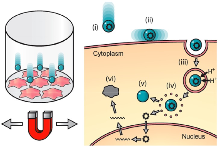
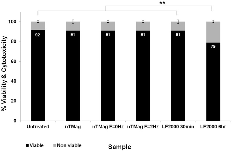
The objective of this work was to examine the potential of oscillating nanomagnetic gene transfection systems (magnefect-nano™) for improving the transfection efficiency of NIH3T3 mouse embryonic fibroblasts (MEFs) in comparison to other non-viral transfection techniques-static magnetofection™ and the cationic lipid agent, Lipofectamine 2000™. Magnetic nanoparticles (MNPs) associated with the plasmid coding for green fluorescent protein (GFP) were used to transfect NIH3T3 cells. The magnefect-nano system was evaluated for transfection efficiency, and any potential associated effects on cell viability were investigated. MNPs associated with the plasmid coding for GFP were efficiently delivered into NIH3T3 cells, and the magnefect-nano system significantly enhanced overall transfection efficiency in comparison to lipid-mediated gene delivery. MNP dosage used in this work was not found to affect the cell viability and/or morphology of the cells. Non-viral transfection using MNPs and the magnefect-nano system can be used to transfect NIH3T3 cells and direct reporter gene delivery, highlighting the wide potential of nanomagnetic gene transfection in gene therapy.

Keywords: DNA; magnetic field; magnetic nanoparticles; nanomagnetic gene transfection; non-viral gene transfection.


Figures

Figure 1
Figure 1
Principle of oscillating nanomagnetic transfection: Plasmid DNA or small interfering RNA (siRNA) is attached to magnetic nanoparticles and incubated with cells in culture (left). An oscillating magnet array below the surface of the cell culture plate pulls the particles into contact with the cell membrane (i) and drags the particles from side-to-side across the cells (ii); mechanically stimulating endocytosis (iii). Once the particle/DNA complex is endocytosed, proton sponge effects rupture the endosome (iv) releasing the DNA (v), which then transcribes the target protein (vi) [26,27].
Figure 2
Figure 2
DNA binding curve showing the nTMag:pEGFP-N1 plasmid ratio of binding.
Figure 3
Figure 3
Fluorescent microscopy images of NIH3T3 cells expressing GFP and correspondingly labeled with Phalloidin for actin stain of the whole cell population. (A,B) Untreated (CL) transfected with 100 nm nTMag MNPs coated with pEGFPN1 plasmid DNA; in the absence of a magnetic field, for 30 min (C,D), in the presence of a static field (nanoTherics static array) for 30 min (E,F) and an oscillating field (nanoTherics magnefect-nano™ array at f = 2 Hz and amplitude = 200 µm), for 30 min (G,H), Lipofectamine 2000™ for 30 min (I,J) and Lipofectamine 2000™ for 6 h (K,L). Cell seeding density was 1 × 10⁴/96 well, incubation period (48 h, 37 °C, 5% CO2) post-transfection and scale bar = 100 μm in (AL). GFP: green fluorescent protein; nTMag MNPs: nanoTherics nTMag magnetic nanoparticles; F: oscillation frequency.
Figure 4
Figure 4
Average of FACS data from transfection efficiency of GFP-expressing NIH3T3 cells transfected with nTMag/GFP-DNA complexes in the absence of a magnetic field, in the presence of the nanoTherics static magnetic array and the nanoTherics magnefect-nano™ oscillating magnetic array (200 µm amplitude, f = 2 Hz) for 30 min, in comparison to lipid-based transfection (LF2000, for 30 min and 6 h). During transfection, cells were incubated at 37 °C and 5% CO2. At 30 min post-transfection, the magnets were removed, and cells were incubated for 48 h before analysis. n = 6 for all samples.FACS: Fluorescence activated cell sorting; GFP: Green fluorescent protein; F: oscillation frequency.
Figure 5
Figure 5
Bar chart showing combined average percentage for viable and non-viable cells following treatment with nTMag MNPs and “no field”, “static field” (f = 0 Hz) and “magnefect-nano” (f = 2 Hz) transfections at 30 min, compared with Lipofectamine 2000 at 30 min and 6 h, at 48 h post-transfection. n = 9 for all groups.

Similar articles

Cited by

References

    1. Cho H.J., Lee T.S., Park J.B., Park K.K., Choe J.Y., Sin D.I., Park Y.Y., Moon Y.S., Lee K.G., Yeo J.H., et al. Disulfiram suppresses invasive ability of osteosarcoma cells via the inhibition of MMP-2 and MMP-9 expression. J. Biochem. Mol. Biol. 2007;40:1069–1076. doi: 10.5483/BMBRep.2007.40.6.1069. - DOI - PubMed
    1. Kersting C., Gebert C., Agelopoulos K., Schmidt H., van Diest P.J., Juergens H., Winkelmann W., Kevric M., Gosheger G., Brandt B., et al. Epidermal growth factor receptor expression in high-grade osteosarcoma is associated with a good clinical outcome. Clin. Cancer Res. 2007;13:2998–3005. doi: 10.1158/1078-0432.CCR-06-2432. - DOI - PubMed
    1. Lamoureux F., Richard P., Wittrant Y., Battaglia S., Pilet P., Trichet V., Blanchard F., Gouin F., Pitard B., Heymann D., Redini F. Therapeutic relevance of osteoprotegerin gene therapy in osteosarcoma: Blockage of the vicious cycle between tumor cell proliferation and bone resorption. Cancer Res. 2007;67:7308–7318. - PubMed
    1. Lu Y. Viral based gene therapy for prostate cancer. Curr. Gene Ther. 2001;1:183–200. doi: 10.2174/1566523013348742. - DOI - PubMed
    1. Bhattarai S.R., Kim S.Y., Jang K.Y., Lee K.C., Yi H.K., Lee D.Y., Kim H.Y., Hwang P.H. Laboratory formulated magnetic nanoparticles for enhancement of viral gene expression in suspension cell line. J. Virol. Methods. 2008;147:213–218. doi: 10.1016/j.jviromet.2007.08.028. - DOI - PubMed

LinkOut - more resources