. 2017 Sep;27(9):1115-1127.
doi: 10.1038/cr.2017.99. Epub 2017 Aug 15.

Ythdc2 is an N6-methyladenosine binding protein that regulates mammalian spermatogenesis

Affiliations

Ythdc2 is an N6-methyladenosine binding protein that regulates mammalian spermatogenesis

Phillip J Hsu et al. Cell Res. 2017 Sep.

Abstract

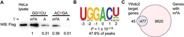
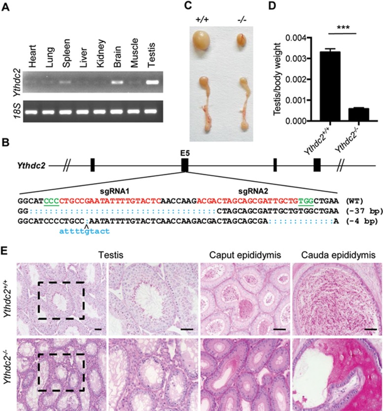
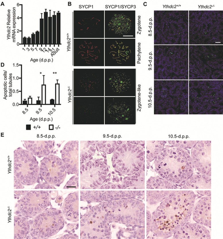
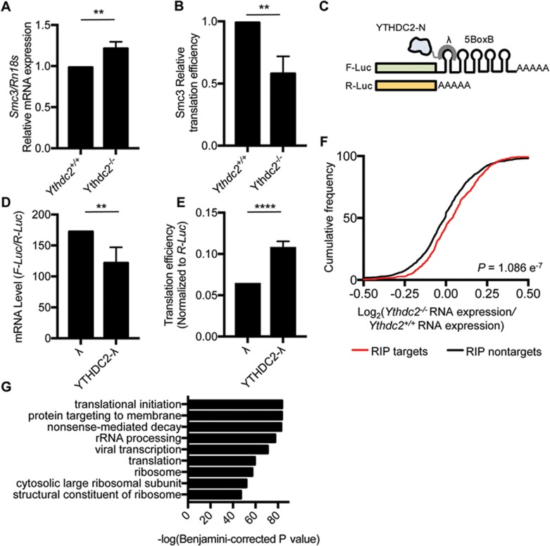
N6-methyladenosine (m6A) is the most common internal modification in eukaryotic mRNA. It is dynamically installed and removed, and acts as a new layer of mRNA metabolism, regulating biological processes including stem cell pluripotency, cell differentiation, and energy homeostasis. m6A is recognized by selective binding proteins; YTHDF1 and YTHDF3 work in concert to affect the translation of m6A-containing mRNAs, YTHDF2 expedites mRNA decay, and YTHDC1 affects the nuclear processing of its targets. The biological function of YTHDC2, the final member of the YTH protein family, remains unknown. We report that YTHDC2 selectively binds m6A at its consensus motif. YTHDC2 enhances the translation efficiency of its targets and also decreases their mRNA abundance. Ythdc2 knockout mice are infertile; males have significantly smaller testes and females have significantly smaller ovaries compared to those of littermates. The germ cells of Ythdc2 knockout mice do not develop past the zygotene stage and accordingly, Ythdc2 is upregulated in the testes as meiosis begins. Thus, YTHDC2 is an m6A-binding protein that plays critical roles during spermatogenesis.


Figures

Figure 1
Figure 1
YTHDC2 binds preferentially to m6A-marked RNA transcripts. (A) In vitro probe pulldown assay in HeLa cells overexpressing FLAG-YTHDC2 showing YTHDC2 binds preferentially to probe with m6A on GGACU. GGYCU Probe sequence: 5′-CGUGGYCUGGCU-B-3′ (Y = m6A or A, B = biotin) ACYGA Probe sequence: 5′-GAUACYGAGAAG-B-3′ (Y = m6A or A, B = biotin). Labels: relative protein expression. (B) Consensus motif of Ythdc2 binding identified by HOMER of CLIP-seq of Ythdc2 in mouse testes. (C) Overlap of Ythdc2 RIP-seq genes and genes containing m6A in young mouse testes. Targets of YTHDC2 are defined as genes enriched in the RIP: Log2(RIP/input) ≥ 1. Non-targets are defined as genes depleted in the RIP: Log2(RIP/input) ≤ −1. Genes with m6A are defined as genes enriched in the m6A IP: Log2(m6A IP/input) ≥ 2.
Figure 2
Figure 2
Ythdc2-deficient mice display defects in testes. (A) RT-PCR analysis of Ythdc2 mRNA levels in various organs of adult mice. (B) Schematic diagram of sgRNAs targeting at Ythdc2 locus. Two founders were generated by micro-injection of sgRNA/Cas9 mRNA into one-cell embryos. PAM sequences are underlined and highlighted in green. sgRNA targeting sites are in red. Mutant sequence are in blue. (C) Testes and epididymis from adult Ythdc2−/− mice are smaller in size than wild type. (D) The testis/body weight ratio was calculated for adult Ythdc2+/+ and Ythdc2−/− mice. Error bars, mean ± sd, n = 4, biological replicates ***P < 0.001 (Student's t-test) (E) Periodic Acid Schiff (PAS) staining of adult testes and epididymis. Scale bar, 50 μm.
Figure 3
Figure 3
Ythdc2-deficient mice display defects in meiotic prophase I. (A) RT-qPCR analysis of Ythdc2 mRNA levels in wild type testes over time, normalized to Rn18s as an internal control. Error bars, mean ± sd, n = 3, technical replicates (Student's t-test). (B) Ythdc2+/+ and Ythdc2−/− spermatocyte chromosome spreads at 15.0 d.p.p. stained for SYCP1 (red) and SYCP3 (green). Scale bar, 10 μm. (C) TUNEL assay of testes sections from 8.5 d.p.p. to 10.5 d.p.p.. DNA was stained with Hoechst (blue). Scale bar, 20 μm. (D) Quantification of apoptotic cells in Ythdc2+/+ (black columns) and Ythdc2−/− (grey columns) testes at 8.5 to 10.5 d.p.p.. Error bars, mean ± sd, n = 3, biological replicates *P < 0.05, *P < 0.01 (Student's t-test). (E) Cleaved Caspase-3 staining of testes sections at 8.5–10.5 d.p.p.. Scale bar, 20 μm.
Figure 4
Figure 4
YTHDC2 affects the translation efficiency and mRNA abundance of its targets. (A) Relative mRNA expression of Smc3, normalized to Rn18s, in 8.5 d.p.p. Ythdc2+/+ and Ythdc2−/− testes. Error bars, mean ± sd, n = 3. **P < 0.01 (Student's t-test). (B) Translation efficiency of Smc3 in 8.5 d.p.p. Ythdc2+/+ and Ythdc2−/− testes, calculated as: translation efficiency = Smc3 protein level / Smc3 mRNA expression level. Smc3 protein level was determined by western blot and normalized to tubulin. Error bars, mean ± sd, n = 3. **P < 0.01 (Student's t-test). (C) Constructs in the tethering reporter assay. Effector: the YTH domain of YTHDC2 was replaced with λ peptide (N-YTHDC2-λ), which binds with high affinity to the BoxB RNA motif. Reporter: 5 BoxB domains were fused to the 3′ UTR of firefly luciferase mRNA (F-Luc-5BoxB). Renilla luciferase (R-Luc) mRNA was used as an internal control to normalize luciferase signals from different samples. (D) mRNA level of F-Luc normalized to R-Luc 4 h post F-Luc induction. Error bars, mean ± sd, n = 4. **P < 0.01 (Student's t-test). (E) Translation efficiency of F-Luc normalized to R-Luc 4 h post F-Luc induction. Error bars, mean ± sd, n = 4. ****P < 0.0001; (Student's t-test). (F) Cumulative frequency of log2-fold changes of RNA expression upon Ythdc2 depletion, in RIP targets and non-targets. P value was calculated using two-sided Mann-Whitney U test. (G) Top GO terms in GO analysis of components of protein complex containing YTHDC2 obtained from tandem affinity purification followed by mass spectrometry.

References

    1. Moore MJ. From birth to death: the complex lives of eukaryotic mRNAs. Science 2005; 309:1514–1518. - PubMed
    1. Desrosiers R, Friderici K, Rottman F. Identification of methylated nucleosides in messenger RNA from Novikoff hepatoma cells. Proc Natl Acad Sci USA 1974; 71:3971–3975. - PMC - PubMed
    1. Zhao BS, Roundtree IA, He C. Post-transcriptional gene regulation by mRNA modifications. Nat Rev Mol Cell Biol 2016; 18:31–42. - PMC - PubMed
    1. Wei C, Moss B. Nucleotide Sequences at the N6-methyladenosine sites of HeLa cell messenger ribonucleic acid. Biochemistry 1977; 16:1672–1676. - PubMed
    1. Wei C, Gershowitz A, Moss B. 5′-Terminal and internal methylated nucleotide sequences in HeLa cell mRNA. Biochemistry 1976; 15:397–401. - PubMed