. 2018 Mar;23(3):521-532.
doi: 10.1038/mp.2017.134. Epub 2017 Aug 15.

Evidence for newly generated interneurons in the basolateral amygdala of adult mice

Affiliations

Evidence for newly generated interneurons in the basolateral amygdala of adult mice

D J Jhaveri et al. Mol Psychiatry. 2018 Mar.

Abstract

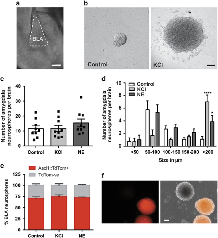
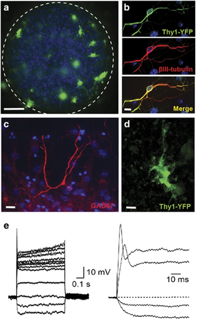
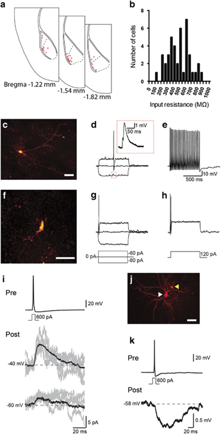
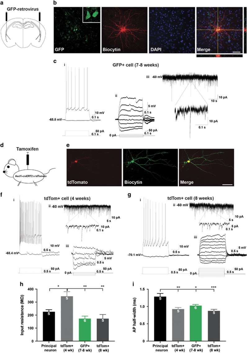
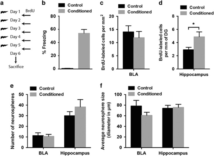
New neurons are continually generated from the resident populations of precursor cells in selective niches of the adult mammalian brain such as the hippocampal dentate gyrus and the olfactory bulb. However, whether such cells are present in the adult amygdala, and their neurogenic capacity, is not known. Using the neurosphere assay, we demonstrate that a small number of precursor cells, the majority of which express Achaete-scute complex homolog 1 (Ascl1), are present in the basolateral amygdala (BLA) of the adult mouse. Using neuron-specific Thy1-YFP transgenic mice, we show that YFP+ cells in BLA-derived neurospheres have a neuronal morphology, co-express the neuronal marker βIII-tubulin, and generate action potentials, confirming their neuronal phenotype. In vivo, we demonstrate the presence of newly generated BrdU-labeled cells in the adult BLA, and show that a proportion of these cells co-express the immature neuronal marker doublecortin (DCX). Furthermore, we reveal that a significant proportion of GFP+ neurons (~23%) in the BLA are newly generated (BrdU+) in DCX-GFP mice, and using whole-cell recordings in acute slices we demonstrate that the GFP+ cells display electrophysiological properties that are characteristic of interneurons. Using retrovirus-GFP labeling as well as the Ascl1CreERT2 mouse line, we further confirm that the precursor cells within the BLA give rise to mature and functional interneurons that persist in the BLA for at least 8 weeks after their birth. Contextual fear conditioning has no effect on the number of neurospheres or BrdU-labeled cells in the BLA, but produces an increase in hippocampal cell proliferation. These results demonstrate that neurogenic precursor cells are present in the adult BLA, and generate functional interneurons, but also show that their activity is not regulated by an amygdala-dependent learning paradigm.


Conflict of interest statement

The authors declare no conflict of interest.

Figures

Figure 1
Figure 1
Presence of precursor cells in the basolateral amygdala (BLA). (a) The BLA was microdissected from 500 μm thick coronal brain slices. (b) Photomicrograph of a neurosphere obtained in control medium versus medium containing 15 mm KCl. (c) Similar numbers of BLA-derived neurospheres per brain were obtained in control, norepinephrine- (10 μm, NE) and KCl-treated wells (n=9). (d) Distribution of neurospheres based on their size in control, NE and KCl conditions. Note the significant increase in the number of neurospheres measuring >200 μm in the NE and KCl treatment conditions compared with the control (n=9, *P<0.05, ****P<0.0001). (e) The majority of neurospheres generated from the BLA are derived from Ascl1-expressing precursor cells (Ascl1::tdTom+) labeled using Ascl1CreERT2; CAGfloxStop-tdTomato mice. (f) Examples of BLA-derived Ascl1::tdTom+ and tdTom− neurospheres. Scale bars, 100 μm.
Figure 2
Figure 2
BLA-derived neurospheres contain functional neurons. (a) Several Thy1- YFP+ cells are present in a basolateral amygdala (BLA)-derived neurosphere. Nuclei are stained with DAPI (blue) Scale bar, 50 μm. (b) Higher magnification of a fixed Thy1-YFP+ neuron (green) co-labeled with βIII-tubulin (red). Scale bar, 10 μm (c) An example of a GAD67+ neuron in a BLA-derived neurosphere. (d, e) Whole-cell recordings from YFP+ cells (scale bar, 10 μm), showing that depolarizing current injections evoke small amplitude action potentials (e).
Figure 3
Figure 3
New neurons are generated in the amygdala nuclei in vivo. (a) BrdU-labeled cells (red, arrows) are present in the basolateral amygdala (BLA) in adult mice. CeA: central amygdala (b) An example of BrdU-labeled cells (red) co-expressing the immature neuronal marker DCX (green) in the BLA. All nuclei are labeled with DAPI (blue). (c) An example of DCX-GFP+ cells (green) in the BLA that are co-labeled with BrdU (red). The merged image and orthogonal views are shown on the far right. (d) DCX-GFP−, BrdU+ cells co-labeled with the mature neuronal marker NeuN in the BLA 4 weeks after the last BrdU injection. (e) A BrdU-labeled cell co-expressing DCX-GFP and NeuN. Scale bars, 50 μm in (a) and 10 μm in (be).
Figure 4
Figure 4
DCX-GFP-expressing cells in the basolateral amygdala (BLA) exhibit the electrophysiological properties of interneurons. (a) Location of recovered cells in the BLA (n=23 cells). (b) Histogram showing the distribution of input resistance of DCX- GFP+ cells (n=42 cells), measured using whole-cell recordings. Recovered cells displayed morphologies ranging from cells with branching dendrites (c), that had interneuron-like fast-spiking firing properties (d, e) and high spontaneous activity (d: inset), to sparsely branching, underdeveloped cells (f), which fired fewer action potentials (g, h). Firing was evoked using increasing steps of current injection (as shown below in g and h). (i) Dual whole-cell recording shows that a DCX-GFP+ cell is capable of forming inhibitory connections onto pyramidal cells (1 paired recording out of 6 dual recordings); an evoked action potential in the presynaptic cell (‘Pre’ recovered cell indicated by white arrowhead in j) evoked an outward postsynaptic current in the postsynaptic cell (‘Post’ recovered cell indicated by yellow arrowhead in j) when voltage-clamped at −40 mV (9.50±0.54 pA; 4.27±0.62 pA at −60 mV, close to the chloride reversal potential). Black traces show the average response with example traces of single episodes in gray. (k) In current clamp this response was an inhibitory postsynaptic potential in the postsynaptic cell (−0.78±0.09 mV). Scale bars, 50 μm in (c), 25 μm in (f) and 50 μm in (j).
Figure 5
Figure 5
Functional properties of new neurons in the basolateral amygdala (BLA) using retrovirus labeling and lineage tracing. (a) A schematic illustrating bilateral injections of green fluorescent protein (GFP)-retrovirus in the BLA of 8-week-old C57BL/6 J mice. (b) Example GFP+ neurons (green) in the BLA recovered from whole-cell patch-clamp recordings and filled with biocytin (red). Note the presence of a doublet. Nuclei are stained with DAPI (blue). Scale bar, 50 μm (c) Whole-cell patch-clamp recordings from one GFP+ cell of the doublet showing (i) a train of action potentials induced by a +190 pA current injection, (ii) an example of spontaneous excitatory potential recording while injecting current steps from −60 to +80 pA, and (iii) an example trace of sEPSCs recorded while holding the cell at −60 mV. (d) Schematic showing tamoxifen injection in Ascl1CreERT2; CAGfloxStop-tdTomato mice. (e) An example of a tdTom+ cell (red) in the BLA, 4 weeks after the last tamoxifen injection, which was recovered with biocytin (green) following whole-cell patch-clamp recordings. Scale bar, 50 μm (f) An example of whole-cell patch-clamp recordings of a tdTom+ cell at 4 weeks post-tamoxifen showing (i) a train of action potentials induced by +190 pA current injection, (ii) an example trace of sEPSCs recorded while holding the cell at −60 mV, and (iii) an example of a spontaneous excitatory potential recording while injecting current steps from −60 to +20 pA. (g) An example of whole-cell patch-clamp recordings from a tdTom+ cell at 8 weeks post-tamoxifen showing (i) a train of action potentials induced by +240 pA current injection, (ii) an example trace of sEPSCs recorded while holding the cell at −60 mV, and (iii) an example of a spontaneous excitatory potential recording while injecting current steps from −60 to +120 pA. (h) Graph showing the input resistances of recorded cells in the BLA. Note that the tdTom+ cells at 4 weeks have higher input resistance than the input resistances of principal neurons (n=5, *P<0.05), retrovirus-GFP+ cells at 7–8 weeks (n=6, **P<0.01), and tdTom+ cells at 8 weeks (n=6, **P<0.01). (i) Graph showing action potential half-widths from recorded cells within the BLA. Principal neurons have a half-width (n=5) that is slower than the half-widths of tdTom+ cells at 4 weeks (n=5, **P<0.01), GFP+ cells at 7–8 weeks (n=6, *P<0.05), and tdTom+ cells at 8 weeks (n=5, ***P<0.001).
Figure 6
Figure 6
Fear learning has no effect on precursor cell activity and cell proliferation in the basolateral amygdala (BLA) (a) Schematic of the contextual fear conditioning protocol and BrdU administration. (b) All animals showed robust context fear conditioning (n=8 in each group). (c, d) There was a significant increase in the number of BrdU-labeled cells in the dentate gyrus (DG) of the hippocampus (f) following fear conditioning, but no change in the number of BrdU-labeled cells in the BLA (e) of conditioned versus control animals (conditioned: n=6, control: n=7; *P<0.05). (e, f) There was no change in the number (e) or size (f) of neurospheres in the BLA or hippocampus following contextual fear conditioning (conditioned: n=13, control: n=12).

Similar articles

Cited by

References

    1. Ming GL, Song H. Adult neurogenesis in the mammalian central nervous system. Annu Rev Neurosci 2005; 28: 223–250. - PubMed
    1. Imayoshi I, Sakamoto M, Ohtsuka T, Takao K, Miyakawa T, Yamaguchi M et al. Roles of continuous neurogenesis in the structural and functional integrity of the adult forebrain. Nat Neurosci 2008; 11: 1153–1161. - PubMed
    1. Rietze RL, Valcanis H, Brooker GF, Thomas T, Voss AK, Bartlett PF. Purification of a pluripotent neural stem cell from the adult mouse brain. Nature 2001; 412: 736–739. - PubMed
    1. Walker TL, White A, Black DM, Wallace RH, Sah P, Bartlett PF. Latent stem and progenitor cells in the hippocampus are activated by neural excitation. J Neurosci 2008; 28: 5240–5247. - PMC - PubMed
    1. Arruda-Carvalho M, Sakaguchi M, Akers KG, Josselyn SA, Frankland PW. Posttraining ablation of adult-generated neurons degrades previously acquired memories. J Neurosci 2011; 31: 15113–15127. - PMC - PubMed

Publication types