. 2017 Dec;158(12):2301-2319.
doi: 10.1097/j.pain.0000000000001002.

CRISPR/Cas9 editing of Nf1 gene identifies CRMP2 as a therapeutic target in neurofibromatosis type 1-related pain that is reversed by (S)-Lacosamide

Affiliations

CRISPR/Cas9 editing of Nf1 gene identifies CRMP2 as a therapeutic target in neurofibromatosis type 1-related pain that is reversed by (S)-Lacosamide

Aubin Moutal et al. Pain. 2017 Dec.

Abstract

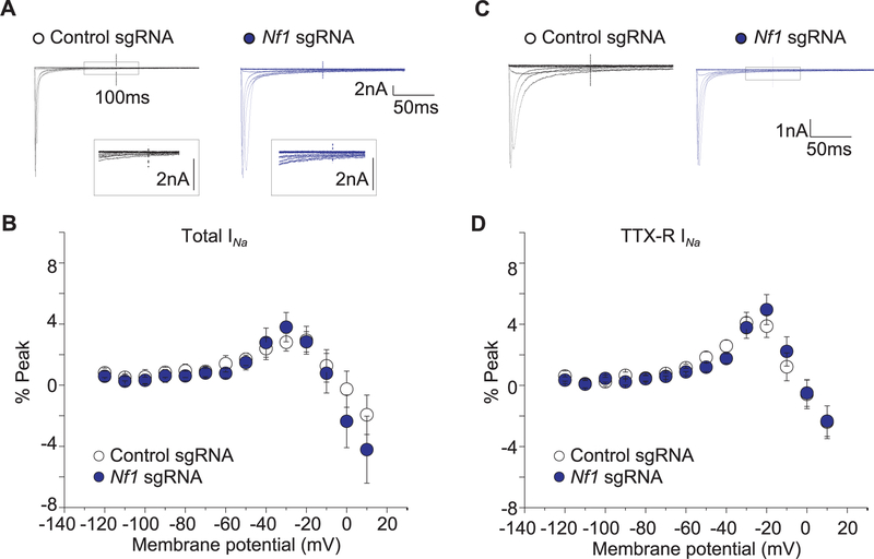
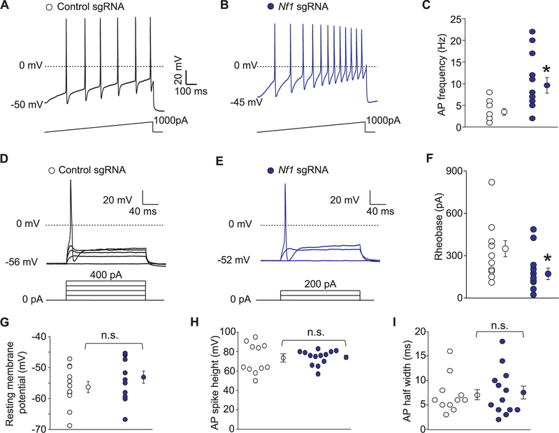
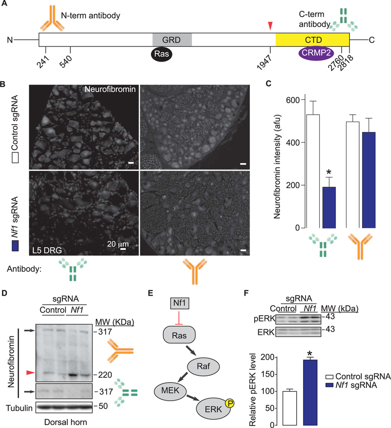
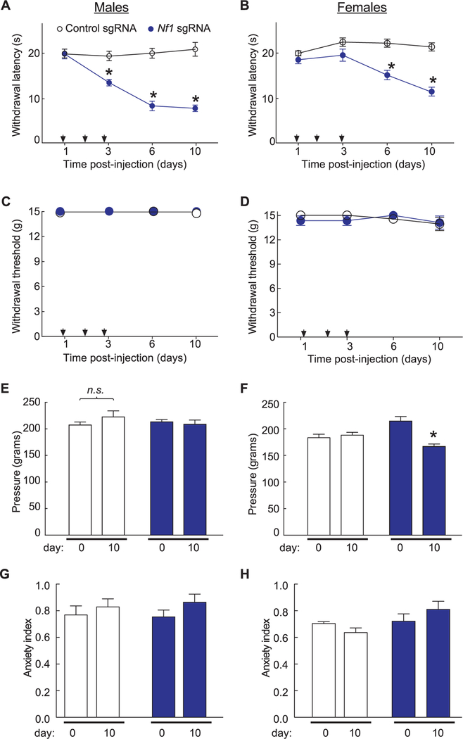
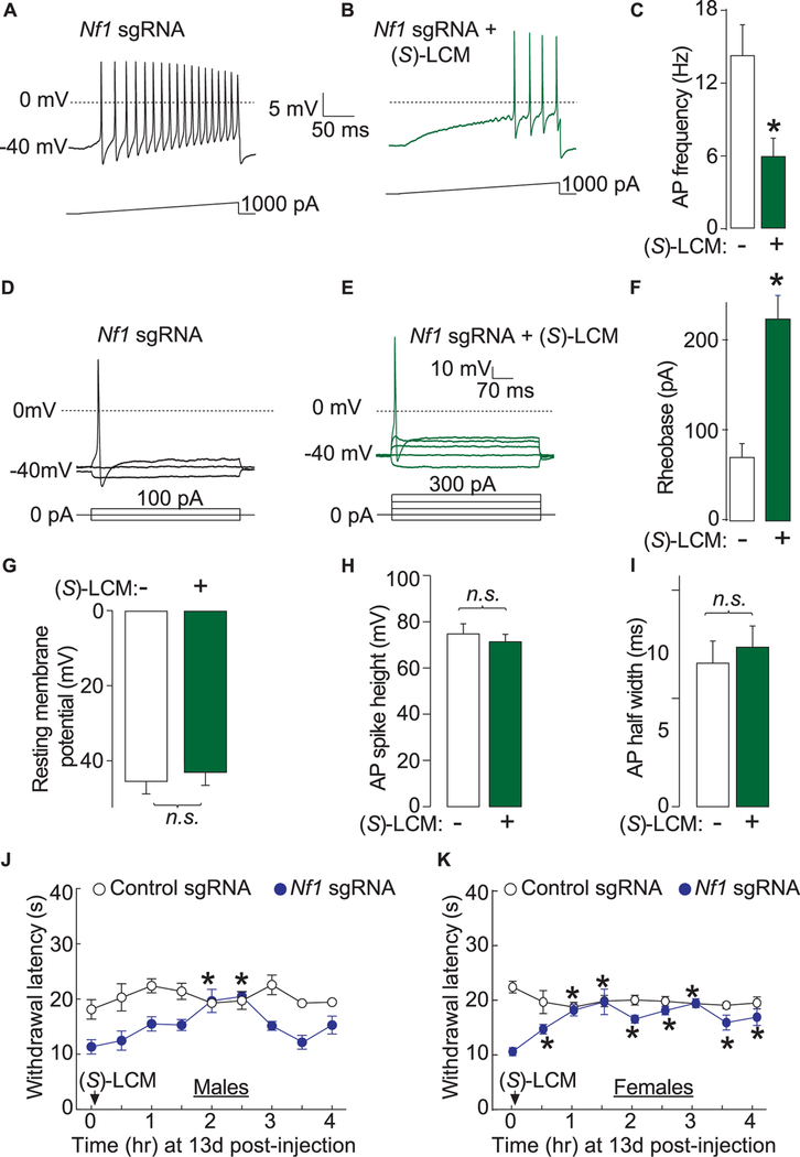
Neurofibromatosis type 1 (NF1) is a rare autosomal dominant disease linked to mutations of the Nf1 gene. Patients with NF1 commonly experience severe pain. Studies on mice with Nf1 haploinsufficiency have been instructive in identifying sensitization of ion channels as a possible cause underlying the heightened pain suffered by patients with NF1. However, behavioral assessments of Nf1 mice have led to uncertain conclusions about the potential causal role of Nf1 in pain. We used the clustered regularly interspaced short palindromic repeats (CRISPR)-associated 9 (CRISPR/Cas9) genome editing system to create and mechanistically characterize a novel rat model of NF1-related pain. Targeted intrathecal delivery of guide RNA/Cas9 nuclease plasmid in combination with a cationic polymer was used to generate allele-specific C-terminal truncation of neurofibromin, the protein encoded by the Nf1 gene. Rats with truncation of neurofibromin, showed increases in voltage-gated calcium (specifically N-type or CaV2.2) and voltage-gated sodium (particularly tetrodotoxin-sensitive) currents in dorsal root ganglion neurons. These gains-of-function resulted in increased nociceptor excitability and behavioral hyperalgesia. The cytosolic regulatory protein collapsin response mediator protein 2 (CRMP2) regulates activity of these channels, and also binds to the targeted C-terminus of neurofibromin in a tripartite complex, suggesting a possible mechanism underlying NF1 pain. Prevention of CRMP2 phosphorylation with (S)-lacosamide resulted in normalization of channel current densities, excitability, as well as of hyperalgesia following CRISPR/Cas9 truncation of neurofibromin. These studies reveal the protein partners that drive NF1 pain and suggest that CRMP2 is a key target for therapeutic intervention.


Conflict of interest statement

Sponsorships or competing interests that may be relevant to content are disclosed at the end of this article.

Conflict of interest statement

The authors have no conflict of interest to declare.

Figures

Figure 1.
Figure 1.
Editing of Nf1 gene in rat dorsal root ganglia (DRG) neurons leads to an increase in calcium currents via voltage-gated N-type calcium (CaV2.2) channels. Exon/intron organization of rat Nf1 gene. Sequence of the single guide RNA (gRNA) against exon 39 is shown. Blue line, protospacer adjacent motif (PAM) sequence. The gRNA (sequence underlined in green) pairs with its DNA target followed by a 59NGG sequence. Cas9 catalyzes double stranded cleavage on the genomic DNA 3 bp before PAM sequence. Nucleotide positions indicated are based on the DNA sequence on the Nf1 gene. kbp: kilo base pair. (B) Representative micrographs of DRG sensory neurons transfected with either pSpCas9(BB)-2A-green fluorescent protein (GFP) (control sgRNA) or pSpCas9(BB)-2A-GFP-Nf1 sgRNA. Green fluorescent protein fluorescence identifies transfected neurons. In this experiment, neuron without GFP has robust expression of neurofibromin, whereas the adjacent neuron with GFP fluorescence (circled) demonstrates significantly decreased neurofibromin expression (marked by an arrow). (C) Representative family of current traces is illustrated for neurons transfected with control or Nf1 sgRNA. (D) Summary of current density (pA/pF) vs membrane potential curves from sensory neurons transfected with control or Nf1 sgRNA. (E) Peak current density, at −10 mV, for the indicated conditions (n = 15 cells for control sgRNA and n = 16 cells for Nf1 sgRNA). N-type calcium currents were pharmacologically isolated with toxins and blockers against the other channel subtypes (see Methods). Asterisks indicate significance compared with control sgRNA transfected cells (*P < 0.05, Student’s t test). (F) Boltzmann fits for activation for DRG treated as indicated. Values for V1/2, the voltage of half-activation, and slope factors (k) were not different between the 2 conditions. Error bars represent mean ± SEM.
Figure 2.
Figure 2.
CRISPR/Cas9 Nf1 gene editing increases Na1 currents in sensory neurons. Representative family of total (A), tetrodotoxin-sensitive (TTX-S) (D), and TTX-resistant (TTX-R) (G) Na+ current traces are illustrated for sensory neurons transfected with pSpCas9(BB)-2A-green fluorescent protein (GFP) plasmid containing either control or Nf1 sgRNAs. Current versus voltage relationships (B, E, and H) and current densities at −20 mV (C, F, and I) for the indicated conditions. Numbers of cells are indicated in parentheses. Asterisks denote statistical significance compared with control sgRNA-transfected cells (P < 0.05, Student’s t test). n.s., not-significant; P > 0.05; unpaired Student’s t test.
Figure 3.
Figure 3.
CRISPR/Cas9 Nf1 gene editing does not alter persistent Na1 currents. (A) Representative family of total Na+ current traces are illustrated for sensory neurons transfected with pSpCas9(BB)-2A-green fluorescent protein (GFP) plasmid containing either control sgRNA or Nf1 sgRNA. The currents were evoked by a series of 200-millisecond voltage pulses that ranged from −120 to +10 mV. The values for the persistent INa were obtained 100 milliseconds after the onset of the prepulse (noted by the vertical dotted line). The peak currents have been truncated for clarity of the persistent INa. Boxed regions illustrate an expanded region of the current traces. (B) Summary for the voltage dependence of the persistent INa measured as the percent of the maximum total transient current for cells transfected with control sgRNA or Nf1 sgRNA. (C) Representative family of Na+ current traces remaining after exposure to TTX are illustrated for sensory neurons transfected with control sgRNA or Nf1 sgRNA. (D) Summary for the voltage dependence of the tetrodotoxinR persistent INa measured as the percent of the maximum total transient current for cells transfected with control sgRNA or Nf1 sgRNA. No significant differences were noted between persistent currents in any conditions (n = 10 cells for control sgRNA and n = 14 cells for Nf1 sgRNA; P > 0.05; unpaired Student’s t test).
Figure 4.
Figure 4.
CRISPR/Cas9 Nf1 gene editing increases excitability in sensory neurons. (A) Representative recordings in response to a step of depolarizing current evoked action potentials (APs) in sensory neurons transfected with plasmids harboring control (A) or Nf1 sgRNAs (B). (C) Summary of the number of APs in the indicated conditions (n = 7 cells for control sgRNA and n = 11 cells for Nf1 sgRNA). Representative recordings in response to various steps of depolarizing current to measure rheobase in sensory neurons transfected with plasmids harboring control (D) or Nf1 sgRNAs (E). Rheobase is the current required for eliciting the first AP. (F) Summary of the measured rheobase in indicated conditions (n = 10 each). Summary of the resting membrane potential (in millivolts, mV) (G), AP spike height (in mV) (H), and AP half width (in milliseconds, ms) (I) in the indicated conditions. Asterisks indicate significance compared with control sgRNA transfected cells (*P < 0.05, Student’s t test). n.s., not-significant; P > 0.05; unpaired Student’s t test. At least 11 to 13 cells were recorded from for the parameters shown in (G-I).
Figure 5.
Figure 5.
CRISPR/Cas9 Nf1 gene editing does not alter K+ currents. Representative IKs (A) currents recorded from a Nf1 edited sensory neuron that exhibited rapid activation with little time-dependent inactivation. The peak (B) and steady-state (C) IKs in sensory neurons transfected with pSpCas9(BB)-2A-green fluorescent protein (GFP) plasmid containing either control sgRNA or Nf1 sgRNA are not different. The steady-state values were measured at the end of the voltage step. The conductance–voltage relations for the peak (D) and the steady-state (E) measurements. The points in (D) have been fitted by the Boltzmann relation and are shown as the continuous lines. The values in each panel of (D) and (E) represent the mean ± SEM obtained from 11 control sgRNA and 14 Nf1 sgRNA. (F) The steady-state inactivation of IKs in neurons from 11 control sgRNA and 14 Nf1 sgRNA. The steady-state inactivation voltage protocol consisted of a step to 160 mV after prepulses to −100 mV. Currents were normalized to the maximal value of G obtained for the −100 mV prepulse. The data points have been fitted by the Boltzmann relation and are shown as a continuous line. Representative IKA (G) currents recorded from a Nf1 edited sensory neuron. The peak (H) IKA in sensory neurons transfected with pSpCas9(BB)-2A-GFP plasmid containing either control sgRNA or Nf1 sgRNA are not different. The conductance–voltage relations for the peak (I). The points in (I) have been fitted by the Boltzmann relation and are shown as the continuous lines. (J) The steady-state inactivation of IK in neurons from 10 control sgRNA and 11 Nf1 sgRNA. The steady-state inactivation voltage protocol consisted of a step to 160 mV after prepulses to −100 mV. Currents were normalized to the maximal value of G obtained for the −100 mV prepulse. The data points have been fitted by the Boltzmann relation and are shown as a continuous line.
Figure 6.
Figure 6.
In vivo CRISPR/Cas9 Nf1 gene editing does not alter expression and function of neurofibromin. (A) Domain scheme of human neurofibromin protein with the catalytic Ras GTPase-activating protein (GAP)–related domain (GRD) and C-terminus domain (CTD) indicated. The location of the epitopes (amino acid numbers from human neurofibromin [Accession#: NP_000,258.1; GeneID4763]) used for raising antibodies against the N-terminus (orange antibody cartoon) or C-terminus (green antibody cartoon) are indicated. Red arrowhead and associated amino acid number denote the site of putative editing (ie, truncation) of neurofibromin. CRMP2 has been reported to bind to the CTD amino acids 2260 to 2818 of neurofibromin. (B) Micrographs of a 10-μm section of adult dorsal root ganglia (DRG) from animals having received intrathecal injection of either Nf1 sgRNA containing or control sgRNA plasmid (day 10) immunostained with neurofibromin antibodies as indicated. Scale bars: 20 μm. (C) Summary of intensity of neurofibromin staining (in arbitrary fluorescence units, afu) in DRG slices from rats injected intrathecally with plasmids harboring control (white bars) or Nf1 (red bars) sgRNAs (n = 8–12 slices from at least 3 independent rats in each condition). Asterisk indicates significance compared with control sgRNA-transfected cells (*P < 0.05, Student’s t test). (D) Western blots of neurofibromin from spinal cord dorsal horn lysates with the N-(top) and C-terminus (bottom) antibodies. β-tubulin is shown as loading control. Arrow represents full-length neurofibromin while the arrowhead denotes edited neurofibromin protein. Two replicates for each condition are shown. (E) Schematic representation of the Ras-ERK pathway. Unabated Ras activation leads to increased ERK phosphorylation. (F) Representative Western blot (top) and summary histograms (bottom) of ERK and phospho-ERK (p-ERK) from spinal cords of rats injected intrathecally with plasmids harboring control or Nf1 sgRNAs (n = 4 each; *P < 0.05, Student’s t test). Two replicates for each condition are shown. Error bars represent mean ± SEM.
Figure 7.
Figure 7.
Comparison of targeted intrathecal editing of Nf1 in vivo reveals sex-specific and stimulus-intensity specific responses. For these experiments, rats received 1 injection (arrows) per day for 3 days of the indicated sgRNA plasmids. Both male (A) and female (B) rats injected with Nf1 sgRNA show a behavioral deficit in response to the radiant heat in the Hargreaves test in comparison to rats injected with control sgRNA (n = 6–8 males each and n = 13–14 females each;*P < 0.05, 1-way analysis of variance [ANOVA]). Male (C) and female (D) rats injected with Nf1 sgRNA show normal responses to von Frey hairs applied using the up-down method in comparison to rats injected with control sgRNA (n = 5–7 males each and n = 13–14 females each). (E) Male rats show normal responses to the Randall-Selitto apparatus applied to the paw indicating no change in mechanical withdrawal thresholds in either sgRNA condition (n = 6–8 male rats each; P > 0.05, 1-way ANOVA). (F) Female rats injected with Nf1 sgRNA show a significant increase in withdrawal threshold in response to the Randall-Selitto test at 10 days after injection compared to rats injected with control sgRNA (n = 19–20 female rats each; *P < 0.05, 2-way ANOVA). Both left (L) and right (R) paws were tested. The anxiety index, an integrated measure of times and entries into the arms of the elevated plus maze, was not different between male (G) and female (H) rats (n = 6–8 each) at baseline (ie, prior to any injections) and 10 days post-injection of control or Nf1 sgRNAs. Error bars represent mean ± SEM.
Figure 8.
Figure 8.
(S)-LCM normalizes Ca21 and Na1 currents after CRISPR/Cas9 Nf1 editing in rats. (A) Schematic showing how convergent signaling involving CRMP2 and neurofibromin controls channel activity. Our previous work identified a direct binding between CaV2.2 and CRMP2 leading to a CRMP2-mediated increase in Ca2+ current density and increased transmitter release in sensory neurons. In addition, CRMP2 was reported to bind to neurofibromin. Loss of neurofibromin increases CRMP2 phosphorylation, which in turn, can increase its association with CaV2.2 and NaV1.7. The direct association of neurofibromin with non-phosphorylated CRMP2 may protect it from phosphorylation. Here, we propose to control the activity of CaV2.2 via an enantiomer of a clinically available small molecule ((S)-LCM) that interferes with the neurofibromin-CRMP2-channel interactions. (B) Representative Western blot (top) and summary histograms (bottom) of Cdk5-phophorylated (ie, p5222) and total CRMP2 from spinal cords of rats isolated 10 days after intrathecal injection with plasmids harboring control or Nf1 sgRNAs (n = 4 each; *P < 0.05, Student’s t test). (C) Peak CaV2.2 current density, at −10 mV, in dorsal root ganglia (DRG) neurons transfected by Nf1 sgRNA containing plasmid and treated with either 10 µM (S)-LCM or DMSO 0.04%. Line shows peak CaV.2.2 current level in DRG neurons transfected with the empty plasmid. (D) Peak current density for Total, tetrodotoxin-sensitive (TTX-S) or TTX-resistant (TTX-R) VGSC in DRG neurons transfected by Nf1 sgRNA containing plasmid and treated with either 10 µM (S)-LCM or DMSO 0.04%. Line shows peak current level in DRG neurons transfected with the empty plasmid. Representative micrographs (E) of and summary (F) of the relative membrane localization of CaV2.2 and NaV1.7 (compared to cytosol) in DRG neurons transfected by Nf1 sgRNA containing plasmid and treated with either 10 µM (S)-LCM (n = 11) or DMSO 0.04% (n = 12). Asterisks indicate significance compared with control sgRNA transfected cells (*P < 0.05, Student’s t test).
Figure 9.
Figure 9.
(S)-LCM reverses excitability and thermal hyperalgesia after CRISPR/Cas9 Nf1 editing in rats. Representative recordings in response to a step of depolarizing current evoked action potentials (APs) in sensory neurons transfected with neurons transfected by Nf1 sgRNA containing plasmid and treated with either DMSO 0.04% (A) or 10 μM (S)-LCM (B). (C) Summary of the number of APs in the indicated conditions (n = 11 cells for Nf1 sgRNA 1 DMSO 0.04% and n = 12 cells for Nf1 sgRNA +10 μM (S)-LCM). Representative recordings in response to various steps of depolarizing current to measure rheobase in sensory neurons transfected with neurons transfected by Nf1 sgRNA containing plasmid and treated with either DMSO 0.04% (D) or 10 μM (S)-LCM (E). Rheobase is the current required for eliciting the first AP. (F) Summary of the measured rheobase in indicated conditions (n = 11 each). Summary of the resting membrane potential (in millivolts, mV) (G), AP spike height (in mV) (H), and AP half width (in milliseconds, ms) (I) in the indicated conditions. Asterisks indicate significance compared with control sgRNA transfected cells (*P < 0.05, Student’s t test). n.s., not-significant; P > 0.05; unpaired Student’s t test. At least 11 to 14 cells were recorded from for the parameters shown in (G-I). (G) Thermal hyperalgesia (paw withdrawal latency) of male (J) and female (K) rats, 13 days after in vivo transfection with control or Nf1 sgRNA containing plasmids. Male rats (n = 6–8) were orally administered 30 mg/kg of (S)-LCM and their paw withdrawal latency was followed for 4 hours. *P < 0.05 vs control sgRNA, Student’s t test. Error bars represent mean ± SEM.

Similar articles

Cited by

References

    1. Bicudo NP, de Menezes Neto BF, da Silva de Avo LR, Germano CM, Melo DG Quality of life in adults with neurofibromatosis 1 in Brazil. J Genet Couns 2016;25:1063–74. - PubMed
    1. Brittain JM, Duarte DB, Wilson SM, Zhu W, Ballard C, Johnson PL, Liu N, Xiong W, Ripsch MS, Wang Y, Fehrenbacher JC, Fitz SD, Khanna M, Park CK, Schmutzler BS, Cheon BM, Due MR, Brustovetsky T, Ashpole NM, Hudmon A, Meroueh SO, Hingtgen CM, Brustovetsky N, Ji RR, Hurley JH, Jin X, Shekhar A, Xu XM, Oxford GS, Vasko MR, White FA, Khanna R. Suppression of inflammatory and neuropathic pain by uncoupling CRMP-2 from the presynaptic Ca(2)(1) channel complex. Nat Med 2011;17: 822–9. - PMC - PubMed
    1. Brittain JM, Piekarz AD, Wang Y, Kondo T, Cummins TR, Khanna R. An atypical role for collapsin response mediator protein 2 (CRMP-2) in neurotransmitter release via interaction with presynaptic voltage-gated calcium channels. J Biol Chem 2009;284:31375–90. - PMC - PubMed
    1. Brittain JM, Wang Y, Eruvwetere O, Khanna R. Cdk5-mediated phosphorylation of CRMP-2 enhances its interaction with CaV2.2. FEBS Lett 2012;586:3813–18. - PubMed
    1. Caterina MJ, Schumacher MA, Tominaga M, Rosen TA, Levine JD, Julius D. The capsaicin receptor: a heat-activated ion channel in the pain pathway. Nature 1997;389:816–24. - PubMed