. 2017 Aug 15;10(3):72.
doi: 10.3390/ph10030072.

Folate Receptor-Positive Gynecological Cancer Cells: In Vitro and In Vivo Characterization

Affiliations

Folate Receptor-Positive Gynecological Cancer Cells: In Vitro and In Vivo Characterization

Klaudia Siwowska et al. Pharmaceuticals (Basel). .

Abstract

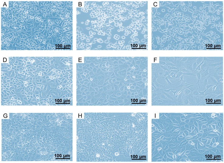
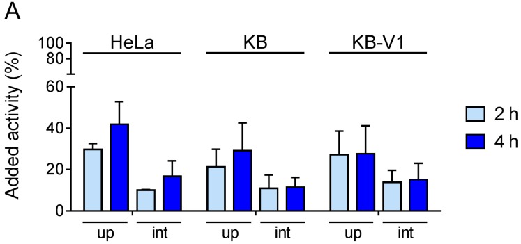
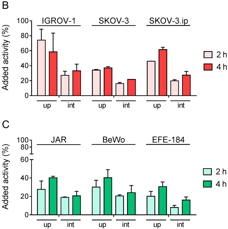
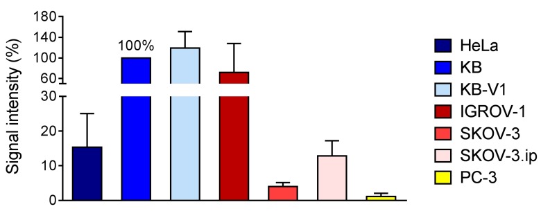
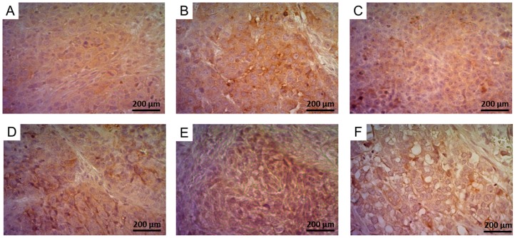
The folate receptor (FR) is expressed in a variety of gynecological cancer types. It has been widely used for tumor targeting with folic acid conjugates of diagnostic and therapeutic probes. The cervical KB tumor cells have evolved as the standard model for preclinical investigations of folate-based (radio) conjugates. In this study, a panel of FR-expressing human cancer cell lines-including cervical (HeLa, KB, KB-V1), ovarian (IGROV-1, SKOV-3, SKOV-3.ip), choriocarcinoma (JAR, BeWo) and endometrial (EFE-184) tumor cells-was investigated in vitro and for their ability to grow as xenografts in mice. FR-expression levels were compared in vitro and in vivo and the cell lines were characterized by determination of the sensitivity towards commonly-used chemotherapeutics and the expression of two additional, relevant tumor markers, HER2 and L1-CAM. It was found that, besides KB cells, its multiresistant KB-V1 subclone as well as the ovarian cancer cell lines, IGROV-1 and SKOV-3.ip, could be used as potentially more relevant preclinical models. They would allow addressing specific questions such as the therapeutic efficacy of FR-targeting agents in tumor (mouse) models of multi-resistance and in mouse models of metastases formation.

Keywords: IGROV-1; KB-V1; SKOV-3; SKOV-3.ip; cervical cancer; choriocarcinoma, KB; endometrial cancer; folate receptor; folic acid; ovarian cancer.


Conflict of interest statement

There is no conflict of interest.

Figures

Figure 1
Figure 1
Microscopic images of (A) HeLa cells; (B) KB cells and (C) KB-V1 cells; (D) IGROV-1 cells; (E) SKOV-3 cells and (F) SKOV-3.ip cells; (G) JAR cells; (H) BeWo cells and (I) EFE-184 cells. Magnification 20×.
Figure 2
Figure 2
Quantification of signal intensity obtained from western blot for FR-expression in cervical, ovarian, choriocarcinoma and endometrial cancer cell lines. The value obtained for KB cells was set as 100% and the percentage of the signals of the other cell lines was calculated for each single western blot (n = 5–6) and expressed as the average ± standard deviation.
Figure 3
Figure 3
Total uptake (up) and internalization (int) of 177Lu-folate in (A) cervical cancer cells; (B) ovarian cancer cells; (C) choriocarcinoma cells and endometrial cancer cell.
Figure 3
Figure 3
Total uptake (up) and internalization (int) of 177Lu-folate in (A) cervical cancer cells; (B) ovarian cancer cells; (C) choriocarcinoma cells and endometrial cancer cell.
Figure 4
Figure 4
Quantification of in vitro autoradiography results in tumor tissues. Values obtained for KB cell line were set as 100% and compared with the other tissues.
Figure 5
Figure 5
Immunohistochemistry results showing FR-expression in (A) HeLa; (B) KB; (C) KB-V1; (D) IGROV-1; (E) SKOV-3 and (F) SKOV-3.ip tumors. Tissue images are shown in magnification 40×.
Figure 6
Figure 6
Tumor-to-kidney ratios of accumulated radioactivity in tumor-bearing mice 4 h and 24 h after injection of the radiofolate. The tumor-to-kidney ratios of all groups of mice bearing FR-positive tumor types (KB, KB-V1 or IGROV-1, respectively) were significantly different (p < 0.05) than the tumor-to-kidney ratio in PC-3 tumor-bearing mice. An exception was the tumor-to-kidney ratio of SKOV-3.ip tumor-bearing mice which was significantly different (p < 0.05) from the ratios in PC-3 tumor-bearing mice only at 4 h p.i. but not (p > 0.05) at 24 h p.i. of the radiofolate.

Similar articles

Cited by

References

    1. Garin-Chesa P., Campbell I., Saigo P.E., Lewis J.L., Old L.J., Rettig W.J. Trophoblast and ovarian cancer antigen LK26—Sensitivity and specificity in immunopathology and molecular identification as a folate-binding protein. Am. J. Pathol. 1993;142:557–567. - PMC - PubMed
    1. Parker N., Turk M.J., Westrick E., Lewis J.D., Low P.S., Leamon C.P. Folate receptor expression in carcinomas and normal tissues determined by a quantitative radioligand binding assay. Anal. Biochem. 2005;338:284–293. doi: 10.1016/j.ab.2004.12.026. - DOI - PubMed
    1. Low P.S., Kularatne S.A. Folate-targeted therapeutic and imaging agents for cancer. Curr. Opin. Chem. Biol. 2009;13:256–262. doi: 10.1016/j.cbpa.2009.03.022. - DOI - PubMed
    1. Holm J., Hansen S.I., Hoiermadsen M., Bostad L. A high-affinity folate binding-protein in proximal tubule cells of human kidney. Kidney Int. 1992;41:50–55. doi: 10.1038/ki.1992.7. - DOI - PubMed
    1. Birn H., Spiegelstein O., Christensen E.I., Finnell R.H. Renal tubular reabsorption of folate mediated by folate binding protein 1. J. Am. Soc. Nephrol. 2005;16:608–615. doi: 10.1681/ASN.2004080711. - DOI - PubMed