. 2017 Aug 15;12(8):e0181350.
doi: 10.1371/journal.pone.0181350. eCollection 2017.

Increased signaling by the autism-related Engrailed-2 protein enhances dendritic branching and spine density, alters synaptic structural matching, and exaggerates protein synthesis

Affiliations

Increased signaling by the autism-related Engrailed-2 protein enhances dendritic branching and spine density, alters synaptic structural matching, and exaggerates protein synthesis

Asma Soltani et al. PLoS One. .

Abstract

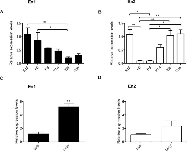
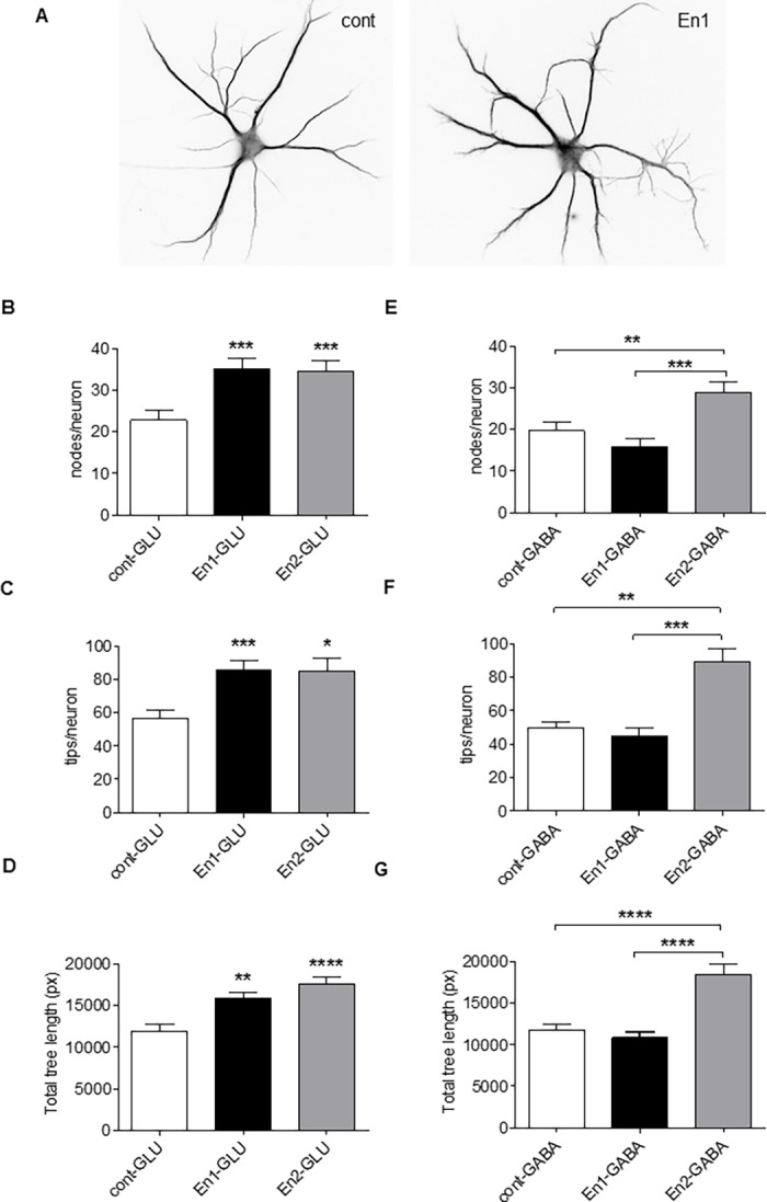
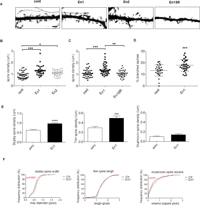
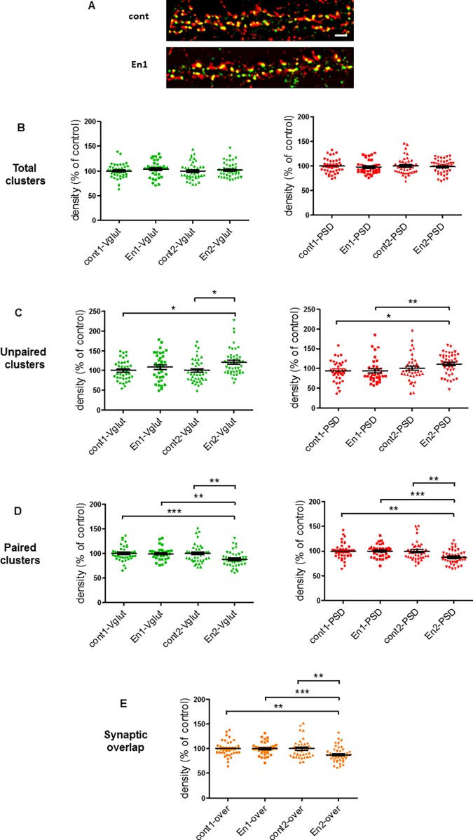
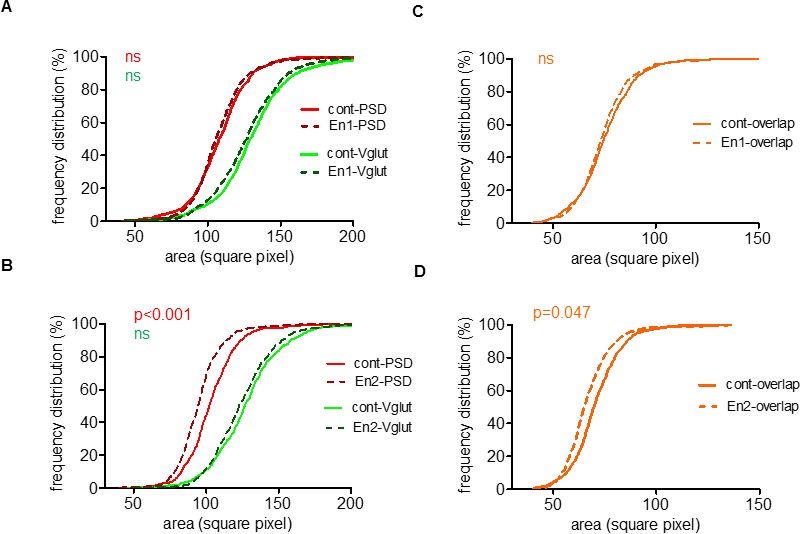
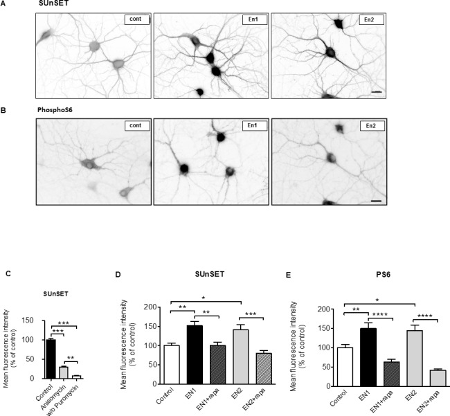
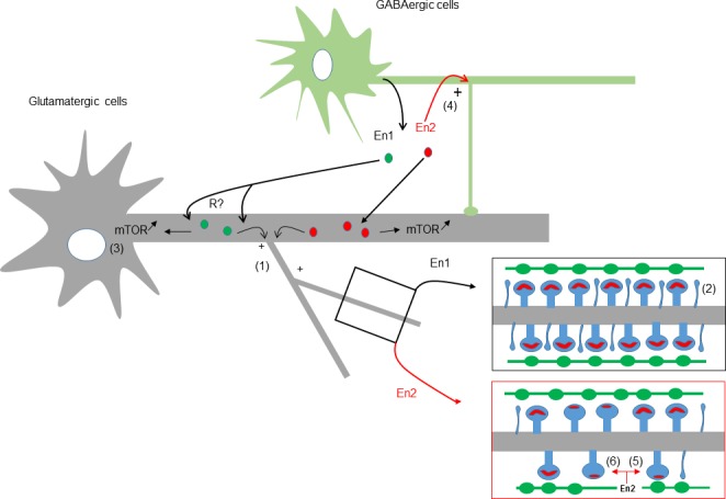
Engrailed 1 (En1) and 2 (En2) code for closely related homeoproteins acting as transcription factors and as signaling molecules that contribute to midbrain and hindbrain patterning, to development and maintenance of monoaminergic pathways, and to retinotectal wiring. En2 has been suggested to be an autism susceptibility gene and individuals with autism display an overexpression of this homeogene but the mechanisms remain unclear. We addressed in the present study the effect of exogenously added En2 on the morphology of hippocampal cells that normally express only low levels of Engrailed proteins. By means of RT-qPCR, we confirmed that En1 and En2 were expressed at low levels in hippocampus and hippocampal neurons, and observed a pronounced decrease in En2 expression at birth and during the first postnatal week, a period characterized by intense synaptogenesis. To address a putative effect of Engrailed in dendritogenesis or synaptogenesis, we added recombinant En1 or En2 proteins to hippocampal cell cultures. Both En1 and En2 treatment increased the complexity of the dendritic tree of glutamatergic neurons, but only En2 increased that of GABAergic cells. En1 increased the density of dendritic spines both in vitro and in vivo. En2 had similar but less pronounced effect on spine density. The number of mature synapses remained unchanged upon En1 treatment but was reduced by En2 treatment, as well as the area of post-synaptic densities. Finally, both En1 and En2 elevated mTORC1 activity and protein synthesis in hippocampal cells, suggesting that some effects of Engrailed proteins may require mRNA translation. Our results indicate that Engrailed proteins can play, even at low concentrations, an active role in the morphogenesis of hippocampal cells. Further, they emphasize the over-regulation of GABA cell morphology and the vulnerability of excitatory synapses in a pathological context of En2 overexpression.


Conflict of interest statement

Competing Interests: The authors have declared that no competing interests exist.

Figures

Fig 1
Fig 1. Expression of Engrailed 1 and 2 in the hippocampus.
(A-D) RT-qPCR using RNAs extracted from the hippocampus at various embryonic (E) and postnatal days/weeks (P, W) (A, B), or from hippocampal cells at different day in vitro (Div) (C, D). Primers used (primers #1, see Material & Methods) are specific for En1 (A, C) or for En2 (B, D). Normalized values are the mean +/- s.e.m. from a minimum of n = 4 independent experiments for each developmental age and time. Significance of the differences were assessed with a Kruskal-Wallis test, in A (***, p = 0.0006) and B (****, p<0.0001), followed by a Dunn's Multiple Comparison Test. *, p<0.05; **, p<0.01. Detailed P-values are given in supplementary information (S1 Appendix).
Fig 2
Fig 2. Exogenous Engrailed increases dendritic complexity.
(A) Div9 hippocampal cells not-treated (A, left) or daily treated with 150ng/ml of purified En for 6 days (div4-div9, A, right) and immunolabelled with MAP-2. Gabaergic cells were identified by immunolabelling with an anti-GAD67 antibody. (B-G) quantification of dendritic nodes (B, E) dendritic tips (C, F), and dendritic length (D, G) per non-treated pyramidal cells (cont) and pyramidal cells treated with either En1 or En2 (B-D), and per non-treated GABA cells (cont) and GABA cells treated with either En1 or En2 (E-G). Values are the mean +/- s.e.m. of measures from a total of 40–45 pyramidal cells and from a total of 30–40 GABA cells/condition from 3 (En1) and 2 (En2) independent experiments. Significance of the differences were assessed with a Kruskal-Wallis test, in B, D, F, G (****, p< 0.0001), C (***, p = 0.0006), and in E (***, p = 0.0002), followed by a Dunn's Multiple Comparison Test. *, p<0.05; **, p<0.01; ***, p<0.001; ****, p<0.0001. Detailed P-values are given in supplementary information. Bar in A, 35μm.
Fig 3
Fig 3. Engrailed increases immature spine density in vitro.
(A-E) Hippocampal cells were transfected with pEGFP, daily treated with 150ng/ml of Engrailed or En1SR, a membrane non-permeant mutated construct, from div15 to div18 and imaged at div19. (B) Quantification of dendritic spine density after treatment with either En1 or En2, or (C) after treatment with either En1 or En1SR. Significance of the differences were assessed with a Kruskal-Wallis test in B (****, p< 0.0001), and C (***, p = 0.0001), followed by a Dunn's Multiple Comparison Test. *, p<0.05; **, p<0.01; ***, p<0.001. Values are the mean +/- s.e.m. of measures from 3 independent experiments with an average of 39 pyramidal cells per condition. (D) Quantification of branched spines after treatment with En1. ***, p = 0.0004 (t-test). (E,F) Morphometric analysis of dendritic spines under control conditions or after En1 treatments. (E) Spines were categorized using Imaris software as described in Methods. En1 increased the density of “stubby”, ****, p<0.0001 and “thin” spines, ***, p = 0.0002 (t-test) but not the one of “mushroom” spines, p = 0.1874 (Mann-Whitney test). (F) En1 increases the maximum diameter of stubby spines (KS test, D = 0.159, p<0.0001), does not increase thin spine length (KS test, D = 0.06, p = 0.054), and slightly increases mushroom spine volume (KS test, D = 0.119, p = 0.01). Values in D-F are the mean +/- s.e.m. of measures from 2 independent experiments each cumulating an average of 30 pyramidal cells per condition. Bar in A, 5μm.
Fig 4
Fig 4. En2 reduces synaptic pairing.
(A) Confocal images of hippocampal cell dendrites double stained for PSD95 and vGlut1 without treatment (cont) or after daily treatment (div15-div18) of hippocampal cells with 150ng/ml of either En1 or En2. (B-E) Quantification of vGlut1 and PSD95 cluster densities (in green and red, respectively) in cells treated as in A (per μm2). Shown are analysis for total clusters (B), single clusters (i.e. vGlut1 and PSD95 not paired with each other, C), paired clusters (i.e. vGlut1 and PSD95 puncta whose area overlapped more than 0.25 μm2 as defined in S1 Appendix, D) and overlap (yellow clusters, i.e. immunofluorescent overlap resulting from pairing of vGlut1 and PSD95, shown in brown in E). Values are the mean +/- s.e.m. of 44 measures from 3 independent experiments. Kruskal-Wallis test in C (**, p = 0.0099), and D, E (***, p = 0.0001), followed by a Dunn's Multiple Comparison Test. *, p<0.05; **, p<0.01; ***, p<0.001. Detailed P-values are given in supplementary information. Bar in A, 5μm.
Fig 5
Fig 5. Engrailed-2 reduces synaptic cluster area.
(A-D) Frequency distribution of the area of vGlut1 (green) and PSD95 (red) (A, B) and vGlut1/PSD95 overlap (i.e. synaptic overlap, brown) (C, D) clusters analyzed in Fig 4. Kolmogorov-Smirnov test on two samples (http://www.physics.csbsju.edu/stats/KS-test.html): A, cont-PSD vs En1-PSD (D = 0.072, p = 0.178), cont-Vglut vs En1-Vglut (D = 0.084, p = 0.055); B, cont-PSD vs En2-PSD (D = 0.183, ***p<0.0001), cont-Vglut vs En2-Vglut (D = 0.055, p = 0.568); C, cont-overlap vs En1-overlap (D = 0.074, p = 0.208); D, cont-overlap vs En2-overlap, (D = 0.112, *, p = 0.047).
Fig 6
Fig 6. Engrailed increases mTORC1 activity and protein synthesis in hippocampal cells.
(A) De novo protein synthesis in hippocampal neurons visualized with SUnSET. DIV18-20 neurons were analyzed without treatment (cont) or after a 1-hr treatment with either En1 or En2. Shown are inverted grayscale images. (B) Phosphorylation of S6 (Ser240/244) assayed by immunofluorescence in untreated hippocampal neurons (cont) or in neurons treated as in A. (C) The SunSET signal was strictly dependent on the presence of puromycin and strongly suppressed by anisomycin indicating that it was reporting de novo protein synthesis. (D) Quantification of SUnSET images. (E) Quantification of phospho-S6 images. Values in D and E are the mean (+/- s.e.m.) fluorescence intensity in neurons from N = 19–22 images from 2 independent experiments. D and E, One-way ANOVA (****, p< 0.0001) followed by a Tukey’s Multiple Comparison Test. C, Kruskal-Wallis test (**** p< 0.0001) followed by Dunn's Multiple Comparison Test. *, p<0.05; **, p<0.01; ***, p<0.001; ****, p<0.0001. Detailed P-values are given in supplementary information. Bar in A and B, 20μm.
Fig 7
Fig 7. Modelling of the effects of En1 and En2 on hippocampal cells.
Coarse hypothetical scheme of the Engrailed signaling pathway in the normal and autistic brain. Engrailed proteins are known to be mainly expressed by GABA neuronal cells [23], and to signal between cells upon secretion (reviewed in [10]). In normal conditions (black lines), low levels of endogenous secreted En1 (from GABA cell for instance) could participate to dendritogenesis (1), spinogenesis (2) (insert), and mTOR signaling (3) in neighbor glutamatergic neurons. In a pathological context, such as autism (red lines), a rise of En2 expression and secretion would generate additional effects, i.e. the increase in dendritic complexity of GABA cells (4), a decrease of clustered PSD95 (5), and the mispairing of glutamatergic synapses (6) (insert). Engrailed proteins require cellular internalization to be active, but functional priming at the cell membrane through an uncharacterized protein “R” is not excluded.

References

    1. Abrahams BS, Geschwind DH. Advances in autism genetics: on the threshold of a new neurobiology. Nat Rev Genet. 2008; 9: 341–355. doi: 10.1038/nrg2346 - DOI - PMC - PubMed
    1. LeBlanc JJ, and Fagiolini M. Autism: A « Critical Period » Disorder? Neural Plast. 2011; 2011: 1–17. - PMC - PubMed
    1. Penzes P, Cahill ME, Jones KA, VanLeeuwen J-E, Woolfrey KM. Dendritic spine pathology in neuropsychiatric disorders. Nat Neurosci. 2011; 14: 285–293. doi: 10.1038/nn.2741 - DOI - PMC - PubMed
    1. Bourgeron T. A synaptic trek to autism. Curr Opin Neurobiol. 2009; 19: 231–234. doi: 10.1016/j.conb.2009.06.003 - DOI - PubMed
    1. Ebert DH, and Greenberg ME. Activity-dependent neuronal signalling and autism spectrum disorder. Nature 2013; 493: 327–337. doi: 10.1038/nature11860 - DOI - PMC - PubMed

MeSH terms

LinkOut - more resources