. 2017 Aug 1;58(10):4355–4365.
doi: 10.1167/iovs.17-22016.

Viral Retinopathy in Experimental Models of Zika Infection

Affiliations

Viral Retinopathy in Experimental Models of Zika Infection

Zhenyang Zhao et al. Invest Ophthalmol Vis Sci. .

Erratum in

  • Erratum.
    [No authors listed] [No authors listed] Invest Ophthalmol Vis Sci. 2017 Sep 1;58(11):4799. doi: 10.1167/iovs.17-22948a. Invest Ophthalmol Vis Sci. 2017. PMID: 28973336 Free PMC article. No abstract available.

Abstract

Purpose: Emerging evidence has shown that both congenital and adult Zika virus (ZIKV) infection can cause eye diseases. The goals of the current study were to explore mechanisms and pathophysiology of ZIKV-induced eye defects.

Methods: Wild-type or A129 interferon type I receptor-deficient mice were infected by either FSS13025 or Mex1-7 strain of ZIKV. Retinal histopathology was measured at different time points after infection. The presence of viral RNA and protein in the retina was determined by in situ hybridization and immunofluorescence staining, respectively. Growth curves of ZIKV in permissive retinal cells were assessed in cultured retinal pigment epithelial (RPE) and Müller glial cells.

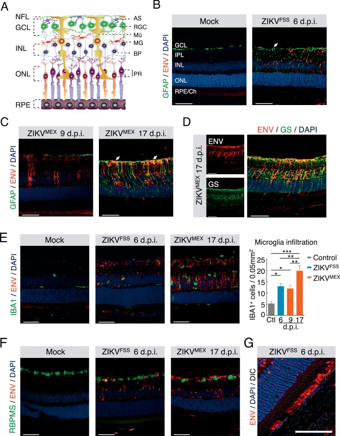
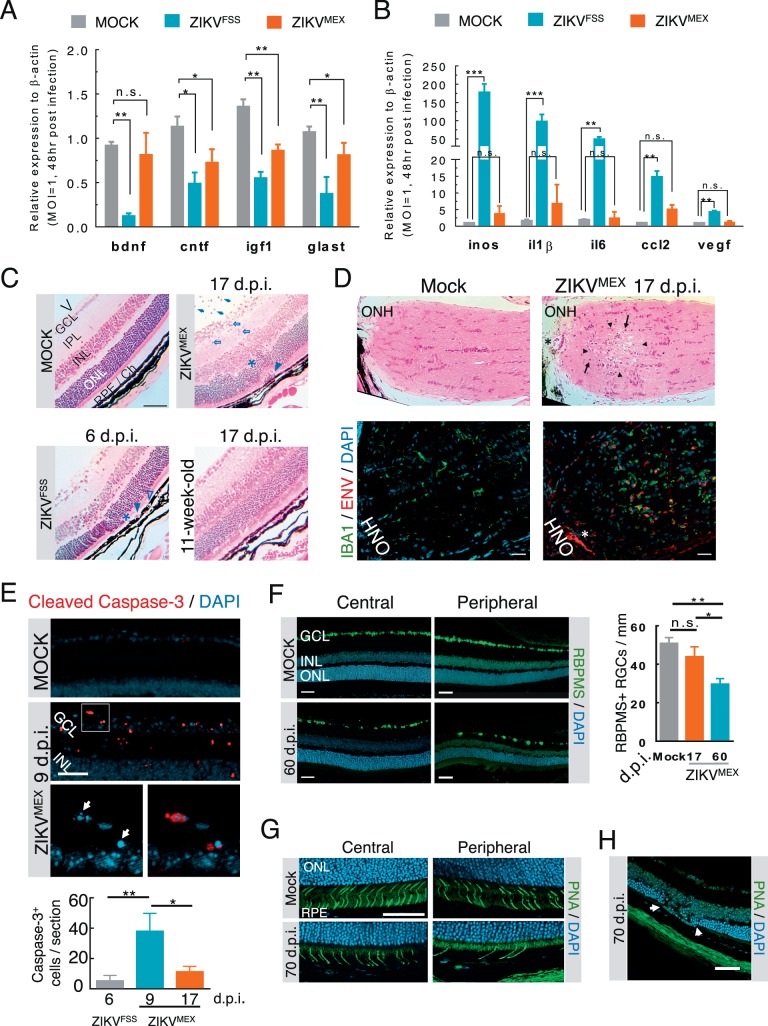
Results: ZIKV-infected mice developed a spectrum of ocular pathologies that affected multiple layers of the retina. A primary target of ZIKV in the eye was Müller glial cells, which displayed decreased neurotrophic function and increased expression of proinflammatory cytokines after infection. ZIKV also infected RPE; and both the RPE and Müller cells expressed viral entry receptors TYRO3 and AXL. Retinitis, focal retinal degeneration, and ganglion cell loss were observed after the clearance of viral particles.

Conclusions: Our data suggest that ZIKV can infect infant eyes with immature blood-retinal barrier and cause structural damages to the retina. The ocular findings in microcephalic infants may not be solely caused by ZIKV-induced impairment of neurodevelopment.


Figures

Figure 1
Figure 1
ZIKV infection of A129 and wild-type mice at weaning. (A, B, D) Immunostaining of viral ENV at indicated times after inoculation by either the ZIKVFSS or ZIKVMEX in A129 mice. Arrows, retinal vessels; open arrows, choroidal vessels; asterisks, RPE infection. IR, iris; CB, ciliary body; GCL, ganglion cell layer; IPL, inner plexiform layer; INL, inner nuclear layer; ONL, outer nuclear layer; CH, choroid. (C) Viral titer from eye and serum of the same animal. Each data point on plot C represents one infected animal. (E) ENV staining of retina from wild-type mice of indicated postnatal (P) age infected with ZIKVFSS. Scale bars: lower magnification, 200 μm (B, D); 50 μm (A, C, E).
Figure 2
Figure 2
Cell type specificity of ZIKV infection in the retina. (A) Retinal structure outlined by a schematic illustration. AS, astrocyte; RGC, retinal ganglion cell; Mü, Müller cell; MG, microglia; BP, bipolar cell; PR, photoreceptors. (B–F) Dual immunostaining of ZIKV ENV with cell type–specific markers, including GFAP for astrocytes and activated Müller cells (B, C), GS for total Müller cells (D), IBA1 for microglia (E), and RBPMS for ganglion cells (F). (G) ENV staining in the RPE. Quantification of infiltrated microglia was performed at different time points after infection. Each time point represents at least three infected animals. *P < 0.05; **P < 0.01. 1-way ANOVA and Bonferroni post hoc test. Scale bars: 50 μm.
Figure 3
Figure 3
ZIKV RNA in retinal Müller cells. (A) Dual staining of ZIKV polyprotein (red) and GS (blue) RNA by in situ hybridization, in retina of a 3-week-old A129 mice infected by ZIKVMEX at 17 dpi. Fluorescent images of the boxed area were enlarged to illustrate the colocalization. (B) Schematic illustration of magnetic activated cell sorting of Müller cells from ZIKV-infected retina. RT-PCR analyses of ZIKV RNA and Müller cell marker in both isolated Müller cells and flow-through fraction. RNA was isolated from A129 mice infected by ZIKVFSS at 6 dpi. Scale bars: 200 μm (A); inset = 50 μm.
Figure 4
Figure 4
ZIKV infection of cultured retinal cells. (A) Immunostaining of viral ENV or NS1 in cultured human fetal RPE cells at 2 dpi by ZIKVFSS. Bottom: reconstitutions of series of Z-stack images at higher magnification. Blue: nuclei. (B) Immunostaining of primary retinal Müller cells infected with ZIKVFSS. (C) Western blot analyses of expression of viral proteins, AXL and TYRO3 receptors in infected cells. (D) Growth curves of ZIKVFSS or ZIKVMEX in RPE and Müller cells. (E) RT-PCR analyses of mRNA expression of potential viral entry receptors in retinal and RPE tissues from 1-month-old A129 mice (top), or cultured primary mice retinal Müller and microglial cells (bottom). (F) Quantitative RT-PCR analyses of viral RNA in Müller cells transfected with siRNA targeting TYRO3 and/or AXL. Data presented are the average from three separate experiments. Scale bars: 20 μm (A, upper), 5 μm (A, lower); 50 μm (B).
Figure 5
Figure 5
Functional consequences of ZIKV infection of the retina. (A, B) Quantitative RT-PCR analyses of neurotrophic and proinflammatory gene expression in cultured Müller cells infected with either ZIKVFSS or ZIKVMEX (n = 4). (C) H&E-stained retinal histology sections from infected A129 mice. The age of the animals was either 3 or 11 weeks at time of viral inoculation. V, vitreous cavity. Arrows: intravitreal infiltrates; open arrows: retinitis with cell infiltration; asterisks: outer nuclear layer damage; arrowheads: subretinal inflammatory cells; open arrowheads: RPE defects. (D) H&E-stained sections of the optic nerve head (ONH). Immediately neighboring sections were stained for viral ENV and IBA1+ cell infiltration. Arrowheads: necrotic lesion; arrows: mononuclear cell infiltrate; asterisk: same blood vessel on adjacent sections. (E) Representative micrograph of cleaved caspase-3 staining. The boxed area at the top was enlarged to show nuclear condensation and fragmentation (arrows). Caspase-3-positive cells were quantified on six slides from each animal (n = 4 each group). (F) Immunostaining of central and peripheral retinal ganglion cells from mock- or ZIKVMEX-infected mice at 60 dpi. Quantitative data were based on the average number of RBPMS-positive cells on 10 retinal sections (n = 3 each group). (G) Staining of cone cells by peanut agglutinin (PNA) at central and peripheral retina. (H) Focal photoreceptor degeneration. Scale bars: 100 μm (C, D); 50 μm (E–H). Data were analyzed by 1-way ANOVA with Turkey post hoc test. *P < 0.05, **P < 0.001.

References

    1. Schuler-Faccini L, Ribeiro EM, Feitosa IM,et al. Possible association between Zika virus infection and microcephaly -- Brazil, 2015. MMWR Morb Mortal Wkly Rep. 2016; 65: 59– 62. - PubMed
    1. de Paula Freitas B, de Oliveira Dias JR, Prazeres J,et al. Ocular findings in infants with microcephaly associated with presumed Zika virus congenital infection in Salvador, Brazil. JAMA Ophthalmol. 2016; 134: 529– 535. - PMC - PubMed
    1. Ventura CV, Maia M, Travassos SB,et al. Risk factors associated with the ophthalmoscopic findings identified in infants with presumed Zika virus congenital infection. JAMA Ophthalmol. 2016; 134: 912– 918. - PubMed
    1. Furtado JM, Esposito DL, Klein TM, Teixeira-Pinto T, da Fonseca BA. . Uveitis associated with Zika virus infection. N Engl J Med. 2016; 375: 394– 396. - PubMed
    1. Ventura CV, Maia M, Bravo-Filho V, Gois AL, Belfort R Jr.. Zika virus in Brazil and macular atrophy in a child with microcephaly. Lancet. 2016; 387: 228. - PubMed

Publication types

MeSH terms