Review
. 2017 Aug 7;38(30):2333-2342.
doi: 10.1093/eurheartj/ehx343.

Cardiomyocyte renewal in the human heart: insights from the fall-out

Affiliations
Review

Cardiomyocyte renewal in the human heart: insights from the fall-out

Eniko Lázár et al. Eur Heart J. .

Abstract

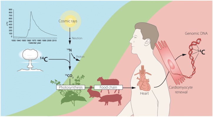
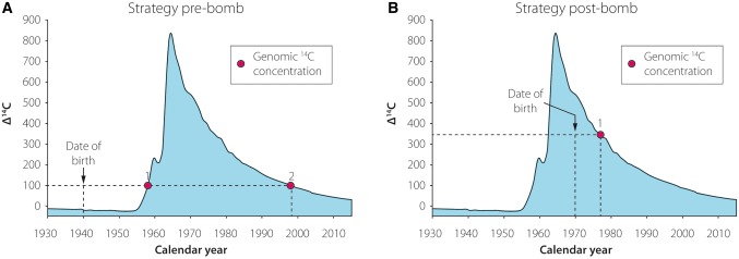
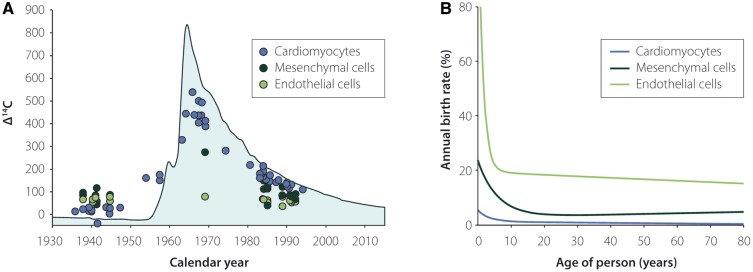
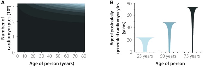
The capacity of the mammalian heart to regenerate cardiomyocytes has been debated over the last decades. However, limitations in existing techniques to track and identify nascent cardiomyocytes have often led to inconsistent results. Radiocarbon (14C) birth dating, in combination with other quantitative strategies, allows to establish the number and age of human cardiomyocytes, making it possible to describe their age distribution and turnover dynamics. Accurate estimates of cardiomyocyte generation in the adult heart can provide the foundation for novel regenerative strategies that aim to stimulate cardiomyocyte renewal in various cardiac pathologies.

Keywords: Cardiomyocyte proliferation; Dynamics of renewal; Retrospective radiocarbon dating.


Figures

Figure 1
Figure 1
Retrospective radiocarbon (14C) birth dating of human cells in the heart. Although 14C is naturally generated in the atmosphere by the action of cosmic rays on nitrogen, above-ground nuclear tests conducted during the Cold War significantly contributed to its environmental level. In the atmosphere, 14C reacts with oxygen to form CO2, which then gets incorporated into plants through photosynthesis. The carbon content of plants is transferred into the human body via the food chain such that the 14C levels in tissues mirror the atmospheric 14C concentrations. With every cell division, 14C is integrated into newly synthetized DNA in amounts proportional to its atmospheric level at any given time. Due to the stability of nuclear DNA, the measured genomic 14C concentration can be used to establish the age of human cardiomyocytes.
Figure 2
Figure 2
Strategy for radiocarbon (14C) birth dating of distinct cell populations. (AB) 14C content in the genomic DNA of the cell population of interest is compared with the recorded atmospheric 14C levels, as shown by the blue graph. The date of birth of the studied individual is indicated by a vertical dashed bar, while the horizontal dashed line corresponds to the 14C value measured in the DNA samples. The age of the sampled cell population can be inferred from the intersection of the horizontal line and the atmospheric 14C curve. (A) Genomic 14C values gained from individuals born before the nuclear bomb tests correspond to two distinct time points (Points 1 and 2). Since the incorporation of environmental 14C might have occurred during the rise and/or the fall of the curve, the precise age of the cell population cannot be determined with certainty based exclusively on these results. (B) However, in individuals born after the nuclear tests, the measured 14C levels unambiguously determine the age of the cell population (Point 1).
Figure 3
Figure 3
Retrospective radiocarbon (14C) birth dating and mathematical modelling of the turnover of different cell types in the heart. (A) Genomic 14C values of cardiomyocytes (blue dots), cardiac endothelial (light green dots), and cardiac mesenchymal cells (dark green dots) from different individuals are plotted against the person’s date of birth. Data points lying on the atmospheric 14C curve indicate no renewal of the cells after birth. The degree of deviation from the atmospheric 14C curve indicates the intensity of DNA synthesis and turnover in the post-natal period, childhood, and adulthood. (B) Applying mathematical modelling, the annual renewal rates of the investigated cell populations can be calculated. While endothelial (light green line) and mesenchymal cells (dark green line) are rapidly exchanged in young adults, with birth rates of 20% per year for the former and 5% per year for the latter, human cardiomyocytes (blue line) are replaced with annual rates of approximately 1% per year at the age of 20, declining to rates of < 0.5% per year in elderly individuals. This figure was adapted from Bergmann et al.
Figure 4
Figure 4
Cell generation and renewal dynamics in human cardiomyocytes. The number and cell turnover dynamics of human cardiomyocytes were established using a mathematical model combining results from radiocarbon (14C) birth dating and stereological strategies. (A) The number of human cardiomyocytes is already set during the perinatal period. Significant exchange of post-natal cardiomyocytes can mainly be observed during the first decades of life. Distinct shades of grey indicate cardiomyocyte subpopulations born during different decades of life. The black area shows cardiomyocytes that were already present at the time of birth. (B) Age distribution of post-natally generated cardiomyocytes in 25-, 50-, and 75-year-old individuals. This figure was adapted from Bergmann et al.
Figure 5
Figure 5
The human heart throughout life. Overview of age-related changes in cardiomyocyte number, multinucleation, polyploidy, and cardiomyocyte renewal in the human heart.

References

    1. Writing Group M, Mozaffarian D, Benjamin EJ, Go AS, Arnett DK, Blaha MJ, Cushman M, Das SR, de Ferranti S, Despres JP, Fullerton HJ, Howard VJ, Huffman MD, Isasi CR, Jimenez MC, Judd SE, Kissela BM, Lichtman JH, Lisabeth LD, Liu S, Mackey RH, Magid DJ, McGuire DK, Mohler ER 3rd, Moy CS, Muntner P, Mussolino ME, Nasir K, Neumar RW, Nichol G, Palaniappan L, Pandey DK, Reeves MJ, Rodriguez CJ, Rosamond W, Sorlie PD, Stein J, Towfighi A, Turan TN, Virani SS, Woo D, Yeh RW, Turner MB; American Heart Association Statistics C, Stroke Statistics S. Heart disease and stroke statistics-2016 update: a report from the American Heart Association. Circulation 2016;133:e38–e60. - PubMed
    1. Townsend N, Nichols M, Scarborough P, Rayner M.. Cardiovascular disease in Europe—epidemiological update 2015. Eur Heart J 2015;36:2696–2705. - PubMed
    1. Ohira T, Iso H.. Cardiovascular disease epidemiology in Asia: an overview. Circ J 2013;77:1646–1652. - PubMed
    1. Task Force on the management of STseamiotESoC, Steg PG, James SK, Atar D, Badano LP, Blomstrom-Lundqvist C, Borger MA, Di Mario C, Dickstein K, Ducrocq G, Fernandez-Aviles F, Gershlick AH, Giannuzzi P, Halvorsen S, Huber K, Juni P, Kastrati A, Knuuti J, Lenzen MJ, Mahaffey KW, Valgimigli M, van 'T Hof A, Widimsky P, Zahger D.. ESC Guidelines for the management of acute myocardial infarction in patients presenting with ST-segment elevation. Eur Heart J 2012;33:2569–2619. - PubMed
    1. Schachinger V, Erbs S, Elsasser A, Haberbosch W, Hambrecht R, Holschermann H, Yu J, Corti R, Mathey DG, Hamm CW, Suselbeck T, Werner N, Haase J, Neuzner J, Germing A, Mark B, Assmus B, Tonn T, Dimmeler S, Zeiher AM.. Improved clinical outcome after intracoronary administration of bone-marrow-derived progenitor cells in acute myocardial infarction: final 1-year results of the REPAIR-AMI trial. Eur Heart J 2006;27:2775–2783. - PubMed