. 2017 Aug 15;6(8):1209-1218.
doi: 10.1242/bio.026070.

Adaptive camouflage: what can be learned from the wetting behaviour of the tropical flat bugs Dysodius lunatus and Dysodiusmagnus

Affiliations

Adaptive camouflage: what can be learned from the wetting behaviour of the tropical flat bugs Dysodius lunatus and Dysodiusmagnus

Florian Hischen et al. Biol Open. .

Abstract

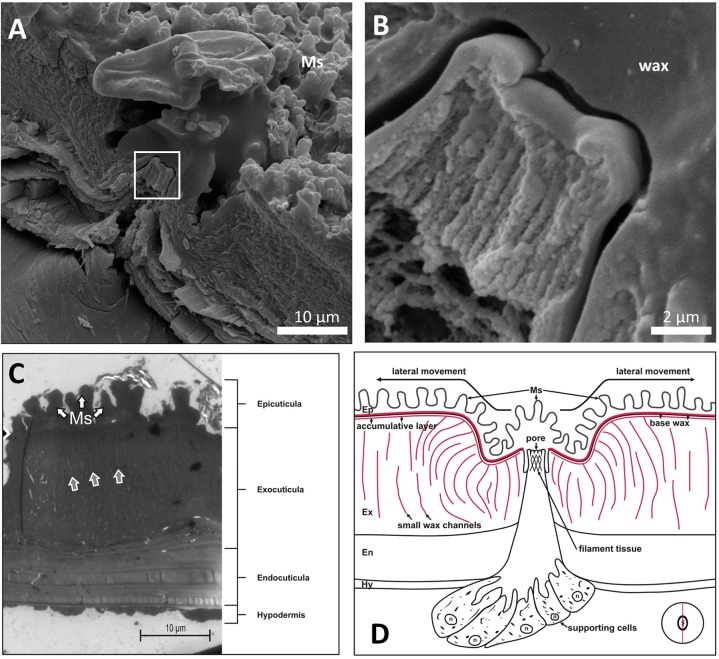
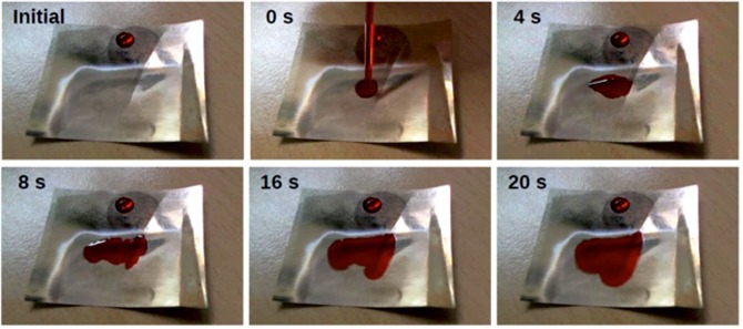
The neotropical flat bug species Dysodius lunatus and Dysodius magnus show a fascinating camouflage principle, as their appearance renders the animal hardly visible on the bark of trees. However, when getting wet due to rain, bark changes its colour and gets darker. In order to keep the camouflage effect, it seems that some Dysodius species benefit from their ability to hold a water film on their cuticle and therefore change their optical properties when also wetted by water. This camouflage behaviour requires the insect to have a hydrophilic surface and passive surface structures which facilitate the liquid spreading. Here we show morphological and chemical characterisations of the surface, especially the cuticular waxes of D. magnus Scanning electron microscopy revealed that the animal is covered with pillar-like microstructures which, in combination with a surprising chemical hydrophilicity of the cuticle waxes, render the bug almost superhydrophilic: water spreads immediately across the surface. We could theoretically model this behaviour assuming the effect of hemi-wicking (a state in which a droplet sits on a rough surface, partwise imbibing the structure around). Additionally the principle was abstracted and a laser-patterned polymer surface, mimicking the structure and contact angle of Dysodius wax, shows exactly the behaviour of the natural role model - immediate spreading of water and the formation of a thin continuous water film changing optical properties of the surface.

Keywords: Bug biomimetics; Camouflage; Laser structuring; Liquid-surface interaction; Microstructures; Reflectance; Wetting.


Conflict of interest statement

Competing interestsThe authors declare no competing or financial interests.

Figures

Fig. 1.
Fig. 1.
Bark bugs and their camouflage on dry and wet bark. A dry, adult specimen of Dysodius lunatus is depicted in (A) while a dry, adult specimen of Dysodius magnus is shown in (B). (C) Clearly, the camouflage of the dry animal on dry bark is excellent (in this case the bark of Sequiadendron giganteum). However, if the bark is wetted, the dry animal can be seen easily (D). (E) If, on the other hand, the animal is wetted itself, the colour changes to darker shades and the camouflage is excellent again. (F) The wet animal could of course easily be seen on dry bark. Images C-F were obtained at the Institute for Biomedical Mechatronics, JKU Linz, to reproduce the findings of Silberglied and Aiello (1980) in a better quality.
Fig. 2.
Fig. 2.
Time course of wetting of Dysodius magnus. A droplet of approximately 5 μl was applied onto the connexival plates. The water spread, finally covering the dorsal abdomen of the bug. Two effects can be observed: (1) the water spreads onto the surface of the conexival plates with local velocities of up to 0.1- 2 mm/s, and (2) the water enters the intersegmental sutures in between the connexival plates (indicated by arrows). In the channels the transport is much faster than the spreading on the surface. However, the water occasionally leaves the channels and spreads on the surface at the corresponding positions.
Fig. 3.
Fig. 3.
Reflective behaviour of bug and bark. (A) Reflectance (in arbitrary units, a.u.) of the surface of a dry and a wetted specimen of Dysodius magnus, measured in the integrated sphere versus the wavelength. (B) Relative scattering reduction (in percent) of bug and bark when comparing dry state versus wet state, plotted against the wavelength.
Fig. 4.
Fig. 4.
SEM-images of the dorsal surface of Dysodius magnus. (A) Overview of the dorsal abdomen. Already at this magnification a microstructure at the connexival plates (co) and the glabrous areas (ga) can be seen. Between the plates, intersegmental sutures occur. (B) Microstructures are naps of about 2 μm diameter, spaced by about 4 μm. In between circular structures can be seen (arrow). (C) After removal of cuticular waxes by hot 10% KOH the microstructures are gone and the circular structures (arrow) become clearly visible. (D) One circular structures at higher magnification can be seen.
Fig. 5.
Fig. 5.
Contact angle measurement of wax surfaces adsorbed on glass. The removed and readsorbed wax from Dysodius magnus (A) exhibits a contact angle of 44° and is therefore hydrophilic, bees’ wax (B) clearly shows a hydrophobic behaviour with a contact angle of 108°. If however the bees’ wax is modified by adding eruccamide (C) or other fatty acids or soaps (not shown), the contact angle can be reduced again. In the example of eruccamide the contact angle dropped to 29°. For comparison the untreated glass (D) exhibits an contact angle of 65°.
Fig. 6.
Fig. 6.
Morphology of the wax secreting glands. (A) Freeze fracture through one of the circular structures depicted in Fig. 4, which are presumably wax glands. In between the superficial microstructures (Ms) a rim with a filamentous tissue in the middle can be seen. (B) Higher magnification of the area marked with the rectangle in A. (C) TEM image of an ultra-thin section through the cuticula in the vicinity of a gland. A typical layered structure can be seen [white arrows showing the microstructure (Ms), also called naps]. In the exocuticula small channels can be seen (grey arrows). (D) Proposed morphology of the wax glands. The insert shows the cutting direction.
Fig. 7.
Fig. 7.
Surface morphology of PET-foil. The untreated foil (A) was found to be virtually flat. If the PET was treated by a pulsed UV laser (B), individual naps can be induced. These naps are with respect to the surface area amplification very similar to the microstructures observed on Dysodius magnus and D. lunatus, resulting in similar values for the Wenzel roughness (r is approx. 1.4 for the bugs and 1.2 for the PET foil and thus determining the needed contact angles for Cassie-Baxter impregnating with 50.5° and 48.2°, respectively).
Fig. 8.
Fig. 8.
Time series of the behaviour of dyed demineralized water on a PET-foil mimicking the surface topography of Dysodius lunatus and D. magnus. The PET was partially laser-structured. The glossy part (upper, left and right margins) is untreated, while the opaque part in the lower centre is structured by laser processing as shown in Fig. 7B. Initially a drop of dyed water was placed on the unstructured PET. Obviously the droplet sits stable with a contact angle of about 40° (± 3.5, n=10). If dyed water is applied onto the laser-structured part, the water immediately begins to spread with an apparent contact angle below 10°. At the waterfront, a margin with slightly different colour can be observed. We assume that here the water is in between the microstructures. The PET-foil piece has a width and length of 40 mm.
Fig. 9.
Fig. 9.
Reflection coefficient dependent on the angle of incidence. The functions were calculated according to eqn. 6, using values for bees’ wax and water covered bees’ wax as a model. Clearly, the wetted surface has a lower reflection coefficient for all angles of incidence and thus would appear darker.

References

    1. Ångström A. (1925). The Albedo of various surfaces of ground. Geogr. Ann. 7, 323 10.2307/519495 - DOI
    1. Arenholz E., Svorcik V., Kefer T., Heitz J. and Bäuerle D. (1991). Structure formation in UV-laser ablated poly-ethylene-terephthalate (PET). Appl. Phys. A 53, 330-331. 10.1007/BF00357196 - DOI
    1. Blomquist G. and Jackson L. (1979). Chemistry and biochemistry of insect waxes. Prog. Lipid Res. 17, 319-345. 10.1016/0079-6832(79)90011-9 - DOI - PubMed
    1. Bormashenko E. Y. (2013). Wetting of Real Surfaces. Berlin; New York: De Gruyter.
    1. Chandra D. and Yang S. (2011). Dynamics of a droplet imbibing on a rough surface. Langmuir 27, 13401-13405. 10.1021/la202208x - DOI - PubMed

LinkOut - more resources