. 2017 Aug 15;8(4):e01218-17.
doi: 10.1128/mBio.01218-17.

Defining the DNA Binding Site Recognized by the Fission Yeast Zn2Cys6 Transcription Factor Pho7 and Its Role in Phosphate Homeostasis

Affiliations

Defining the DNA Binding Site Recognized by the Fission Yeast Zn2Cys6 Transcription Factor Pho7 and Its Role in Phosphate Homeostasis

Beate Schwer et al. mBio. .

Abstract

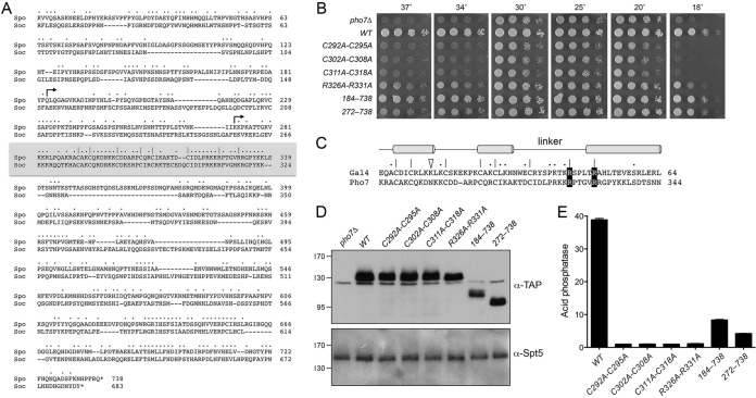
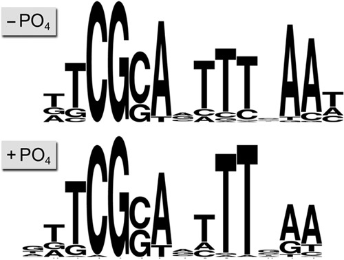
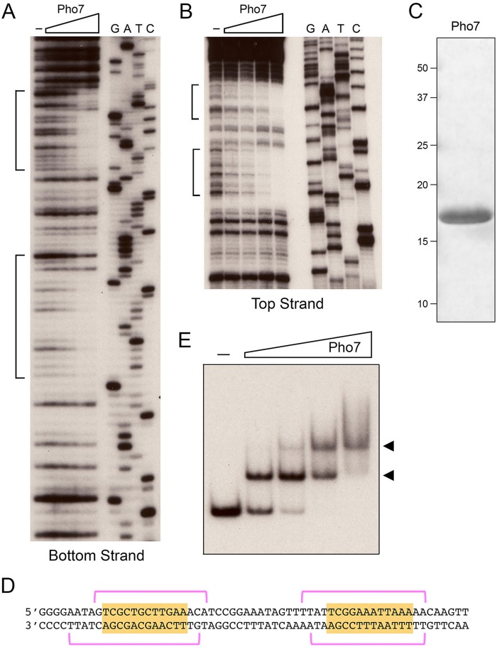
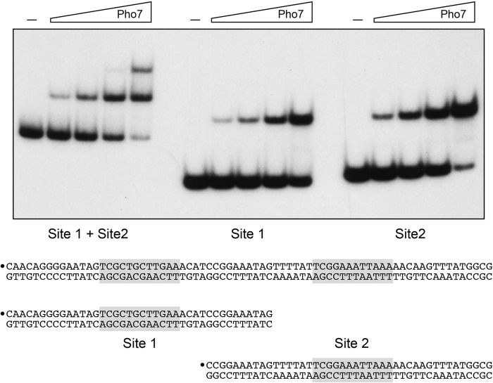
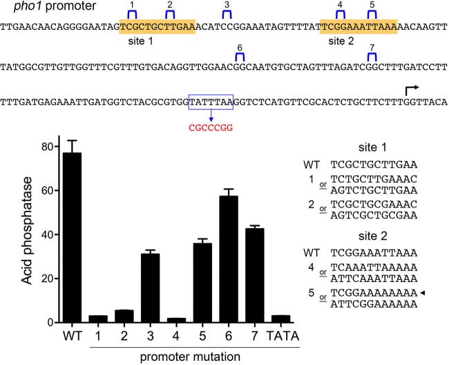
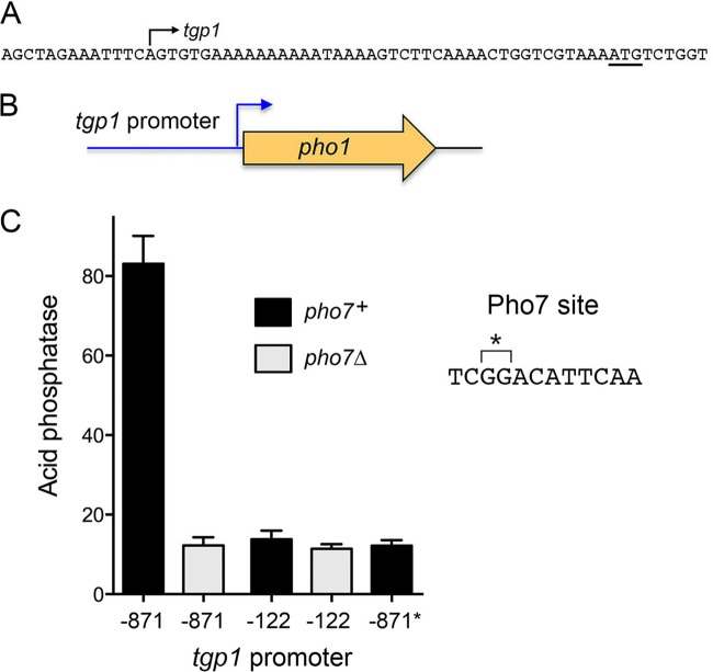
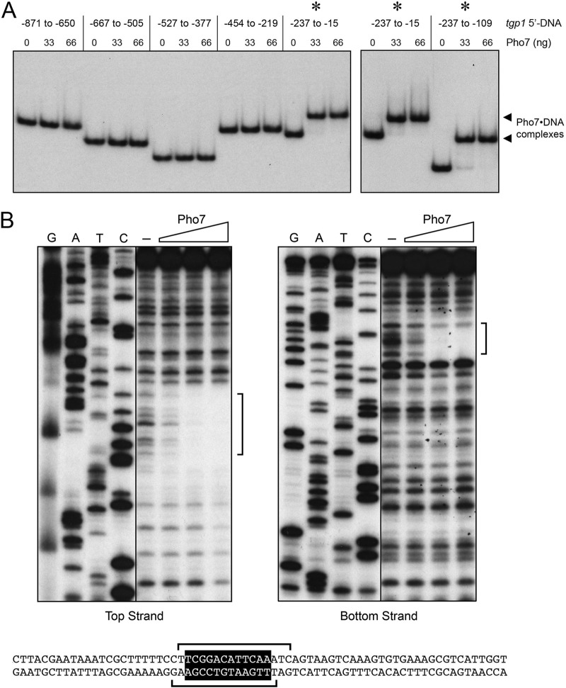
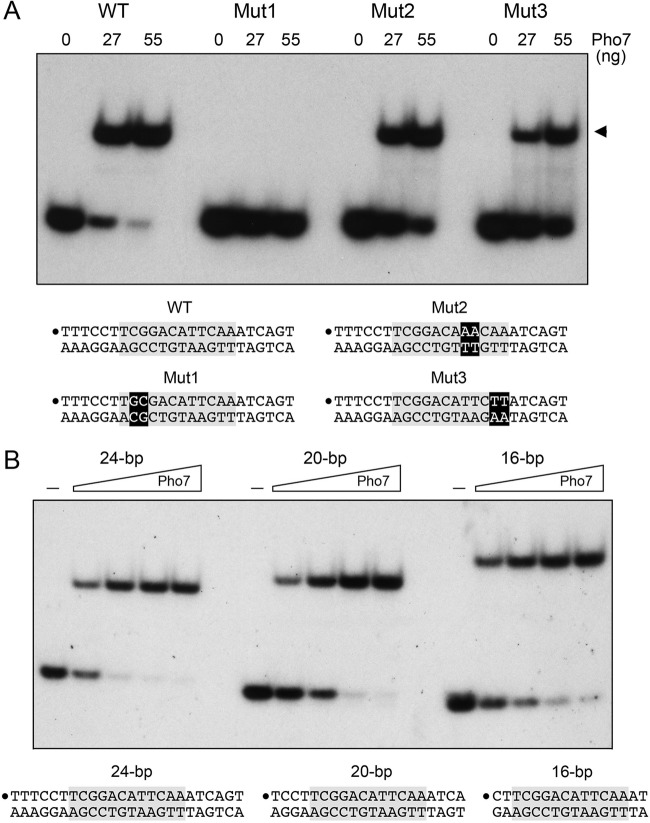
Fission yeast phosphate homeostasis entails transcriptional induction of genes encoding phosphate-mobilizing proteins under conditions of phosphate starvation. Transcription factor Pho7, a member of the Zn2Cys6 family of fungal transcription regulators, is the central player in the starvation response. The DNA binding sites in the promoters of phosphate-responsive genes have not been defined, nor have any structure-function relationships been established for the Pho7 protein. Here we narrow this knowledge gap by (i) delineating an autonomous DNA-binding domain (DBD) within Pho7 that includes the Zn2Cys6 module, (ii) deploying recombinant Pho7 DBD in DNase I footprinting and electrophoretic mobility shift assays (EMSAs) to map the Pho7 recognition sites in the promoters of the phosphate-regulated pho1 and tgp1 genes to a 12-nucleotide sequence motif [5'-TCG(G/C)(A/T)xxTTxAA], (iii) independently identifying the same motif as a Pho7 recognition element via in silico analysis of available genome-wide ChIP-seq data, (iv) affirming that mutations in the two Pho7 recognition sites in the pho1 promoter efface pho1 expression in vivo, and (v) establishing that the zinc-binding cysteines and a pair of conserved arginines in the DBD are essential for Pho7 activity in vivoIMPORTANCE Fungi respond to phosphate starvation by inducing the transcription of a set of phosphate acquisition genes that comprise a phosphate regulon. Pho7, a member of the Zn2Cys6 family of fungal transcription regulators, is the central player in the phosphate starvation response in fission yeast. The present study identifies a 12-nucleotide Pho7 DNA binding motif [5'-TCG(G/C)(A/T)xxTTxAA] in the promoters of phosphate-regulated genes, pinpoints DNA and protein features important for Pho7 binding to DNA, and correlates them with Pho7-dependent gene expression in vivo The results highlight distinctive properties of Pho7 vis-a-vis other fungal zinc binuclear cluster transcription factors as well as the divergent cast of transcription factors deployed for phosphate homeostasis in fission yeast versus budding yeast.

Keywords: DNA binding; fission yeast; phosphate homeostasis; transcriptional regulation.


Figures

FIG 1
FIG 1
Features of Pho7 essential for its activity in vivo. (A) The amino acid sequence of S. pombe Pho7 (Spo) is aligned to the sequence of the homologous protein from S. octosporus (Soc). Gaps in the alignment are denoted by dashes. Positions of side-chain identity/similarity are denoted by dots. The putative Zn2Cys6 DNA-binding domain is highlighted in the gray box. Conserved pairs of cysteines and arginines in the DBD that were targeted for alanine substitution are indicated by |. The margins of the N-terminal deletions are indicated by arrowheads. (B) S. pombe strains deleted of pho7 (pho7Δ) or bearing the pho7 alleles as indicated were spot tested for growth at the temperatures specified. (C) Similarity of the Zn2Cys6 DNA-binding domains of S. cerevisiae Gal4 and S. pombe Pho7. The secondary structure elements of Gal4 are indicated above the amino acid sequence, with α-helices depicted as horizontal cylinders. The signature zinc-binding cysteines are indicated by |. Conserved arginines that in Gal4 contact the DNA phosphodiester backbone are shown in white font on a black background. Other positions of side-chain identity/similarity are denoted by dots. The Gal4 lysine that contacts adjacent guanine nucleobases in the DNA target site is indicated by a triangle. (D) Western blots of whole-cell extracts prepared from S. pombe strains with the indicated pho7 alleles. The blots were probed with antibodies recognizing the TAP tag or the Spt5 protein. (E) Acid phosphatase activity of cells bearing the indicated pho7-TAP alleles assayed 5 h after transfer of logarithmically growing cells to medium lacking phosphate.
FIG 2
FIG 2
In silico identification of a candidate Pho7 recognition element. Motifs were identified via analysis of previously reported Pho7-TAP ChIP-seq data (1) from phosphate-replete and phosphate-starved cells as described in the text.
FIG 3
FIG 3
Recombinant Pho7 Zn2Cys6 DBD binds and footprints the pho1 promoter. (A and B) DNase I footprint analyses of the bottom strand (A) and top strand (B) of the pho1 promoter are shown. Binding reaction mixtures contained 0.75 pmol of 5′ 32P-labeled DNA and either no added Pho7 DBD (lanes –) or increasing amounts of Pho7 DBD (13, 17, 22, 33, and 66 ng in panel A or 20, 27, 40, and 80 ng in panel B). The DNase I digestion products were analyzed by denaturing PAGE in parallel with a series of DNA-directed primer extension reactions (using a 32P-labeled primer identical to the labeled 5′ end of the footprinted DNA) that contained mixtures of standard and chain-terminating nucleotides. (The chain terminator is specified above the lanes.) The margins of the footprints are indicated by brackets. (C) SDS-PAGE analysis of recombinant Pho7 DBD. The Coomassie blue-stained gel is shown. The positions and sizes (in kilodaltons) of marker polypeptides are indicated on the left. (D) Nucleotide sequence of the top and bottom strands of the pho1 promoter from −196 to −136 upstream of the transcription start site. The DNase I footprints on each strand are denoted by magenta brackets. A conserved dodecamer sequence within the footprints is highlighted in gold. (E) Gel shift assay on Pho7 binding to the pho1 promoter. Reaction mixtures (10 µl) containing 1 pmol of a 160-nt 32P-labeled DNA fragment, 0.34 µg poly(dI-dC), and either no protein (–) or increasing amounts of Pho7 DBD (8.3, 17, 33, or 66 ng) were incubated for 10 min at room temperature and then analyzed by native PAGE. An autoradiograph of the gel is shown. The arrowheads on the right indicate positions of DNA-protein complexes.
FIG 4
FIG 4
Pho7 DBD binds independently to two target sites in the pho1 promoter. The sequences of three pho1 promoter DNAs containing both Pho7 binding sites or individual Pho7 binding sites are shown at the bottom. The consensus motifs are shaded in gray, and the 5′ 32P labels on the top strands are indicated by large dots. Reaction mixtures (10 µl) containing 1 pmol 32P-labeled DNA, 0.34 µg poly(dI-dC), and either no protein (–) or increasing amounts of Pho7 DBD (8.3, 17, 33, or 66 ng) were incubated for 10 min at room temperature. The mixtures were analyzed by native PAGE. An autoradiograph of the gel is shown.
FIG 5
FIG 5
Effect of Pho7 site mutations in the pho1 promoter on Pho1 expression in vivo. The sequence of the pho1 promoter from −206 to +7 relative to the pho1 transcription start site (denoted by an arrowhead) is shown at the top. Pho7 binding sites 1 and 2 are shaded in gold, and dinucleotides that were deleted in promoter mutations 1 to 7 are marked by blue brackets above the sequence. A putative TATA element (outlined in blue) was replaced by the sequence shown in red. The sequences at right in the bottom panel show the two possibilities of how the dinucleotide deletions alter the Pho7 binding motifs for site 1 and site 2. The arrowhead indicates the more likely scenario of how promoter mutation 5 alters the Pho7 binding site. Reporter plasmids with wild-type and the mutated pho1 promoters were introduced into a strain with the endogenous pho1 gene deleted. Plasmid-containing cells were grown logarithmically in YES medium and assayed for acid phosphatase activity.
FIG 6
FIG 6
Plasmid reporter of tgp1 promoter function. The DNA sequence from positions −13 to +51 relative to the tgp1 transcription start site (indicated by the arrow) is shown. The tgp1 ATG translation start site is underlined. (B) In the tgp1-pho1 reporter plasmids, the pho1 ORF was fused immediately downstream of a fragment of genomic DNA encompassing the tgp1 transcription start site plus 42 bp of downstream sequences and 871 bp (−871) or 122 bp (−122) of upstream tgp1 sequences. (C) Acid phosphatase activity of S. pombe pho1Δ strains bearing the indicated tgp1-pho1 reporter plasmids during logarithmic growth in YES medium. The strains were either pho7+ or pho7Δ as specified. A dinucleotide deletion (*) in the Pho7 binding site, indicated by a bracket in the sequence at right, was introduced in the context of the −871 tgp1-pho1 reporter (−871*).
FIG 7
FIG 7
Pho7 binding site in the tgp1 promoter. (A) EMSAs using 32P-labeled DNA fragments embracing the indicated sequences of the tgp1 locus upstream of the tgp1 transcription start site (defined as +1). The nucleotide margins of the DNA segments are indicated at the top. Reaction mixtures (10 µl) containing 0.6 pmol labeled DNA, 340 ng poly(dI-dC), and 0, 33, or 66 ng of Pho7 DBD were incubated for 10 min at room temperature. The mixtures were analyzed by native PAGE. An autoradiograph of the gel is shown. The asterisks above the gel denote tgp1 promoter segments that are bound by Pho7; the arrowheads at right mark the positions of the DNA-protein complexes. (B) DNase I footprinting analyses of the top strand (left panel) and bottom strand (right panel) of the tgp1 promoter are shown along with the respective sequencing ladders. Binding reaction mixtures contained 1 pmol of 5′ 32P-labeled DNA and either no added Pho7 DBD (lanes –) or increasing amounts of Pho7 DBD (12.5, 25, or 50 ng). The margins of the footprints are indicated by brackets. The tgp1 sequence from −216 to −148 upstream of the tgp1 transcription start site is shown at the bottom. The Pho7 binding motif is shown in white font on a black background. The brackets denote the DNase I footprints on the top and bottom strands.
FIG 8
FIG 8
Effect of nucleotide substitutions and deletion of flanking nucleotides on Pho7 binding to its DNA target. EMSAs were performed using the indicated DNA duplexes encompassing the Pho7 binding site in the tgp1 promoter. Reaction mixtures (10 µl) containing 32P-labeled DNAs (1 pmol), 340 ng poly(dI-dC), and Pho7 DBD as specified were incubated for 10 min at room temperature and then analyzed by native PAGE. Autoradiographs of the gels are shown. The sequences of the DNAs are indicated below the gels; the Pho7 binding motif is shaded gray, and the 5′ 32P labels on the top strand are indicated by large dots. (A) Effect of nucleotide changes. The two-nucleotide substitutions in Mut1, Mut2, and Mut3 are denoted in white font on a black background. Reaction mixtures contained 0, 27, or 55 ng Pho7 DBD as indicated. (B) Effect of deleting flanking nucleotides. Reaction mixtures contained either no added Pho7 DBD (lanes –) or increasing amounts of Pho7 DBD (8.3, 17, 33, or 66 ng from left to right in the titration series).

References

    1. Carter-O’Connell I, Peel MT, Wykoff DD, O’Shea EK. 2012. Genome-wide characterization of the phosphate starvation response in Schizosaccharomyces pombe. BMC Genomics 13:697. doi: 10.1186/1471-2164-13-697. - DOI - PMC - PubMed
    1. Henry TC, Power JE, Kerwin CL, Mohammed A, Weissman JS, Cameron DM, Wykoff DD. 2011. Systematic screen of Schizosaccharomyces pombe deletion collection uncovers parallel evolution of the phosphate signal pathways in yeasts. Eukaryot Cell 10:198–206. doi: 10.1128/EC.00216-10. - DOI - PMC - PubMed
    1. Schwer B, Sanchez AM, Shuman S. 2015. RNA polymerase II CTD phospho-sites Ser5 and Ser7 govern phosphate homeostasis in fission yeast. RNA 21:1770–1780. doi: 10.1261/rna.052555.115. - DOI - PMC - PubMed
    1. MacPherson S, Larochelle M, Turcotte B. 2006. A fungal family of transcriptional regulators: the zinc cluster proteins. Microbiol Mol Biol Rev 70:583–604. doi: 10.1128/MMBR.00015-06. - DOI - PMC - PubMed
    1. Chatterjee D, Sanchez AM, Goldgur Y, Shuman S, Schwer B. 2016. Transcription of lncRNAprt, clustered prt RNA sites for Mmi1 binding, and RNA polymerase II CTD phospho-sites govern the repression of pho1 gene expression under phosphate-replete conditions in fission yeast. RNA 22:1011–1025. doi: 10.1261/rna.056515.116. - DOI - PMC - PubMed

MeSH terms