

Exercise training prevents skeletal muscle plasma membrane cholesterol accumulation, cortical actin filament loss, and insulin resistance in C57BL/6J mice fed a western-style high-fat diet
- PMID: 28811359
- PMCID: PMC5582260
- DOI: 10.14814/phy2.13363
Exercise training prevents skeletal muscle plasma membrane cholesterol accumulation, cortical actin filament loss, and insulin resistance in C57BL/6J mice fed a western-style high-fat diet
Abstract
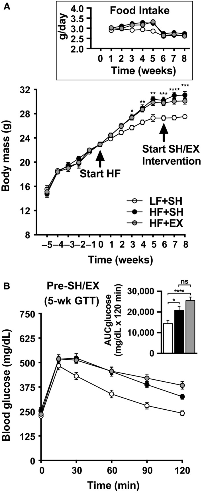
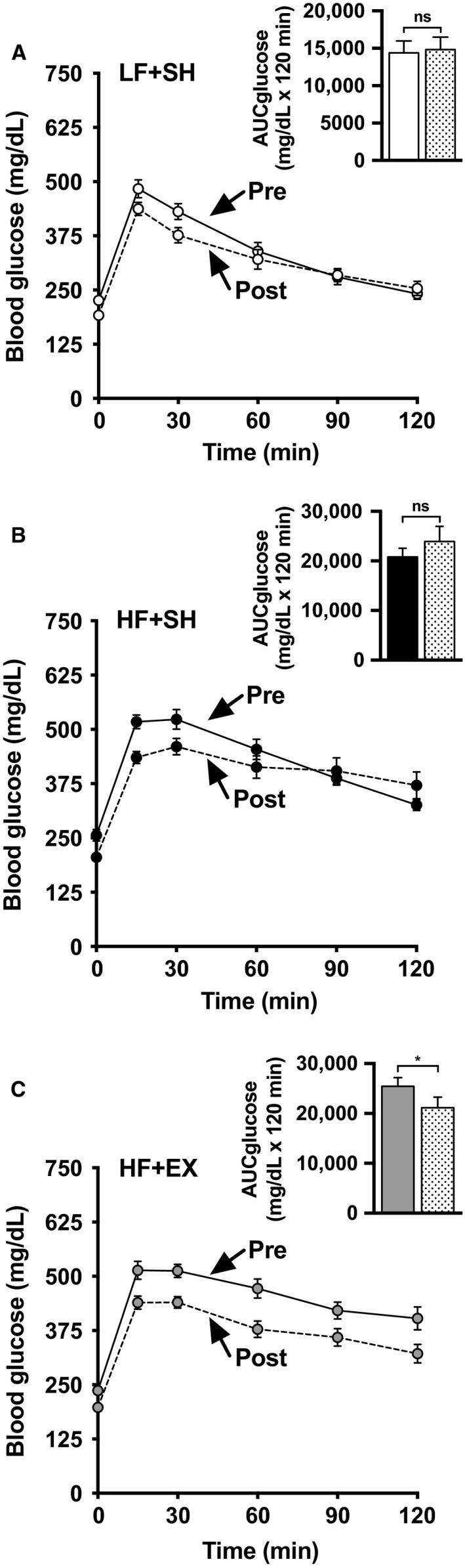
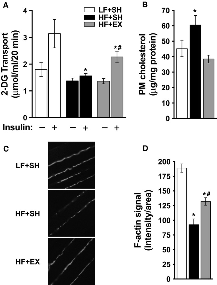
Insulin action and glucose disposal are enhanced by exercise, yet the mechanisms involved remain imperfectly understood. While the causes of skeletal muscle insulin resistance also remain poorly understood, new evidence suggest excess plasma membrane (PM) cholesterol may contribute by damaging the cortical filamentous actin (F-actin) structure essential for GLUT4 glucose transporter redistribution to the PM upon insulin stimulation. Here, we investigated whether PM cholesterol toxicity was mitigated by exercise. Male C57BL/6J mice were placed on low-fat (LF, 10% kCal) or high-fat (HF, 45% kCal) diets for a total of 8 weeks. During the last 3 weeks of this LF/HF diet intervention, all mice were familiarized with a treadmill for 1 week and then either sham-exercised (0 m/min, 10% grade, 50 min) or exercised (13.5 m/min, 10% grade, 50 min) daily for 2 weeks. HF-feeding induced a significant gain in body mass by 3 weeks. Sham or chronic exercise did not affect food consumption, water intake, or body mass gain. Prior to sham and chronic exercise, "pre-intervention" glucose tolerance tests were performed on all animals and demonstrated that HF-fed mice were glucose intolerant. While sham exercise did not affect glucose tolerance in the LF or HF mice, exercised mice showed an improvement in glucose tolerance. Muscle from sham-exercised HF-fed mice showed a significant increase in PM cholesterol, loss of cortical F-actin, and decrease in insulin-stimulated glucose transport compared to sham-exercised LF-fed mice. These HF-fed skeletal muscle membrane/cytoskeletal abnormalities and insulin resistance were improved in exercised mice. These data reveal a new therapeutic aspect of exercise being regulation of skeletal muscle PM cholesterol homeostasis. Further studies on this mechanism of insulin resistance and the benefits of exercise on its prevention are needed.
Keywords: Actin; cholesterol; exercise; insulin.
© 2017 The Authors. Physiological Reports published by Wiley Periodicals, Inc. on behalf of The Physiological Society and the American Physiological Society.
Figures
References
-
- Bhonagiri, P. , Pattar G. R., Habegger K. M., McCarthy A. M., Tackett L., and Elmendorf J. S.. 2011. Evidence coupling increased hexosamine biosynthesis pathway activity to membrane cholesterol toxicity and cortical filamentous actin derangement contributing to cellular insulin resistance. Endocrinology 152:3373–3384. - PMC - PubMed
-
- Brault, M. , Ray J., Gomez Y. H., Mantzoros C. S., and Daskalopoulou S. S.. 2014. Statin treatment and new‐onset diabetes: a review of proposed mechanisms. Metabolism 63:735–745. - PubMed
MeSH terms
Substances
Grants and funding
LinkOut - more resources
Full Text Sources
Other Literature Sources
Medical
Research Materials
Miscellaneous
