. 2017 Aug 15;7(1):8126.
doi: 10.1038/s41598-017-05326-9.

RGD delivery of truncated coagulase to tumor vasculature affords local thrombotic activity to induce infarction of tumors in mice

Affiliations

RGD delivery of truncated coagulase to tumor vasculature affords local thrombotic activity to induce infarction of tumors in mice

Rana Jahanban-Esfahlan et al. Sci Rep. .

Abstract

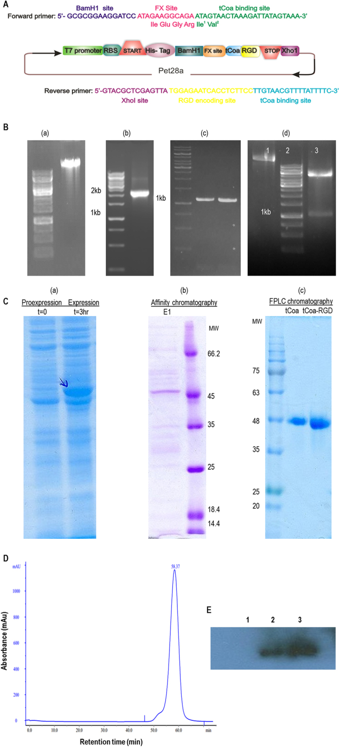
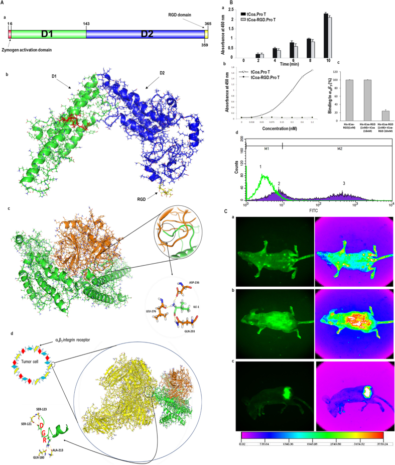
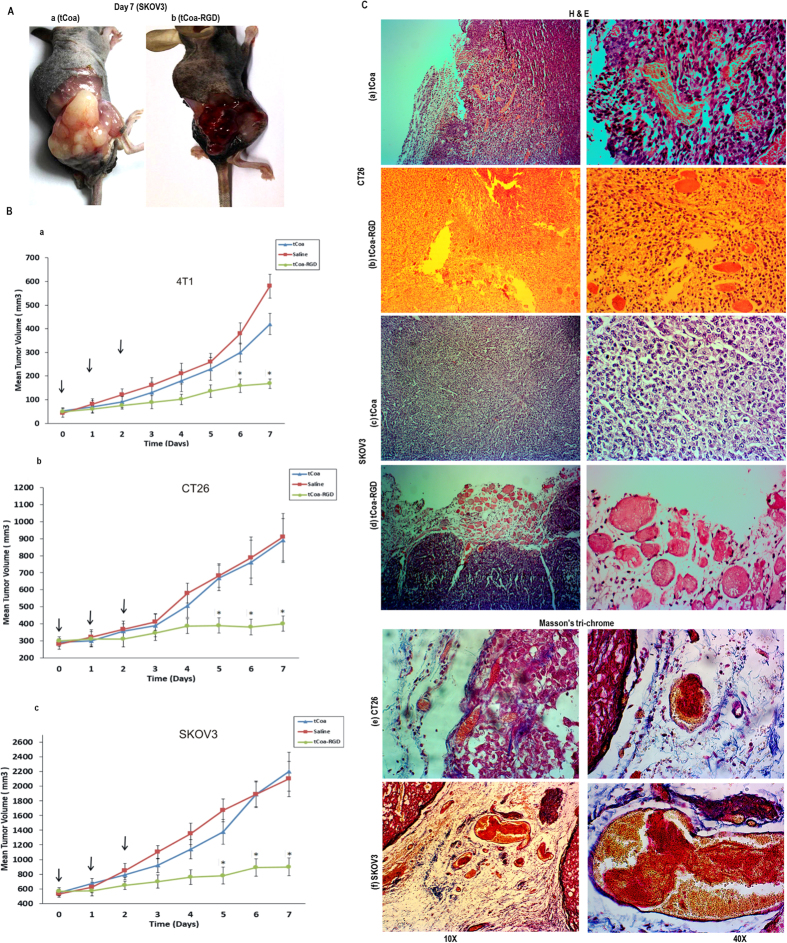
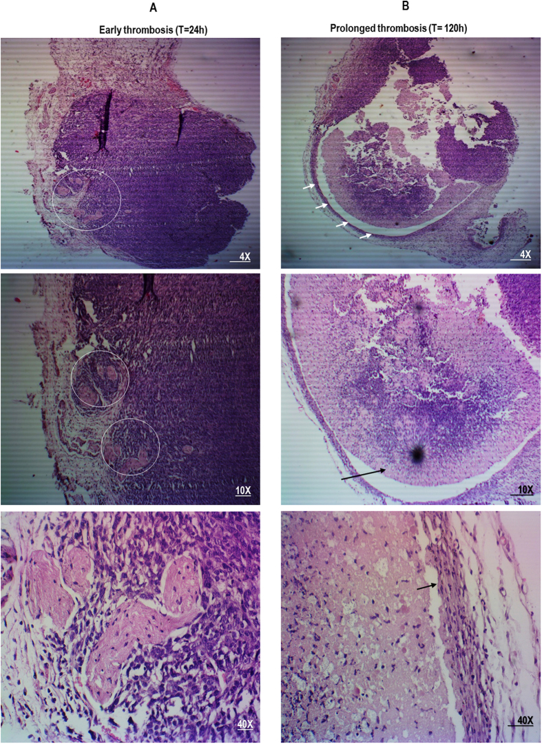
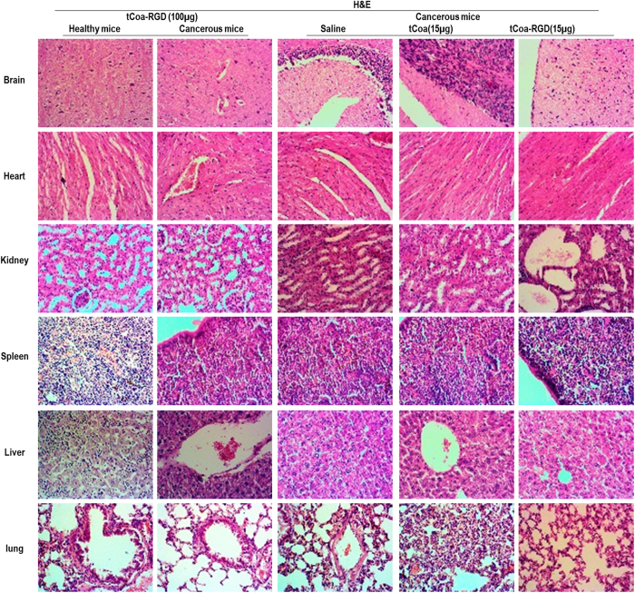
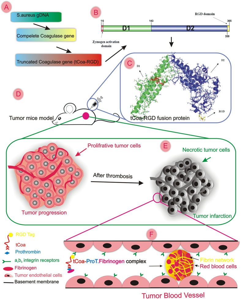
Induction of thrombosis in tumor vasculature represents an appealing strategy for combating cancer. Herein, we combined unique intrinsic coagulation properties of staphylocoagulase with new acquired functional potentials introduced by genetic engineering, to generate a novel bi-functional fusion protein consisting of truncated coagulase (tCoa) bearing an RGD motif on its C-terminus for cancer therapy. We demonstrated that free coagulase failed to elicit any significant thrombotic activity. Conversely, RGD delivery of coagulase retained coagulase activity and afforded favorable interaction of fusion proteins with prothrombin and αvβ3 endothelial cell receptors, as verified by in silico, in vitro, and in vivo experiments. Although free coagulase elicited robust coagulase activity in vitro, only targeted coagulase (tCoa-RGD) was capable of producing extensive thrombosis, and subsequent infarction and massive necrosis of CT26 mouse colon, 4T1 mouse mammary and SKOV3 human ovarian tumors in mice. Additionally, systemic injections of lower doses of tCoa-RGD produced striking tumor growth inhibition of CT26, 4T1 and SKOV3 solid tumors in animals. Altogether, the nontoxic nature, unique shortcut mechanism, minimal effective dose, wide therapeutic window, efficient induction of thrombosis, local effects and susceptibility of human blood to coagulase suggest tCoa-RGD fusion proteins as a novel and promising anticancer therapy for human trials.


Conflict of interest statement

The authors declare that they have no competing interests.

Figures

Figure 1
Figure 1
The design of the coagulase gene construct, isolation, cloning, expression, purification and identification of the corresponding fusion proteins. (A) The design of tCoa-RGD gene constructs for cloning. The addition of Factor X site to the 5′ end of coagulase retained the order of Ile-1-Val-2, which is critical for full coagulase activity. Factor Xa cleaves after the arginine residue in its preferred cleavage site Ile-(Glu or Asp)-Gly-Arg. (B) (a) genomic DNA extraction, (b) amplification of complete coagulase gene (~2 Kbp), (c) amplification of 1.2 Kbp gene constructs, and (d) cloning of tCoa-RGD into the pet 28-a vector: (1) undigested plasmid, (2) size marker, and (3) double digested (XhoI, BamH1) plasmid. (C) SDS-PAGE analysis of tCoa- RGD (a) pro-expression (time = 0) and expression (time = 3 h), (b) after purification with NiNTA chromatography, (c) after purification with FPLC. (D) FPLC analysis of purified tCoa-RGD. The peck corresponds to a ~45 kDa single protein with ~60 min retention time. (E) Western blotting analysis of tCoa-RGD: (1) before protein induction, (2) after protein induction, and (3) after purification, Abbreviations: T = time, E1 = elute1, MW = molecular weight.
Figure 2
Figure 2
Functional studies of tCoa-RGD fusion proteins. (A) Modeling, docking, and MD simulation. (a) Amino acid sequence of tCoa-RGD, its domains, and 3D structure. (b) tCoa-RGD has a helical structure within its two domains. (c) The 3D structure of tCoa-RGD in complex with prothrombin after equilibration in MD simulation. Insertion of tCoa-N-terminal into prothrombin is highlighted by a circle. IVTKDY hexapeptide at the N-terminus of protein interacts strongly with zymogen activation domain of prothrombin in a “molecular sexuality mechanism.” (d) The 3D structure of tCoa-RGD-prothrombin in complex with the extracellular domain of αvβ3 integrins after equilibration in MD simulation. The interaction of RGD domain with integrin residues is highlighted with a circle. (B) Functional studies to determine coagulase activity of the fusion proteins (a), and retention of antibody activity by tCoa-RGD to αvβ3 integrins by ELISA (b, c) and FACS (d). (a) Retention of coagulase activity was determined by the capability of tCoa and tCoa-RGD to convert fibrinogen into fibrin through the conformational activation of ProT. tCoa-RGD presented coagulase activity comparable to that of tCoa at each time point, whereas ProT alone did not potentiate coagulation and there was no detectable absorbance. (b) In vitro binding studies demonstrated that tCoa-RGD specifically bind to immobilized αvβ3 integrins in a concentration dependent manner, with the highest binding at 1.2 nM concentration. (c) In presence of His-tag removed tCoa-RGD but not tCoa, binding of tCoa-RGD to the αvβ3 integrins was significantly inhibited (>70%). (d) FACS analysis verified differential binding of tCoa (peck 1) and tCoa-RGD (peck 2, 3) on endothelial cells in suspension. In this histogram, M1 marker covers the negative cells presenting no/none-specific binding while M2 marker comprises the positive cells. Accordingly, specific binding of tCoa-RGD to the αvβ3 integrins was determined 64.24% (peck 3). None-specific binding for tCoa and tCoa-RGD was measured 98% and 36.05%, respectively. Correspondingly, the measured fluorescence intensity for tCoa-RGD was eight times higher than that of tCoa. (C) Tracing of fluorescently labeled drugs in vivo. tCoa-RGD labeled with FITC injected to (a) healthy mice with no tumor; while (b) tCoa and (c) tCoa-RGD injected to mice bearing SKOV3 ovarian carcinoma xenografts. tCoa-RGD fusion protein but not tCoa showed specific accumulation at the subcutaneously implanted tumor site in C57Bl/6 nude mice.
Figure 3
Figure 3
Efficacy of tCoa-RGD fusion proteins in mice. (A) Representative photographs of mice bearing malignant ovarian cancer (SKOV3) xenografts treated with tCoa (a) or tCoa-RGD (b) at the end of treatment. (B) Tumor growth inhibition studies to demonstrate therapeutic efficacy of tCoa-RGD fusion proteins. (a) 4T1, (b) CT26 and (c) SKOV3 tumor-bearing mice were injected i.v with one of the following: saline, 15 µg tCoa, 15 µg tCoa-RGD. The treatment was repeated 24 hours and 48 hours after the first injection (the arrows). In all three type of solid tumors, a significant tumor growth inhibition was demonstrated for a group of animals that were injected with three consecutive doses of tCoa-RGD, compared to tCoa and saline groups (*P < 0.05). (C), Histological staining of CT26 colon carcinoma and SKOV3 human ovarian carcinoma tumors treated with tCoa and tCoa-RGD coaguligand. Figures a–e show H&E staining of (a,b) CT26 and (c,d) SKOV3, as well as Masson’s trichrome staining of (e) CT26 and (f) SKOV3 tumor sections. (a,c) Histological tumor sections of mice treated with tCoa showed no sign of thrombosis, blood vessels were either intact, containing red blood cells (a), or invisible (c), meanwhile tumor cells appeared vital. (b) Injection of tCoa-RGD resulted in thrombosis of the blood vessels distinguishing with blurred outlines, which was accompanied by disintegration and necrosis of tumoral cells (the white arrows). An example of thrombosed vessel is shown with an arrow and the thrombotic area is highlighted with a circle. (d) Likewise, in SKOV3 tumor xenografts, tCoa-RGD resulted in massive occlusion of the tumor blood vessels, including the rim area. Induction of thrombosis was highlighted by the formation of apparent mesh networks of fibrin (an example of fibrin mesh network is shown with an arrow). Induction of complete thrombosis was affirmed by deposition of fibrin and RBCs in (e) CT26 and (f) SKOV3 tumor histological sections. Red staining represents fibrins while yellow staining indicates the RBCs.
Figure 4
Figure 4
The effect of sustained induction of thrombosis by tCoa-RGD fusion proteins on 4T1 mouse mammary carcinoma tumors. Histological changes are depicted on day one and day five after the first injection. For more clarification, the tumor cross-sections are shown with three different magnifications. (A) Injection of tCoa fusion proteins into animal bearing mouse mammary solid tumors resulted in induction of thrombosis. Thrombotic vessels with packed erythrocytes and deposition of fibrin were apparent in stained tumor sections following injection of tCoa-RGD within 24 hours. Examples of thrombosed vessels are shown with a circle. (B) Induction of long-termed thrombosis by injection of three consecutive doses of tCoa-RGD at 24-hour intervals produced massive necrosis and destruction of tumor cells within the whole tumor region. Note the separation of tumor cells from each other and also complete detachment from the tumor basal membrane (the arrow).
Figure 5
Figure 5
Toxicological studies in mice. This figure shows the H&E staining of organs from the healthy mice and cancerous mice treated with high dose of tCoa-RGD (100 µg). H&E staining of organs from the mice bearing human ovarian tumors treated with saline (200 µl), tCoa (15 µg), and tCoa-RGD (15 µg) is also represented. Injection of therapeutic doses, as well as high doses of the tCoa-RGD fusion protein, resulted in no sign of necrosis or thrombosis in histological analysis of vital organs including brain, heart, kidney, liver, spleen and lung, indicating the high selectivity of tCoa-RGD fusion proteins. Magnification (40x).
Figure 6
Figure 6
tCoa-RGD fusion proteins induce infarction of tumors in mice. This figure illustrates the main steps including design (A), molecular modeling (B), production (C) and the pre-clinical evaluation of the novel fusion proteins (D). It also highlights the unique mechanism of coagulase (E,F). RGD directed targeting of truncated coagulase to tumor neovasculature produces significant thrombosis and subsequent infarction and destruction of the tumor cells (E). Coagulase mediated coagulation is safe and efficient; this process requires the participation of only two partners, prothrombin, and fibrinogen. RGD directed delivery of coagulase to αvβ3 integrin receptors on tumor endothelial cells affords the appropriate spatial localization of bacterial proteins to induce an efficient coagulation within minutes. This figure is drawn by one of our authors, it’s original and there is no need for permission.

References

    1. Jahanban-Esfahlan, R., de la Guardia, M., Ahmadi, D. & Yousefi, B. Modulating tumor hypoxia by nanomedicine for effective cancer therapy. J. Cell. Physiol, doi:10.1002/jcp.25859 (2017). - PubMed
    1. Liu C, et al. Prostate-specific membrane antigen directed selective thrombotic infarction of tumors. Cancer. Res. 2002;62:5470–5475. - PubMed
    1. Bieker R, et al. Infarction of tumor vessels by NGR-peptide-directed targeting of tissue factor: experimental results and first-in-man experience. Blood. 2009;113:5019–5027. doi: 10.1182/blood-2008-04-150318. - DOI - PubMed
    1. Hu P, et al. Comparison of three different targeted tissue factor fusion proteins for inducing tumor vessel thrombosis. Cancer. Res. 2003;63:5046–5053. - PubMed
    1. Huang FY, et al. A fusion protein containing murine vascular endothelial growth factor and tissue factor induces thrombogenesis and suppression of tumor growth in a colon carcinoma model. J. Zhejiang. Univ. Sci. B. 2008;9:602–609. doi: 10.1631/jzus.B0820120. - DOI - PMC - PubMed

Publication types

MeSH terms